. 2025 Aug 3;6(8):e70315.
doi: 10.1002/mco2.70315. eCollection 2025 Aug.

The Biological Characteristics and Mouse Model of Lassa Virus From the First Imported Case in China

Affiliations

The Biological Characteristics and Mouse Model of Lassa Virus From the First Imported Case in China

Yanan Zhou et al. MedComm (2020). .

Abstract

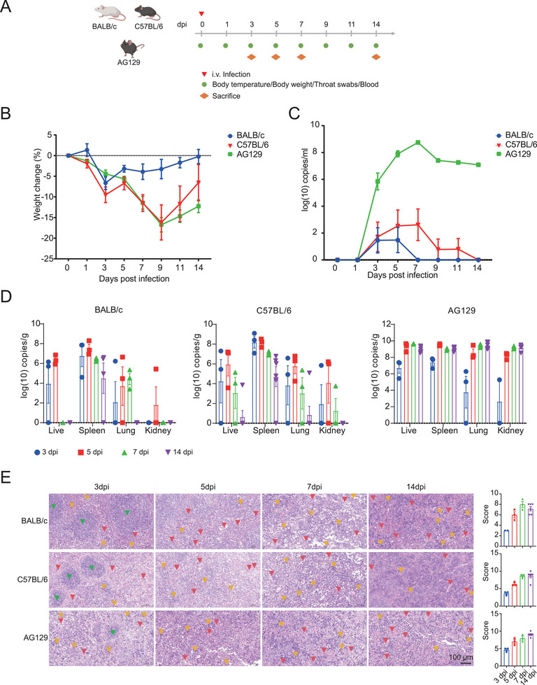
Lassa fever (LF) is a fatal hemorrhagic disease caused by the Lassa virus (LASV), which mainly spreads in Africa. As China's interactions with Africa become more frequent, the risk of LF being imported into China also rises, making the study of LASV increasingly urgent. In this study, the Lineage IV LASV strain was successfully isolated from the first imported case in China. Compared with the LASV genome, the isolated strain may exhibit greater infectivity and interspecies transmission capabilities. We successfully established BALB/c, C57BL/6, and AG129 mouse infection models and found that intranasal inoculation was the most stable infection method. Select the anti-LASV drug LHF-535 for preliminary evaluation, further confirming the stability of the model. In summary, the isolated strain exhibits enhanced transmission capabilities and may spread between mice via the respiratory tract, meriting greater attention and emphasis. This study will bridge the gap in China's independent P4-level pathogen isolation, meet national biosafety and strategic needs, and provide certain support for LASV research.

Keywords: Lassa virus; biological characteristics; mouse infection model; virus isolation.


Conflict of interest statement

The authors declare no conflicts of interest.

Figures

FIGURE 1
FIGURE 1
Isolation and Identification of LASV. (A) LF case situation and isolation process of LASV; (B) Ct values of LASV case samples (n = 1); (C) Ct values of blood and tissue samples from AG129 mice (n = 1) infected with LASV after isolation; (D) Cytopathic effects (CPE) of LASV infection in Vero and Vero E6 cells. Images were captured using a Leica DMiL microscope (10× objective); (E) Transmission electron microscopy observation of Lassa virus particles. Ct values = 0 indicate that it is below the detection limit, and the viral nucleic acid was not detected.
FIGURE 2
FIGURE 2
The LASV strain is shown to be lineage IV, and changes in B cell epitopes in the viral GPC protein. (A) Genome structure of the isolated strain, (B) A bootstrap phylogenetic tree of the isolated strain; Isolated‐L (left), Isolated‐S (right); (C) Amino acid variation of the glycoprotein of the isolated strain.
FIGURE 3
FIGURE 3
Structural simulation of the binding between different LASV strains and receptors. (A) Structural simulation of the binding between the NC strain GPC and mouse α‐DG; (B) Structural simulation of the binding between the NC strain GPC and human α‐DG; (C) Structural simulation of the binding between the SLE strain GPC and mouse α‐DG; (D) Structural simulation of the binding between the SLE strain GPC and human α‐DG; (E) Structural simulation of the binding between the isolated strain GPC and mouse α‐DG; (F) Structural simulation of the binding between the isolated strain GPC and human α‐DG. The color scheme represents deep blue for GPC, sky blue for α‐DG, and red for interaction sites. Iptm indicates interaction score (higher scores denote stronger interactions), ptm represents structural prediction accuracy (higher accuracy indicates more precise predictions), and the sum of these two values reflects the strength of the final receptor‐ligand binding ability.
FIGURE 4
FIGURE 4
Intravenous tail infection method for infecting BALB/c, C57BL/6, and AG129 mice with LASV. (A) Schematic design of the intravenous tail infection of BALB/c, C57BL/6, and AG129 mice. Body temperature, weight, blood, and throat swabs were collected every other day postinfection. Mice were dissected on days 3, 5, 7, and 14 postinfection. (B) Changes in body weight of BALB/c, C57BL/6, and AG129 mice (n = 6) postinfection. (C) Changes in viral load in the blood of BALB/c, C57BL/6, and AG129 mice (n = 6) postinfection. (D) Viral load in liver, spleen, lung, and kidney tissues of BALB/c, C57BL/6, and AG129 mice dissected on days 3, 5, 7 (n = 3), and 14 (n = 6) postinfection. (E) The spleen tissue's pathological sections and pathological scores of BALB/c, C57BL/6, and AG129 mice dissected on Days 3, 5, 7 (n = 3), and 14 (n = 6) postinfection (the red markings indicate the site of hemorrhage, the yellow markings denote areas of inflammatory cell infiltration, and the green markings highlight the germinal centers of the spleen. Splenic hemorrhage and inflammatory cell infiltration are diffuse, with only partial locations marked in the figure).
FIGURE 5
FIGURE 5
Intranasal, intramuscular (i.m.), and intraperitoneal (i.p.) infection of BALB/c, C57BL/6, and AG129 mice with LASV. (A) Schematic design of intranasal, i.m., and i.p. infection of BALB/c, C57BL/6, and AG129 mice. Body temperature, weight, blood, and throat swabs were collected every other day postinfection. Mice were dissected on day 14 postinfection. (B) Changes in body weight of BALB/c, C57BL/6, and AG129 mice (n = 3) postinfection. (C) Changes in viral load in the blood of BALB/c, C57BL/6, and AG129 mice (n = 3) postinfection. (D) Viral load in liver, spleen, lung, and kidney tissues of BALB/c, C57BL/6, and AG129 mice (n = 3) dissected on Day 14 postinfection. (E) The spleen tissue's pathological sections and pathological scores of BALB/c, C57BL/6, and AG129 mice (n = 3) dissected on Day 14 postinfection (the red markings indicate the site of hemorrhage, while the yellow markings denote areas of inflammatory cell infiltration. Splenic hemorrhage and inflammatory cell infiltration are diffuse, with only partial locations marked in the figure).
FIGURE 6
FIGURE 6
LHF‐535 drug treatment of LASV intranasal infection in BALB/c, C57BL/6, and AG129 mice. (A) Schematic design of LHF‐535 drug treatment for LASV infection in BALB/c, C57BL/6, and AG129 mice. Blood and throat swabs were collected daily postinfection. Medication was administered by gavage at a dose of 10 mg/kg daily. Mice were dissected on Day 5 postinfection. (B) Changes in viral load in the blood of BALB/c, C57BL/6, and AG129 mice (n = 5) after LHF‐535 drug treatment. (C) Changes in viral load in the throat swabs of BALB/c, C57BL/6, and AG129 mice (n = 5) after LHF‐535 drug treatment. (D) Changes in viral load after LHF‐535 drug treatment in BALB/c, C57BL/6, and AG129 mice (n = 5) post‐dissection. p < 0.05 was considered significant. The significance levels are indicated as follows: *p < 0.05; **p < 0.01; ***p < 0.001; and ****p < 0.0001.

Similar articles

References

    1. Monath T. P., Newhouse V. F., Kemp G. E., Setzer H. W., and Cacciapuoti A., “Lassa Virus Isolation From Mastomys natalensis Rodents During an Epidemic in Sierra Leone,” Science 185, no. 4147 (1974): 263–265. - PubMed
    1. Olayemi A., Cadar D., Magassouba N., et al., “New Hosts of the Lassa Virus,” Scientific Reports 6 (2016): 25280. - PMC - PubMed
    1. Akpede G. O., Asogun D. A., Okogbenin S. A., et al., “Caseload and Case Fatality of Lassa Fever in Nigeria, 2001–2018: A Specialist Center's Experience and Its Implications,” Frontiers in Public Health 7 (2019): 170. - PMC - PubMed
    1. Buba M. I., Dalhat M. M., Nguku P. M., et al., “Mortality Among Confirmed Lassa Fever Cases During the 2015–2016 Outbreak in Nigeria,” American Journal of Public Health 108, no. 2 (2018): 262–264. - PMC - PubMed
    1. Asogun D. A., Günther S., Akpede G. O., Ihekweazu C., and Zumla A., “Lassa Fever: Epidemiology, Clinical Features, Diagnosis, Management and Prevention,” Infectious Disease Clinics of North America 33, no. 4 (2019): 933–951. - PubMed

LinkOut - more resources