Review
. 2025 Jul 26:34:102139.
doi: 10.1016/j.mtbio.2025.102139. eCollection 2025 Oct.

Bioactive LDH nanoplatforms for cancer therapy: Advances in modulating programmed cell death

Affiliations
Review

Bioactive LDH nanoplatforms for cancer therapy: Advances in modulating programmed cell death

Li Wang et al. Mater Today Bio. .

Abstract

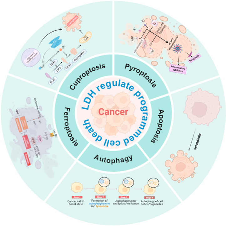
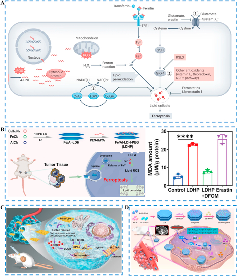
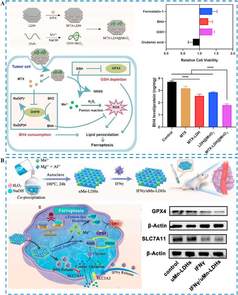
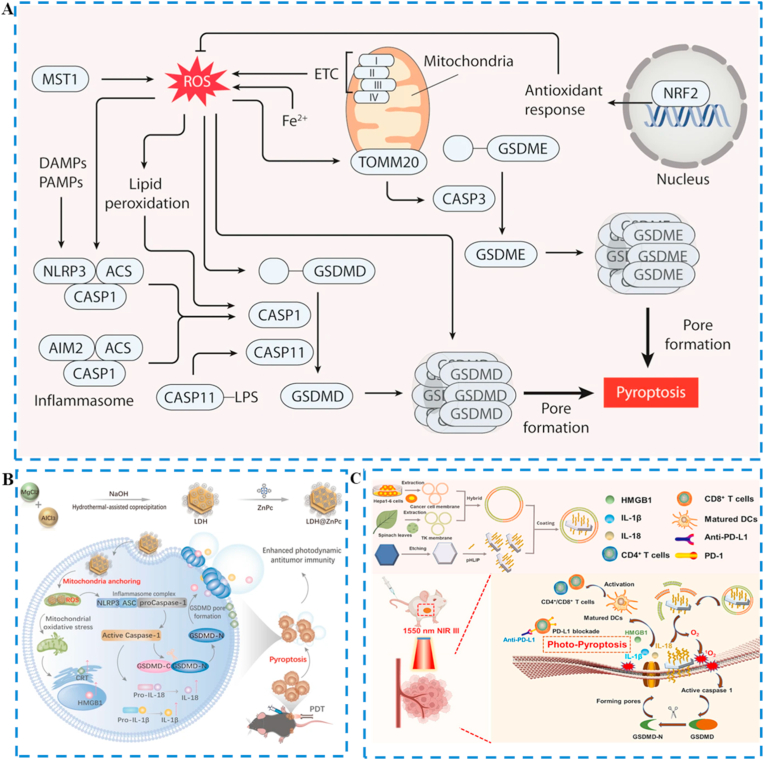
In recent years, the rapid advancement of nanotechnology and tumor biology has significantly expanded the application of nanomaterials in cancer therapy, particularly through the induction of programmed cell death (PCD) in cancer cells. Layered double hydroxides (LDH), a class of two-dimensional inorganic nanomaterials, have attracted considerable attention due to its tunable structures, excellent biocompatibility, and superior drug delivery capabilities. Emerging research has highlighted the great potential of LDH in modulating various forms of PCD. In this review, we provide a comprehensive overview of recent progress in the use of LDH to regulate different PCD pathways in cancer cells, including apoptosis, autophagy, ferroptosis, cuproptosis and pyroptosis. It emphasizes the underlying mechanisms of action, material design strategies, and the application of LDH in precise cancer therapy. Finally, this review is concluded with perspectives on the key challenges and bottlenecks of bioactive LDH in cancer therapy, providing potential solutions and outlining future perspectives.

Keywords: Cancer therapy; Layered double hydroxides; Nanomaterials; Programmed cell death.


Conflict of interest statement

The authors declare that they have no known competing financial interests or personal relationships that could have appeared to influence the work reported in this paper.

Figures

Image 1
Graphical abstract
Fig. 1
Fig. 1
LDH-based nanomaterials enhance cancer therapy through regulating various programmed cell death pathway, including apoptosis, autophagy regulation, and the induction of ferroptosis, cuproptosis, and pyroptosis.
Fig. 2
Fig. 2
ROS induces apoptosis in cancer cells. (A) Schematic illustration of the synthesis and functional mechanism of Zn-CoMo-LDH nanosheets, highlighting their role in inducing apoptosis in cancer cells. Reproduced with permission [44]. Copyright from Wiley-VCH GmbH, 2024. (B) Schematic illustration showing the preparation of LA&LDH and their in situ activation by the TME leading to cancer cell apoptosis and tumor eradication under NIR-II laser irradiation. Reproduced with permission [45]. Copyright from Wiley-VCH GmbH, 2023. (C) Schematic illustration of the preparation of CoBiFe-LDH-PEG nanosheets and their application in SDT to induce apoptosis in cancer cells. Reproduced with permission [46]. Copyright from Wiley-VCH GmbH, 2024.
Fig. 3
Fig. 3
Drugs induce apoptosis in cancer cells. (A) Schematic illustration of the preparation of LDH-based nanomaterials and their mechanism of inducing cancer cell apoptosis. Reproduced with permission [49]. Copyright from Wiley-VCH GmbH, 2019. (B) Schematic illustration of the preparation and co-delivery of 5FU-ABX-loaded albumin-stabilized LDH nanoparticles (BLDH/5FU-ABX) for inducing apoptosis in colorectal cancer cells. Reproduced with permission [50]. Copyright from Elsevier Ltd, 2021. (C) Schematic illustration of the synthesis of nanoparticles, and their targeted mechanism toward hepatocellular carcinoma (HCC) cells, leading to apoptosis induced by FT-BL@P. Reproduced with permission [51]. Copyright from Wiley-VCH GmbH, 2025. (D) Schematic illustration of the preparation of ATX/LDH, which significantly elevates oxidative stress and induces apoptosis in cancer cells. Reproduced with permission [52]. Copyright from Tsinghua University Press and Springer-Verlag GmbH Germany, part of Springer Nature, 2022.
Fig. 4
Fig. 4
LDH activation of autophagy for enhanced tumor therapy. (A) Schematic illustration of the mechanism by which celastrol-modified LDH exerts therapeutic effects against bone tumors. Reproduced with permission [68]. Copyright from American Chemical Society, 2023. (B) Schematic illustration of the autophagy-inducing mechanism of Cur/LDH. Reproduced with permission [69]. Copyright from American Scientific Publishers, 2016.
Fig. 5
Fig. 5
LDH-mediated inhibition of autophagy for enhanced tumor therapy. (A) Hydrolysis of LDH nanoparticles releases OH, which neutralize extracellular H+ in the tumor microenvironment, inhibiting the acidification of autophagolysosomes in tumor cells. This disruption of autophagy promotes tumor cell death. Reproduced with permission [72]. Copyright from American Chemical Society, 2022. (B) Schematic diagram of the mechanism of MG-LAAO killing tumor cells. Reproduced with permission [73]. Copyright from Wiley-VCH GmbH, 2025. (C) Synthesis of Zn-LDH by substituting Mg2+ with Zn2+ in the brucite layers of LDH through a metal ion exchange method. The Zn-LDH internalized by tumor cells can inhibit the acidification and degradation of autophagosomes to block the autophagy pathway. Reproduced with permission [75]. Copyright from Wiley-VCH GmbH, 2022. (D) Schematic diagram of the synthesis of LDH@miR-141-3p and miR-141-3p mediated breast cancer resistance to paclitaxel (PTX) by inhibiting autophagy through downregulation of RAB10, increased breast cancer cell sensitivity to PTX treatment. Reproduced with permission [76]. Copyright from American Chemical Society, 2025.
Fig. 6
Fig. 6
Iron-based LDH induce ferroptosis by enhancing the Fenton reaction in tumor cells. (A) Cellular mechanisms of ferroptosis. Reproduced with permission [79]. Copyright from Springer Nature 2024. (B) Mechanism schematics of cancer immunotherapy by Fe/Al-LDH-induced tumor ferroptosis. Reproduced with permission [81]. Copyright from Wiley-VCH GmbH, 2024. (C) Illustration of ferroptosis therapy for the treatment of kidney cancer using Fe/Ni-LDH. Reproduced with permission [82]. Copyright from Elsevier Ltd, 2023. (D) Schematic illustration of the construction and theranostic mechanism of the siR/IONs@LDH nanoplatform, Reproduced with permission [83]. Copyright from American Chemical Society, 2023.
Fig. 7
Fig. 7
Nonferrous based LDH induce ferroptosis in tumor cells by the Fenton-like reaction and GSH depletion. (A) Schematic illustration of the synthesis and antitumor performance of MTX-LDH@MnO2 nanoplatform. Reproduced with permission [88]. Copyright from Elsevier Ltd. 2023. (B) The synthetic process of ultrathin IFN-γ/uMn-LDHS and its therapeutic mechanism for co-enhancement of ferroptosis and antitumor. Reproduced with permission [89]. Copyright from Wiley-VCH GmbH, 2023.
Fig. 8
Fig. 8
(A) Reactive oxygen species induced pyroptosis in tumor cells. Reproduced with permission [101]. (B) Illustration of LDH@ZnPc featured with simultaneous mitochondria anchoring and pyroptosis abilities for potent photodynamic therapy and enhanced antitumor therapy. Reproduced with permission. Reproduced with permission [102]. (C) Schematic diagram of the production of acid-etched layered double hydroxides nanosheets based smart nanoplatforms (STEP) and its mediated pyroptosis. Reproduced with permission [103]. Copyright from Elsevier Ltd. 2024.
Fig. 9
Fig. 9
Regulating ion homeostasis to induce pyroptosis in tumor cells. (A) Inducting pyroptosis by disrupting multiorganelle to break the cytoprotection. Reproduced with permission [104]. Copyright from American Chemical Society, 2024. (B) The mechanisms of pyroptosis by ion homeostasis. Reproduced with permission [105]. (C) Schematic illustration of the pyroptosis pathway induced by R@AZOH. Reproduced with permission [106]. Copyright from American Chemical Society, 2025. (D) The disrupts zinc homeostasis, achieving a positive feedback loop of pyroptosis. Reproduced with permission [107]. Copyright from American Chemical Society, 2024. (E) Schematic illustration of the mechanisms for pyroptosis in cancer cells based on Zn-LDH@Mg implants under an AMF. Reproduced with permission [108]. Copyright from Wiley-VCH GmbH, 2024.

Similar articles

References

    1. Siegel R.L., Miller K.D., Fuchs H.E., Jemal A. Cancer statistics, 2022. CA: A Cancer J. Clin. 2022;72(1):7–33. - PubMed
    1. Zheng K., Song R., Li R., Liu M., Ba Y., Jiang W., Fan K. Nanomaterials for refining tumor microenvironment and enhancing therapy in head and neck squamous cell carcinoma: a review. Oncol. Translat. Med. 2024;10(4):151–161.
    1. Qin Y., Zeng W.-F., Liang W. Development of therapeutic cancer vaccines using nanomicellar preparations. Oncol. Translat. Med. 2023;9(6):265–268.
    1. Soerjomataram I., Bray F. Planning for tomorrow: global cancer incidence and the role of prevention 2020-2070. Nat. Rev. Clin. Oncol. 2021;18(10):663–672. - PubMed
    1. Bai Y., Lam H.C., Lei X. Dissecting programmed cell death with small molecules. Acc. Chem. Res. 2020;53(5):1034–1045. - PubMed

LinkOut - more resources