Review
. 2025 Jul 26:34:102137.
doi: 10.1016/j.mtbio.2025.102137. eCollection 2025 Oct.

Engineering DNA nanopores: from structural evolution to sensing and transport

Affiliations
Review

Engineering DNA nanopores: from structural evolution to sensing and transport

Fengyu Liu et al. Mater Today Bio. .

Abstract

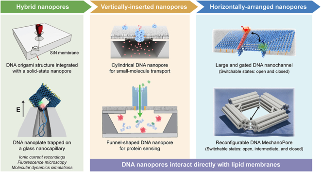
Synthetic nanopores, inspired by natural ion channels and nuclear pore complexes, hold immense potential for elucidating cellular transport mechanisms and enhancing molecular sensing technologies. DNA nanotechnology, particularly DNA origami, stands out as a transformative platform for designing biomimetic nanopores, leveraging its biocompatibility, structural programmability, and mechanical tunability. This review traces the structural evolution of DNA nanopores across three phases: early hybrid designs with solid-state platforms, vertically-inserted nanopores in lipid bilayers, and horizontally-arranged nanopores with advanced functionalities. Unlike prior reviews, we integrate this progression with critical insights into limitations-such as stability, scalability, and noise-while highlighting breakthroughs in single-molecule sensing and controlled transmembrane transport. We conclude by outlining strategies for next-generation DNA nanopores, offering a roadmap for their optimization in synthetic biology and nanomedicine.

Keywords: DNA origami; Molecular sensing; Nanopores; Transmembrane transport.


Conflict of interest statement

The authors declare that they have no known competing financial interests or personal relationships that could have appeared to influence the work reported in this paper.

Figures

Image 1
Graphical abstract
Fig. 1
Fig. 1
Evolutionary trajectory of DNA origami nanopores. This review focuses on three stages of DNA nanopore development: hybrid nanopores, vertically-inserted DNA nanopores, and horizontally-arranged DNA nanopores. Reproduced from Refs. [15,19,21,36,59,77] with permission.
Fig. 2
Fig. 2
Hybrid nanopores consisting of DNA origami structures. (a) Schematic and TEM characterization of the 3D DNA origami structure used for assembling a hybrid nanopore. Reproduced from Ref. [15] with permission. (b) Aperture-customized DNA nanoplates mounted on solid-state nanopores for size-selective macromolecular sensing. Reproduced from Ref. [16] with permission. (c) The ionic current analysis of using glass nanocapillaries to trap DNA nanoplates. Inserts: schematic of the trapping and ejection of DNA nanoplates on the glass nanocapillary with different voltages. Reproduced from Ref. [26] with permission. (d) Linear regression analysis between applied voltage and nanocapillary resistance. Reproduced from Ref. [28] with permission. (e) Bar chart of the normalized frequency of the λ-DNA translocations derived using different experimental setups at 300, 400, and 500 mV of applied voltage. Reproduced from Ref. [28] with permission.
Fig. 3
Fig. 3
Characterization techniques of DNA origami-based hybrid nanopores. (a) Ionic permeability characterization of DNA nanoplates with respect to salt concentration and applied voltage. Corresponding DNA nanoplates are free of central aperture design. Reproduced from Ref. [33] with permission. (b) Experimental design and simulation system for characterizing the conductance properties and structural deformation of DNA origami-involved hybrid nanopores. Top: schematic of the simplified experimental setup. Bottom: local configuration of the DNA nanoplate after 40 μs of coarse-grained Brownian dynamics simulations under a 400 mV bias. Reproduced from Ref. [36] with permission.
Fig. 4
Fig. 4
Vertically-inserted DNA nanopores with narrow lumens. (a) Channel-like DNA nanopore assembled by a barrel-shaped cap (white cylinders) and a long stem (red cylinders). Lower right: Example TEM images of synthetic DNA channels adhering to SUVs. Reproduced from Ref. [17] with permission. (b) Cylindrical DNA nanopore composed of six interconnected duplexes. Below: amplified AFM micrograph of individual DNA nanopores. Scale bar: 20 nm. Reproduced from Ref. [18] with permission. (c) Schematic design and AFM image of the DNA-tile structure consisting of four interconnected duplexes. Reproduced from Ref. [40] with permission. (d) DNA duplex inserted into the lipid bilayer. Bottom: representative AFM images of the DNA duplex structures. Reproduced from Ref. [41] with permission. (For interpretation of the references to colour in this figure legend, the reader is referred to the Web version of this article.)
Fig. 5
Fig. 5
Different gating mechanisms of cylindrical DNA nanopores. (a) 3D representation of the frequently-used cylindrical DNA nanopore. Reproduced from Ref. [44] with permission. (b) Averaged simulation configurations of cylindrical DNA nanopores under different conditions. Left: Simulation model is free of lipid membrane; Middle and Right: simulation models containing lipid membranes were run at different salt concentrations. Reproduced from Ref. [44] with permission. (c) The Molecular valve-equipped nanopore is triggered by specific oligonucleotides (named ‘key’). Reproduced from Ref. [45] with permission. (d) Reversible channel opening and closing of the thermo-sensitive cylindrical DNA nanopore. Reproduced from Ref. [47] with permission. (e) Controllable molecular transport of the light-triggered cylindrical DNA nanopore. Below: the cis-trans isomerization of Azobenzene induced by irradiation at different wavelengths. Reproduced from Ref. [48] with permission. (f) Assembly procedure of the barrel-like DNA nanopore (A•B) from two constituent components to an integral whole. Reproduced from Ref. [49] with permission.
Fig. 6
Fig. 6
Vertically-inserted DNA nanopores with wide lumens. (a) Current traces of T-shaped DNA nanopores recorded in the absence (black) or presence (purple) of double-stranded DNA (527 bp). Reproduced from Ref. [53] with permission. (b) Statistical analyses in the rate constant of pore-mediated EGFR efflux traces. The concentration of funnel-shaped DNA nanopore was 1 pM (larger histogram) and 10 pM (smaller histogram), separately. Reproduced from Ref. [59] with permission. (c) Fluorescence intensity changes of different molecules-filled SUVs docked by plugged DNA nanopore. The black arrow refers to the added time of unplugging strands. Reproduced from Ref. [60] with permission. (d) Top: Insertion mechanism of the extended-DNA-contained nanopore to the tailored lipid vesicles. Bottom: Long-term observation of dye influx into the GUVs via the nanopore with or without the addition of Blocker-DNA. Reproduced from Ref. [61] with permission. (e) Top: Assembly procedure and representative TEM images of the micron-length DNA channel. Bottom: Schematic and fluorescence micrographs of dye influx assay through capped DNA pores into GUVs. Reproduced from Ref. [62] with permission. (For interpretation of the references to colour in this figure legend, the reader is referred to the Web version of this article.)
Fig. 7
Fig. 7
Mimicking nuclear pore complexes with horizontally-arranged nanopores. (a) Structural presentation and time-resolved AFM kymographs of the horizontally-arranged DNA nanopores containing intrinsically-disordered nucleoporins. The pores were pre-attached on top of a supported lipid bilayer before AFM imaging. Reproduced from Ref. [68] with permission. (b) Spatial distribution of FG-Nup densities attached in DNA rings from cryo-electron microscopy and molecular dynamics simulations. Reproduced from Ref. [69] with permission. (c) Schematic representation and fluorescence recovery validation of the DNA origami pores embedding into lipid vesicles. Left corner: Schematics of the cDICE workflow. Right: Diameter of gyration of dextran Dgvs molecular weight Mw. Reproduced from Ref. [20] with permission.
Fig. 8
Fig. 8
Horizontally-arranged DNA nanopores with tunable gating behaviors. (a) Structural models and representative AFM images of the large and gated DNA nanochannel. Reproduced from Ref. [21] with permission. (b) Single-channel current traces of Tri-20-Spike pore before and after the addition of SARS-CoV-2 antibody. Tri-20 was assembled with three subunits (side length: 20 nm). Reproduced from Ref. [22] with permission. (c) Schematics and typical current traces of the semiflexible pore before and after binding protein. Reproduced from Ref. [76] with permission. (d) Reversible conformational changes among three states of the DNA MechanoPore (Top: Schematics; Bottom: TEM images). Reproduced from Ref. [77] with permission. (e) Allosteric mechanism and configurational characterization of the triangular DNA nanopore between expanded and contracted states. Reproduced from Ref. [78] with permission.

Similar articles

References

    1. Howorka S. Building membrane nanopores. Nat. Nanotechnol. 2017;12:619–630. doi: 10.1038/nnano.2017.99. - DOI - PubMed
    1. Ying Y.-L., Hu Z.-L., Zhang S., Qing Y., Fragasso A., Maglia G., Meller A., Bayley H., Dekker C., Long Y.-T. Nanopore-based technologies beyond DNA sequencing. Nat. Nanotechnol. 2022;17:1136–1146. doi: 10.1038/s41565-022-01193-2. - DOI - PubMed
    1. Dekker C. Solid-state nanopores. Nat. Nanotechnol. 2007;2:209–215. doi: 10.1038/nnano.2007.27. - DOI - PubMed
    1. Lee K., Park K.-B., Kim H.-J., Yu J.-S., Chae H., Kim H.-M., Kim K.-B. Recent progress in solid-state nanopores. Adv. Mater. 2018;30 doi: 10.1002/adma.201704680. - DOI - PubMed
    1. He Y., Tsutsui M., Zhou Y., Miao X.-S. Solid-state nanopore systems: from materials to applications. NPG Asia Mater. 2021;13:48. doi: 10.1038/s41427-021-00313-z. - DOI

LinkOut - more resources