Review
. 2025 Jul 22:34:102127.
doi: 10.1016/j.mtbio.2025.102127. eCollection 2025 Oct.

Reconstructing the female reproductive system using 3D bioprinting in tissue engineering

Affiliations
Review

Reconstructing the female reproductive system using 3D bioprinting in tissue engineering

Heesuh Yi et al. Mater Today Bio. .

Abstract

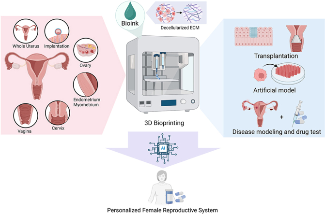
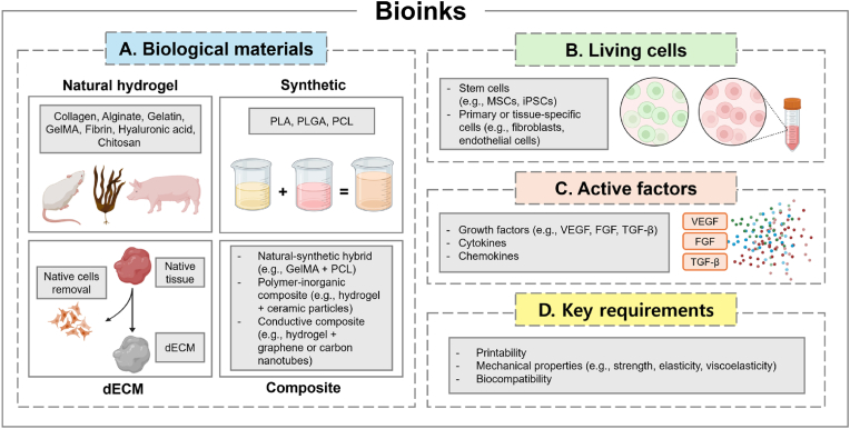
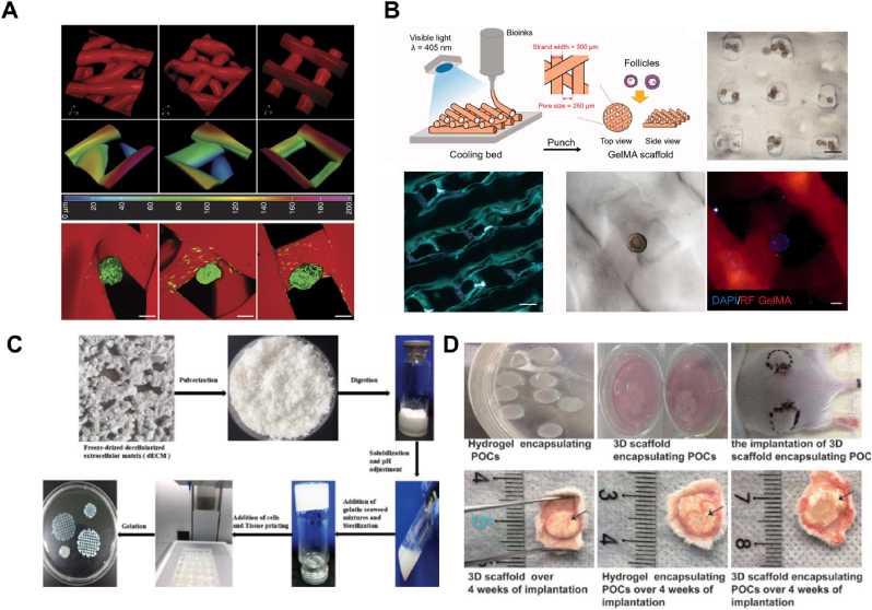
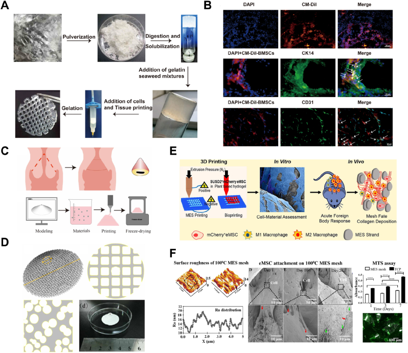
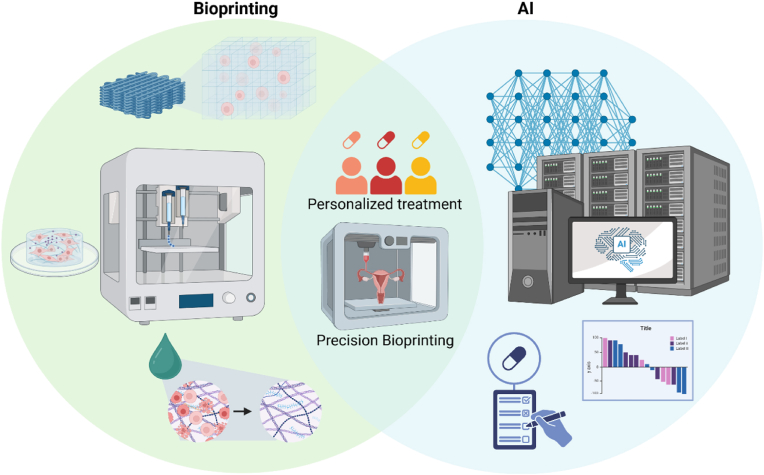
Three-dimensional bioprinting enables the precise fabrication of complex biological tissues through the layer-by-layer deposition of living cells and biomaterials, offering a promising strategy for reconstructing the female reproductive system. This technology has facilitated the development of in vitro models for tissues such as the endometrium, ovary, cervix, and vagina, providing improved structural fidelity and functional relevance. By leveraging bioinks, including decellularized extracellular matrix and advanced bioprinting techniques, researchers can recreate the intricate microarchitectures and vascular networks required for tissue functionality. These bioprinted systems serve as high-fidelity microphysiological systems for studying reproductive health, modeling disease progression, and evaluating therapeutic responses. Moreover, the integration of artificial intelligence into bioprinting workflows enhances reproducibility, scalability, and patient-specific customization. This review summarizes recent advances in reproductive tissue bioprinting and highlights its potential to transform regenerative gynecology and personalized reproductive healthcare.


Conflict of interest statement

The authors declare no competing interests.

Figures

Image 1
Graphical abstract
Fig. 1
Fig. 1
Essential components of bioinks for 3D bioprinting in reproductive tissue engineering. (A) Biological materials comprise natural hydrogels (e.g., collagen, alginate, gelatin, GelMA, fibrin, hyaluronic acid), synthetic polymers (e.g., PLA, PLGA, PCL), decellularized extracellular matrix (dECM) derived from native tissues, and composite formulations that integrate natural-synthetic hybrids or functional additives (e.g., ceramics, carbon nanotubes) to enhance structural and biological performance (B) Living cells, including stem cells (e.g., MSCs, iPSCs) and primary or tissue-specific cells (e.g., fibroblasts, endothelial cells), are incorporated to restore tissue-specific functions and support regeneration. (C) Active factors such as growth factors (e.g., VEGF, FGF, TGF-β), cytokines, and chemokines guide cell behavior within the bioprinted constructs, influencing proliferation, migration, and differentiation. (D) Key requirements for bioinks include printability, tunable mechanical properties (e.g., strength, elasticity, viscoelasticity), and biocompatibility to ensure structural fidelity and tissue maturation.
Fig. 2
Fig. 2
A schematic diagram of a top-down and a bottom-up approach. (A) In the top-down approach, a porous scaffold is first printed, onto which cells are seeded. As the cells proliferate and migrate within the scaffold, they eventually form a tissue construct that mimic native tissue. (B) In the bottom-up approach, cells are directly incorporated into bioink and printed layer by layer to form tissue constructs, allowing for more precise spatial organization of cells and biomaterials. This method eliminates the need for pre-formed scaffolds.
Fig. 3
Fig. 3
Representative 3D bioprinting modalities. (A) Extrusion bioprinting delivers continuous strands of bioink through a nozzle to build structures layer by layer. (B) Inkjet bioprinting forms patterns using bioink droplets ejected in a controlled manner. (C) Laser-assisted bioprinting employs laser energy to deposit cell-laden droplets without direct contact. (D) Vat-polymerization bioprinting utilizes light-mediated curing of bioresins within a vat to fabricate high-resolution constructs.
Fig. 4
Fig. 4
ECM-derived bioinks for reproductive tissue bioprinting. (A) Ovary dECM bioink maintains follicular structure and supports hormone secretion. Representative images show native ovary, unfreeze-dried and freeze-dried dECMs, and scanning electron microscopy (SEM) visualization highlighting ECM components such as collagen and fibronectin. Reproduced with permission from Ref. [57], ©2022 International Journal of Bioprinting. (B) Endometrial dECM bioink derived from porcine uterus supports endometrial regeneration. The schematic illustrates the decellularization process, with comparisons between Endo-UdECM and Whole-UdECM. Reproduced with permission from Ref. [66], ©2023 Advanced Functional Materials. (C) Oviduct dECM bioink retains structural integrity for embryo transport. The decellularization process is depicted alongside control and treated samples, demonstrating preserved morphology across different treatments. Reproduced with permission from Ref. [68], ©2025 Theriogenology. (D) Vaginal dECM bioink enhances epithelial remodeling and structural integrity. Images compare native vaginal tissue with unfreeze-dried and freeze-dried AVM bioinks. Histological staining (H&E, Masson) and SEM images confirm ECM preservation and microstructural characteristics. Reproduced with permission from Ref. [64], ©2021 International journal of biological macromolecules.
Fig. 5
Fig. 5
Schematic overview of 3D bioprinting applications in modeling female reproductive tissues. (A) Bioprinted endometrial constructs incorporating hMSCs within cell-laden hydrogels, designed for in vitro culture and in vivo transplantation to support cyclic regeneration [79]. © 2020 Acta biomaterialia. (B) Myometrial model using magnetically responsive smooth muscle cell-laden constructs to assess contractility and tocolytic drug efficacy. Reproduced from Ref. [83] under an open-access license, © 2017 International Journal of molecular sciences. (C) Cervical reconstruction using 3D-printed polyurethane (PU) scaffolds with tailored porosity and elasticity to match native cervical tissue biomechanics [59]. © 2020 Biomedical materials. (D) Vaginal tissue constructs printed with acellular vaginal matrix (AVM) bioinks and BMSCs, supporting epithelial regeneration and tissue integration [64]. © 2021 International journal of biological macromolecules. (E1) Ovarian scaffold bioprinted with GelMA and commercial ovarian cell lines (COV434, KGN, ID8) to replicate hormone production and stromal structure. (E2) Follicle-laden ovarian scaffold using visible light–crosslinked GelMA bioinks for follicular development and oogenesis. Reproduced from Ref. [65] under an open-access license, © 2021 Climacteric. (F) Implantation platform based on 3D-printed scaffolds for visualizing embryo adhesion and modeling early implantation events in vitro. Reproduced with permission from Ref. [60], © 2022 Journal of cellular physiology.
Fig. 6
Fig. 6
3D printing of the endometrium. (A) A graphical abstract illustrating the 3D printing of an endometrial construct (EC) and its application in vivo. (B) Characterization of the printed layers using bright-field and SEM imaging. (C) A schematic diagram showing the partial full-thickness uterine excision and the implantation procedures for either a 3D bioprinted EC or a non-printed graft. Reproduced with permission from Ref. [58] ©2023 Acta biomaterialia. (D) Overview of the experimental design for a 3D-printed granulate colony-stimulating factor-loaded sustained-release microsphere system. (E) Structure and morphology of the microsphere-scaffold visualized under SEM. Reproduced with permission from Ref. [61], ©2022 Biomaterials science. (F) Characterization of the 3D printed hydrogel scaffold loaded with hiMSCs, as seen under SEM. Reproduced with permission from Ref. [79], © 2020 Acta biomaterialia. (G) A schematic diagram illustrating the construction of 3D printed hydrogel loaded with cells. (H) Macroscopic view of the printed porous hydrogel. (I) Immunofluorescence images showing human amnion mesenchymal stem cells in the printed scaffolds. Reproduced with permission from Ref. [71], ©2021 ACS omega.
Fig. 7
Fig. 7
3D printing of embryo implantation and the whole uterus. (A) A schematic illustration of a study where embryos were cultured using controlled contact angles and embryo-scaffold interactions via 3D printing. Reproduced with permission from Ref. [60], © 2022 Journal of cellular physiology. (B) A schematic flow diagram of 3D printed magnetic ring structures used to evaluate a uterine contractility assay. The diagram also shows the analysis of myometrial smooth muscle cells printed at varying densities and their contraction response to indomethacin. Reproduced from Ref. [83] under an open-access license, © 2017 International Journal of molecular sciences. (C) Customized 3D physical models of the uterus created for five different cases of uterine cancer surgical removal. The models depict the uterus (transparent), endometrium (light yellow), endometrial cancer (red), and blood vessels (yellow). Reproduced from Ref. [102] under an open-access license, © 2017 Radiological physics and technology. (For interpretation of the references to colour in this figure legend, the reader is referred to the Web version of this article.)
Fig. 8
Fig. 8
3D printing of the ovary. (A) 3D reconstructions from confocal fluorescence image stacks of 3D printed scaffolds, visualized from various angles to show structural complexity. Reproduced from Ref. [65] under an open-access license, © 2017 Nature communications [78]. (B) A schematic representation of the 3D bioprinting process for ovarian follicles, along with bright-field and confocal fluorescence images of the 3D printed follicular constructs. The cooling bed is used to support the formation of the porous GelMA scaffold. Reproduced from Ref. [65] under an open-access license, © 2021 Climacteric. (C) The biofabrication process of bioink derived from ovarian dECM, followed by the 3D printing of porous scaffold structures. (D) Comparative images of 3D printed scaffolds encapsulating POCs before and after implantation, showing tissue integration and scaffold degradation over several weeks post-implantation. Reproduced from Ref. [57] under an open-access license, © 2022 International journal of bioprinting.
Fig. 9
Fig. 9
3D printing of vagina and cervix. (A) Production process of bioink using decellularized vaginal tissue and the 3D printing of porous cylindrical scaffold structures. (B) Fluorescence images of 3D printed scaffolds encapsulating CM-Dil-labeled BMSCs. Reproduced with permission from Ref. [64], © 2021 International journal of biological macromolecules. (C) A schematic diagram of the Conization surgery procedure and the corresponding 3D printing system used to reconstruct cervical tissue. (D) 3D printed reconstruction of missing cervical tissue following conization surgery. Reproduced with permission from Ref. [59], © 2020 Biomedical materials. (E) Schematic illustration of bioprinting endometrial mesenchymal stem/stromal cells (eMSC) to improve treatments for pelvic organ prolapse (POP). (F) Characterization of melt electrospun (MES) mesh and assessment of eMSC attachment on the 3D printed mesh, illustrating the mesh's structural properties and cell integration. Reproduced with permission from Ref. [82], © 2019 Acta biomaterialia.
Fig. 10
Fig. 10
The schematic illustration of the integration of bioprinting and artificial intelligence (AI) for precision medicine. Bioprinting enables the fabrication of complex tissue structures using bioinks containing cells and biomaterials. AI-driven analysis enhances precision bioprinting by optimizing printing parameters, predicting outcomes, and facilitating personalized treatment strategies. The synergy between AI and bioprinting contributes to advancements in regenerative medicine and patient-specific therapies. Reproduced from under [153] an open-access license, ©2022 Bioprinting.

Similar articles

References

    1. Deol P.S. Blue Rose Publishers; 2023. Anatomy Physiology of Female Reproductive System.
    1. Bulun S.E. In: Williams Textbook of Endocrinology. thirteenth ed. Melmed S., et al., editors. Elsevier; Philadelphia: 2016. Chapter 17 - physiology and pathology of the female reproductive axis; pp. 589–663.
    1. Critchley H.O.D., et al. Physiology of the endometrium and regulation of menstruation. Physiol. Rev. 2020;100(3):1149–1179. - PubMed
    1. Phillippi J., Kantrowitz-Gordon I. Jones & Bartlett Learning; 2023. Varney's Midwifery.
    1. Rankin J. Elsevier; 2024. Physiology in Childbearing - E-Book: Physiology in Childbearing - E-Book.

LinkOut - more resources