. 2025 Jul 17:34:102109.
doi: 10.1016/j.mtbio.2025.102109. eCollection 2025 Oct.

Polydopamine@Zinc oxide coated macroporous membrane for antibacterial protection and early pulp repair in pulpitis

Affiliations

Polydopamine@Zinc oxide coated macroporous membrane for antibacterial protection and early pulp repair in pulpitis

Yumeng Guo et al. Mater Today Bio. .

Abstract

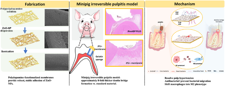
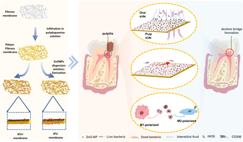
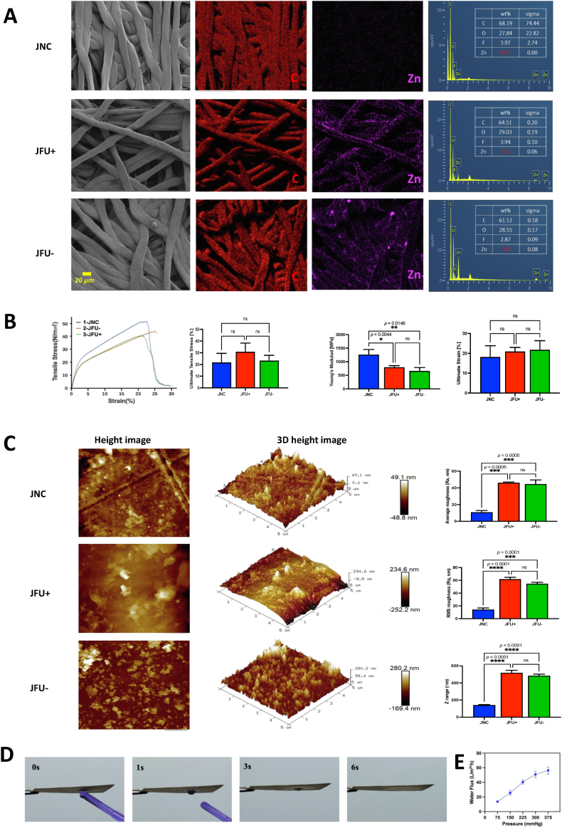
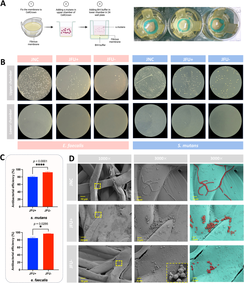
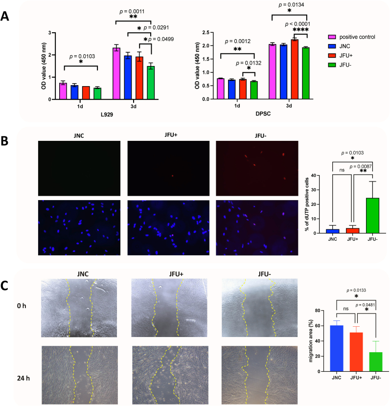
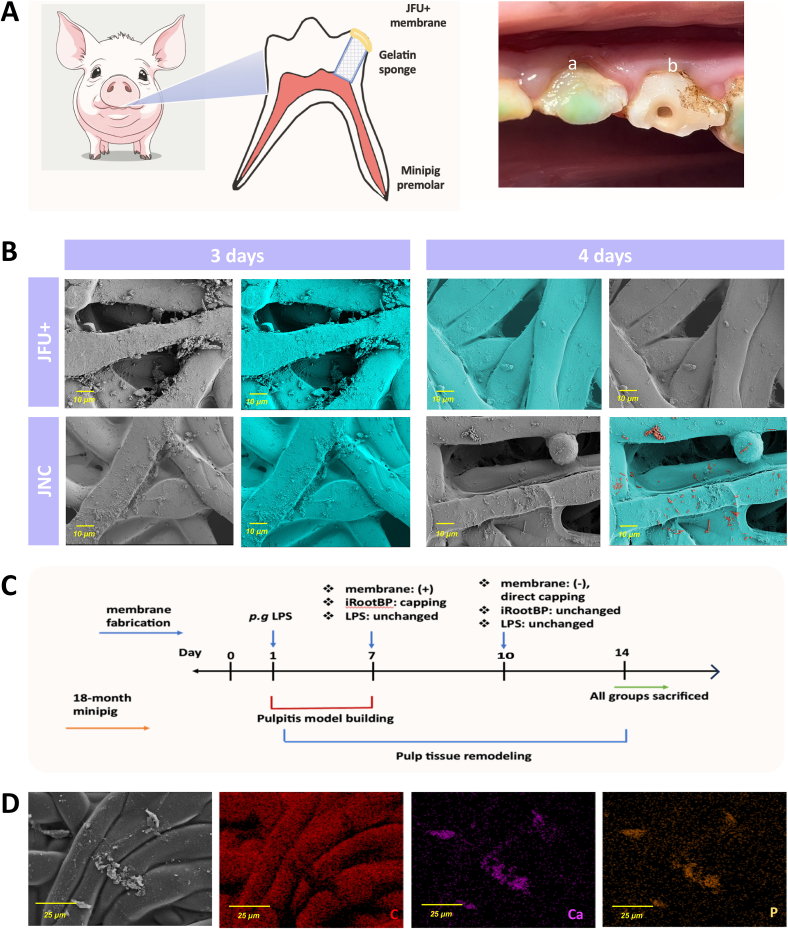
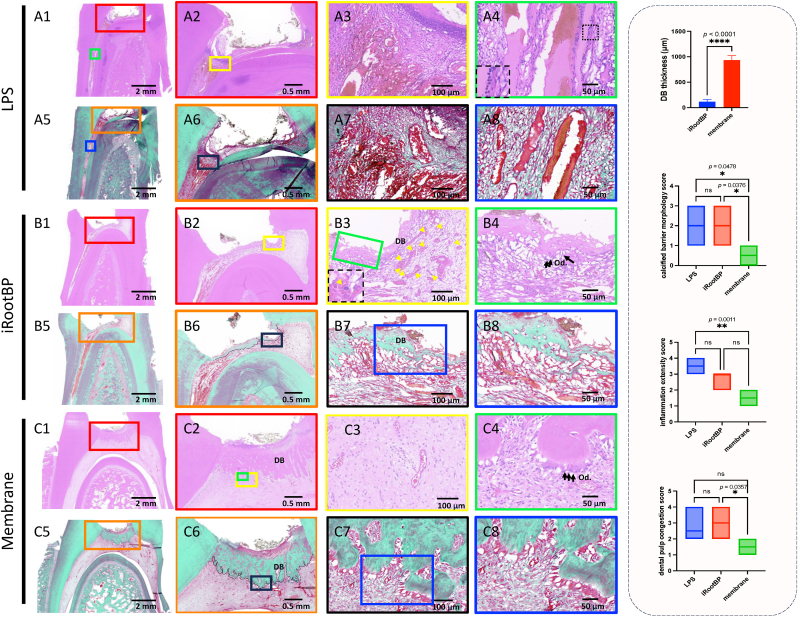
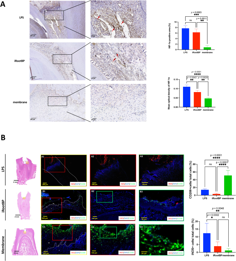
Continuous decompression and drainage are a vital surgical strategy for managing severe tissue infections. In vital pulp therapy (VPT) for irreversible pulpitis, there is a clinical demand for advanced biomaterials capable of effectively sealing the pulp cavity, alleviating pulpal hypertension, preventing bacterial infiltration, and resolving acute pulp inflammation in bacteria-rich environments. In this study, we developed a polydopamine@zinc oxide nanoparticle (ZnO-NP)-coated polytetrafluoroethylene (PTFE) membrane with tunable Zn content ranging from 2.51 to 7.45 wt%. The polydopamine enhanced ZnO-NP adhesion to the PTFE membrane, enabling superior fluid permeability and robust antibacterial efficacy against E. faecalis and S. mutans, while maintaining excellent biocompatibility. In a minipig pulpitis model, the ZnO-coated membrane significantly outperformed iRootBP PLUS by promoting faster dentine bridge formation (934.0 ± 91.3 μm vs. 116.3 ± 45.4 μm), preserving the integrity of the underlying pulp tissue and inducing M2 macrophage polarization.These findings deomstrate that ZnO-functionalized fibrous membrane can address key challenges in VPT by alleviating pulp hypertension, preventing microbial invasion, and simultaneously promoting pulp tissue regeneration. This approach offers a promising strategy to enhance tharepeutic outcomes of vital pulp therapy.

Keywords: Antimicrobial effect; Irreversible pulpitis; Polydopamine; Vital pulp therapy; ZnO-Coated biomaterial.


Conflict of interest statement

The authors declare that they have no known competing financial interests or personal relationships that could have appeared to influence the work reported in this paper.

Figures

Image 1
Graphical abstract
Fig. 1
Fig. 1
Fabrication process of functionalized fibrous membrane. JFU- membrane and JFU+ membrane were prepared via immersion in a polydopamine solution at pH 8.5, followed by coating with 30 nm ZnO-NPs. The membranes were then subjected to sonication for 0 min (JFU-) and 5 min (JFU+), respectively.
Fig. 2
Fig. 2
Characterization of polydopamine@ZnO coated membranes. (A) SEM and EDX mapping images of JNC, JFU+ and JFU- membranes showing surface morphology. The purple coloration highlights Zn distribution on the membrane surfaces, indicating the deposition of ZnO-NPs. (B) Representative uniaxial tensile stress-strain curves of functionalized membranes and associated ultimate tensile stress values, Young's modulus and ultimate strain. (C) Two-dimensional AFM images and corresponding deflection signal images of various membranes. The upper row displays JNC, the middle row shows the JFU+ membrane and the lower row presents the JFU- membrane. Accompanying quantitative analysis of surface roughness parameters. (D) Pictures of the droplet interacting with JFU+ membrane. (E) Water flux measurement through the JFU+ membrane. Bar graphs represent mean ± standard deviation. n = 3, ∗p< 0.05, ∗∗p< 0.01, ∗∗∗p< 0.001, and ∗∗∗∗p< 0.0001. (For interpretation of the references to color in this figure legend, the reader is referred to the Web version of this article.)
Fig. 3
Fig. 3
In vitro qualitative and quantitative characterization of the antibacterial properties of fibrous membranes. (A) Schematic diagram of in vitro bacteria migration assay. (B) Colony forming units (CFUs) of E. faecalis and S. mutans colonies on the surface of the membranes. (C) Quantitative analysis of CFUs of E. faecalis and S. mutans. (D) Morphology of S. mutans adhesion on the fiber of various membranes. Bar graphs represent mean ± standard deviation. n = 3, ∗p < 0.05, and ∗∗∗∗p < 0.0001.
Fig. 4
Fig. 4
In vitro evaluation of various membranes. (A) CCK-8 assay measuring cell viability of L929 cells and DPSCs on various membranes at 1 and 3 days. (B) TUNEL fluorescence staining of DPSCs on various membranes, with corresponding quantitative analysis. (C) Scratch assay of DPSCs treated with different membranes, including quantitative measurement of wound healing.
Fig. 5
Fig. 5
In vivo barrier properties of various membranes. (A) The pattern diagram of JFU+ membrane put on the occlusal surface of minipig premolars. Design of minipig pulp exposure: a 1 mm diameter exposure was created on the occlusal surface, then covered with a gelatin sponge and sealed securely with GIC. a: LPS group; b: membrane group. (B) SEM images of the pulpal side of JNC and JFU+ membranes after placement on premolars for 3 and 4 days. (C) Schematic diagram of the capping experiment. (D) EDS mapping images of deposits on pulp side.
Fig. 6
Fig. 6
Histological analysis of early pulp repair in minipig pulpitis. 7 days after pulp capping, the inflammatory response of dental pulp in minipig premolars. (A1-A4, B1-B4, C1-C4) represent sequential magnifications of HE staining in the LPS, iRootBP PLUS, and membrane groups respectively. (A5-A8, B5-B8, C5-C8) shows sequential magnifications of Goldner's trichrome staining in the corresponding groups. DB, dentine bridge; Od, odontoblast; yellow arrow, inflammatory cell. Semiquantitative analysis of inflammation extensity score, calcified barrier morphology score and dental pulp congestion score is expressed in box plots. n = 6, ∗p< 0.05, ∗∗p< 0.01, ∗∗∗p< 0.001and ∗∗∗∗p< 0.0001. (For interpretation of the references to color in this figure legend, the reader is referred to the Web version of this article.)
Fig. 7
Fig. 7
Membrane treatment alleviated hypoxia and polarized macrophages into M2 phenotype. (A) HIF-1α immunohistochemical staining in pulp tissue sections from the experimental groups. (B) iNOS and CD206 positive macrophages determined by IF staining. Bar graphs represent mean ± standard deviation. n = 6, ∗∗p< 0.01, ∗∗∗p< 0.001 and ∗∗∗∗p< 0.0001.

Similar articles

References

    1. León-López M., Cabanillas-Balsera D., Martín-González J., Montero-Miralles P., Saúco-Márquez J.J., Segura-Egea J.J. Prevalence of root canal treatment worldwide: a systematic review and meta-analysis. Int. Endod. J. 2022;55(11):1105–1127. doi: 10.1111/iej.13822. - DOI - PMC - PubMed
    1. Yu C., Abbott P.V. An overview of the dental pulp: its functions and responses to injury. Aust. Dent. J. 2007;52(1 Suppl):S4–S16. doi: 10.1111/j.1834-7819.2007.tb00525.x. - DOI - PubMed
    1. AlShwaimi E., Majeed A., Ali A.A. Pulpal responses to direct capping with Betamethasone/gentamicin cream and mineral trioxide aggregate: Histologic and micro-computed tomography assessments. J. Endod. 2016;42(1):30–35. doi: 10.1016/j.joen.2015.09.016. - DOI - PubMed
    1. Guan X., Zhou Y., Yang Q., Zhu T., Chen X., Deng S., Zhang D. Vital pulp therapy in permanent teeth with irreversible pulpitis caused by caries: a prospective cohort study. J. Personalized Med. 2021;11(11) doi: 10.3390/jpm11111125. - DOI - PMC - PubMed
    1. Aguilar P., Linsuwanont P. Vital pulp therapy in vital permanent teeth with cariously exposed pulp: a systematic review. J. Endod. 2011;37(5):581–587. doi: 10.1016/j.joen.2010.12.004. - DOI - PubMed

LinkOut - more resources