Review
. 2025 Jul 21:17:1587986.
doi: 10.3389/fnagi.2025.1587986. eCollection 2025.

Insights into targeted ferroptosis in mechanisms, biology, and role of Alzheimer's disease: an update

Affiliations
Review

Insights into targeted ferroptosis in mechanisms, biology, and role of Alzheimer's disease: an update

Bingyuan Zhou et al. Front Aging Neurosci. .

Abstract

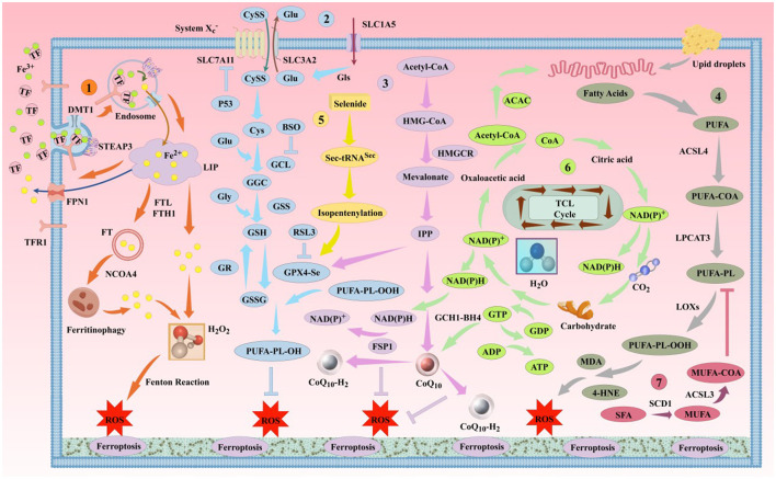
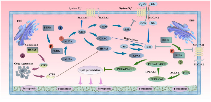
Ferroptosis is a newly discovered form of programmed cell death, primarily caused by an imbalance between iron-dependent oxidative damage and antioxidant defense mechanisms within the cell. It differs from previously reported forms of cell death, such as apoptosis, necrosis, and autophagy, in terms of morphology, biochemistry, and genetics. Alzheimer's disease (AD) is the most common neurodegenerative disorder, characterized by pathological features including neurofibrillary tangles (NFTs), senile plaques (SPs), and abnormal iron deposition, suggesting that ferroptosis may be involved in its disease progression. Although recent studies have made significant progress, the mechanisms underlying neuronal ferroptosis in AD remain incompletely understood. This review, based on elucidating the process and regulatory mechanisms of cellular ferroptosis, explores, and supplements the correlation between iron overload and redox imbalance with the main pathological mechanisms of AD, providing new insights for the treatment of AD and the development of new drugs.

Keywords: Alzheimer's disease; biology; ferroptosis; mechanisms; update.


Conflict of interest statement

The authors declare that the research was conducted in the absence of any commercial or financial relationships that could be construed as a potential conflict of interest.

Figures

Diagram depicting the biochemical pathways leading to ferroptosis, highlighting various steps such as iron metabolism, lipid peroxidation, and the tricarboxylic acid (TCA) cycle. Key elements include iron uptake through TFR1, conversion processes involving DMT1 and FPN, the role of PUFA, and the formation of reactive oxygen species (ROS). The pathway also outlines the impacts of fatty acids, acetyl-CoA, and enzymes like GPX4 and LOXs, illustrating the complex interplay of cellular components in ferroptosis.
Figure 1
The classic mechanism of ferroptosis. Ferroptosis is primarily driven by iron-dependent lipid peroxidation. This process involves several pathways, including the iron metabolism pathway, the Cys-GSH-GPX4 pathway, the mevalonate pathway, and the lipid metabolism pathway, among others. DMT1, divalent metal ion transporter 1; STEAP3, prostate 6 transmembrane epithelial antigen 3; NCOA4, nuclear receptor coactivator 4; SLC7A11, solute carrier family 7 member 11; SLC3A2, solute carrier family 3 member 2; SLC1A5, solute carrier family 1 member 5; GLS, glutaminase; P53, tumor protein 53; BSO, buthionine sulfoximine; GCL, glutamate-cysteine ligase; GGC, γ-L-glutamyl-L-cysteine; GSS, glutathione synthetase; GR, glutathione reductase; GSSG, reduced glutathione; HMGCR, 3-hydroxy-3-methylglutaryl-CoA reductase; IPP, isoprenoid diphosphate; ACAC, acetyl-CoA carboxylase; LPCAT3, lysophosphatidylcholine acyltransferase 3; LOX, lipoxygenase; ACSL3, acyl-CoA synthetase long-chain family member 3; SCD1, stearoyl-CoA desaturase 1; BH4, tetrahydrobiopterin; GCH1, human GTP cyclohydrolase 1.
Diagram illustrating ferroptosis pathways and associated diseases. Left section shows factors like oxidative stress, iron homeostasis, epigenetics, and autophagy leading to ferroptosis. Right section highlights diseases linked to ferroptosis in the brain and body, such as brain injury, Parkinson's disease, ischemia, heart failure, and liver fibrosis. Central human figure with organs is included for context.
Figure 2
Ferroptosis is related to the occurrence of multiple major diseases.
Diagram illustrating pathways related to ferritinophagy, circadian autophagy, chaperone-mediated autophagy, and lipophagy leading to lipid peroxidation and ferroptosis. Key proteins and processes include NCOA4, SQSTM1, ARNTL, GPX4, ROS formation, and EGLN2/HIF1A signaling, which are depicted through arrows and labeled components.
Figure 3
The Classic Autophagy Regulatory Mechanism of Ferroptosis. (1). Ferritinophagy; (2). Lipophagy; (3). Clock Autophagy; (4). CMA ARNTL, aryl hydrocarbon receptor nuclear translocator-like protein; SQSTM1/P62, sequestosome 1; HSPA8, heat shock protein family A member 8; HSPA5, heat shock protein family A member 5; HSP90, heat shock protein 90; LAMP-2a, lysosomal-associated membrane protein 2A; RAB7A, Ras-related protein Rab-7a.
Diagram illustrating cellular processes related to ferroptosis. It shows the pathways involving PERK, eIF2a, ATF4, CHOP, GSH, and IRE1α with interactions through protein phosphorylation and degradation. System Xc–, SLC proteins, and ER stress markers like HSPA5 and ATF6 are depicted. Processes like lipid peroxidation involving PUFA, GPX4, LPCAT3, and ACSL4 are also shown, contributing to ferroptosis. Arrows indicate direction and sequence of reactions within the cell membrane.
Figure 4
ERS and ferroptosis. (1). PERK can oligomerize and undergo trans-autophosphorylation under ERS conditions, inhibiting protein translation by phosphorylating eIF2α, thereby reducing the entry of proteins into the overloaded ER. The phosphorylation of eIF2α by PERK selectively translates the mRNA of ATF4, alleviating ERS. The activation of the PERK pathway in the short term can protect cells by inhibiting protein synthesis and reducing unfolded proteins in the ER. However, the long-term activation of the PERK pathway can damage cell viability. The sustained activation of the PERK pathway can induce the accumulation of CHOP and the expression of CHAC1, inhibiting SLC7A11 and GSH, and promoting the occurrence of ferroptosis. On the other hand, ATF4-mediated HSPA5 expression prevents the degradation of GPX4, inhibiting ferroptosis. (2). Under ERS conditions, ATF6 is released from the HSPA5 complex and transferred to the Golgi apparatus in vesicles. ATF6 is sequentially cleaved by S1P and S2P, releasing transcriptionally active ATF6. Active ATF6 leads to increased HSPA5 expression, which promotes the correct folding and transport of unfolded or misfolded proteins and further affects the sensitivity of cells to ferroptosis by the binding of HSPA5 to GPX4, thereby alleviating ERS and maintaining the normal function of the ER. (3). When unfolded proteins accumulate in the ER, IRE1 undergoes dimerization and trans-autophosphorylation, promoting ER protein folding, secretion, and activation of phospholipid biosynthesis and ER-associated degradation pathways. If ERS continues or worsens, IRE1α is further activated. The mRNA transcripts of GCL subunits (GCL) and SLC7A11 are newly identified targets of IRE1α negative regulation (Jiang et al., 2024). IRE1α determines the sensitivity of cells to RSL3 and ferroptosis by negatively regulating SLC7A11 and GCL. PERK, protein kinase RNA-like endoplasmic reticulum kinase; eIF2α, eukaryotic translation initiation factor; ATF4, transcription factor 4; CHOP, C/EBP homologous protein; S1P, site-1 protease; S2P, site-2 protease; CHAC1, glutathione-specific gamma-glutamylcyclotransferase 1; P53, cellular tumor antigen p53; ATF6, transcription factor 6; IRE1α, inositol-requiring kinase 1α.
Diagram illustrating the pathways related to ferroptosis in the brain. Key elements include amyloid-beta generation, tau phosphorylation, neuroinflammation, oxidative stress, mitochondrial function, and the cholinergic system, all connecting to ferroptosis.
Figure 5
Six major pathogenesis of ferroptosis and AD.
Illustration of complex biochemical pathways showing the interactions between iron metabolism, oxidative stress, and neurodegeneration. It includes elements like DMT1, FPN1, ROS, Tau, NFT, and their roles in processes such as lipid peroxidation and ferroptosis. Arrows depict pathways, and labels identify key proteins and reactions involved in aging, inflammatory factors, and cellular damage.
Figure 6
Iron participates in the formation of AD's primary pathological mechanisms through multiple pathways. The aggregation of Aβ plaques and hyperphosphorylation of Tau protein are closely associated with ferroptosis. (1). Mechanisms of iron uptake in AD. Neurons uptake iron via the TF/TFR1 complex or through DMT1/PrPC-dependent pathways. TF undergoes autophagy mediated by NCOA4, releasing iron and leading to lethal iron levels and ferroptosis. (2). Mechanisms of iron efflux in AD. FPN1/Cp or FPN1/Heph facilitates iron efflux with the assistance of APP, which stabilizes FPN1 via soluble Tau transport. Aging, inflammation, and OS dysregulate iron transporters, causing iron retention. (3). Role of the GSH/GPX4 pathway in AD ferroptosis. Under certain conditions, reduced GPX4 or GSH in neurons fails to counteract lipid peroxidation. Accumulation of PUFA-OOH to lethal levels via Fenton reactions or ALOX catalysis induces Tau hyperphosphorylation, Aβ formation, and neuronal loss. (4). Molecular mechanisms of Aβ plaque formation and aggregation. Iron overload upregulates FT, FPN1, and APP expression via IRP-IRE interactions while inhibiting normal FUR function, leading to BACE1 upregulation and increased extracellular Aβ deposition and lipid peroxidation. (5). Molecular mechanisms of Tau hyperphosphorylation. Iron promotes Tau hyperphosphorylation, aggregation, NFT formation, and lipid peroxidation. Overexpression of GSK-3β and HO-1 induces Tau phosphorylation, leading to cerebral accumulation and NFT formation. Elevated GPX4 expression inhibits this process. Reduced soluble Tau increases brain iron deposition by suppressing FPN1 activity, exacerbating ferroptosis. Aβ, β-amyloid; Tau, microtubule-associated protein; APP, amyloid precursor protein; PrPC, prion protein; Cp, ceruloplasmin; Heph, hephaestin; FUR, furin protease; IRP, iron regulatory protein; BACE1, β-secretase 1; HO-1, heme oxygenase-1; SP, senile plaques; NFT, neurofibrillary tangles.
Diagram comparing M1 and M2 phenotypes in immune response. The M1 phenotype on the left shows pro-inflammatory pathways involving NF-κB, ROS, TLR, and various cytokines. The M2 phenotype on the right shows anti-inflammatory pathways involving MAPK/ERK, TREM2, and different cytokines. Iron metabolism processes like TFR1, DMT1, FPN1, and LIP are visible with arrows indicating flow directions. Both phenotypes involve interactions with non-transferrin-bound iron (NTBI), lipopolysaccharides (LPS), and other molecular structures. The diagram also highlights the impact of aging, ROS, iron, and pro-inflammatory media on the phenotypic shift.
Figure 7
Iron and iron chelators modulate microglial M1/M2 polarization. The protective M2 microglial phenotype secretes anti-inflammatory mediators and neurotrophic factors, clearing or sequestering Aβ and Tau to protect neurons. M2 microglia upregulate DMT1 and FT expression, enhancing NTBI uptake, expanding iron storage in FT and MtFt, and compartmentalizing extracellular and intracellular iron. In AD, microglia exposed to elevated iron, LPS, and extracellular Aβ from damaged neurons favor M1 activation. High iron intake and impaired efflux increase the LIP in microglia, activating inflammatory pathways via iron-derived ROS, releasing cytokines, and exacerbating neuroinflammation. Elevated iron upregulates complement C3 and C1q while suppressing the APOE-TREM2 axis, reducing Aβ plaque phagocytosis, damaging neurons, and accelerating AD progression. MtFt, mitochondrial ferritin; LPS, lipopolysaccharide; TGF-β, transforming growth factor-β; TNF-α, tumor necrosis factor-α; IL-1β, interleukin-1β; IL-4, interleukin-4; IL-6, interleukin-6; IL-10, interleukin-10; IL-12, interleukin-12; IL-13, interleukin-13; TLR, toll-like receptor; MAPK, mitogen-activated protein kinase; ERK, extracellular signal-regulated kinase; C1q, complement 1q; C3, complement 3; NTBI, non-transferrin-bound iron; NF-κB, nuclear factor-kappa B; APOE, apolipoprotein E; TREM2, triggering receptor expressed on myeloid cells 2; ASC, apoptosis-associated speck-like protein; NLRP3, NOD-like receptor pyrin domain-containing 3.
Illustration depicting the interaction between neurons, microglia, and astrocytes in the cerebromicrovascular environment. It highlights the roles of selenoproteins (SelR, SelW, SelM, SelK, SelS, SelP) in cellular processes like oxidization, calcium regulation, and inflammation via TNF-alpha and IL-6 signaling. The image includes key components like dendrites, axons, endoplasmic reticulum, and oxidative stress markers (H2O2, ROS).
Figure 8
Major functions of selenoproteins associated with AD ferroptosis mechanisms. (1). SelP can promote the transport of selenium from cerebral blood vessels to brain parenchymal tissues. Its deficiency leads to a lack of selenium and various selenoproteins in brain parenchymal tissues. SelP also has the ability to inhibit the aggregation of Aβ and Tau proteins, thereby inhibiting ferroptosis. (2). SelK can promote the release of Ca2+ from the endoplasmic reticulum and further induce the influx of extracellular Ca2+, enhancing the migration and phagocytic abilities of microglia. (3). SelR has the function of preventing the oxidation of the amino acid residue Met35 in MSRA by ROS. (4). SelW primarily acts on the mTORC2/Akt signaling pathway, promoting autophagy in neurons. (5). SelS can reduce the release of L-6 from astrocytes, degrade Aβ in neurons, inhibit Tau protein phosphorylation caused by endoplasmic reticulum stress, and decrease the release of IL-6 and TNF-α in microglia. (6). SelM has the function of regulating calcium homeostasis in neurons. (7). GPX4 and GPX1 can inhibit the production of H2O2 and organic peroxides in neurons, astrocytes, and microglia. (8). TRXR1 can inhibit the production of H2O2 and organic peroxides in neurons. SelP, selenoprotein P; SelK,selenoprotein K; SelR,selenoprotein R; SelW, selenoprotein W; SelS, selenoprotein S; SelM, selenoprotein M; GPX1, glutathione peroxidase 1; MSRA, methionine sulfoxide reductase A; mTORC2, mammalian target of rapamycin complex 2; Akt, protein kinase B.
Illustration of mitochondrial processes and signaling pathways. Key components include oxidative stress (ROS), autophagy, ferroptosis, and calcium ion exchange, with interactions involving proteins like PINK1, Parkin, GRP75, and VDAC. This diagram highlights connections with the endoplasmic reticulum and autophagy indicators (LC3), and indicates pathways for protein degradation, represented by ubiquitination processes.
Figure 9
Schematic diagram of the mechanisms of mitochondrial Ca2+ homeostasis and mitophagy in regulating AD ferroptosis. (1). ER stress and Aβ promote mitochondrial Ca2+ influx, leading to mitochondrial Ca2+ overload, ferroptosis, and cellular dysfunction. (2). When mitochondrial damage reduces membrane potential, PINK1 accumulates on the outer mitochondrial membrane (OMM), forming a complex with TOM. PINK1 activates Parkin, which ubiquitinates mitochondrial proteins. Ubiquitinated mitochondria are engulfed by autophagosomes via LC3 complexes, fuse with lysosomes, and are degraded, inhibiting ferroptosis. (3). OMM receptors (e.g., BNIP3, NIX, FUNDC1) can directly bind LC3 to initiate mitophagy independently of the PINK1/Parkin pathway, suppressing ferroptosis. GRP75, glucose-regulated protein 75; IP3R, inositol 1,4,5-trisphosphate receptor; VDAC, voltage-dependent anion channel; MCU, mitochondrial calcium uniporter; OMM, outer mitochondrial membrane; IMM, inner mitochondrial membrane; PINK1, PTEN-induced putative kinase 1; Parkin/PARK2, Parkinson disease protein 2; TOM, translocase of outer mitochondrial membrane; LC3, microtubule-associated protein 1 light chain 3; BNIP3, BCL2/adenovirus E1B 19 kDa protein-interacting protein 3; NIX, BNIP3-like; FUNDC1, FUN14 domain-containing protein 1; LETM1, mitochondrial proton/calcium exchanger protein; MPTP, mitochondrial permeability transition pore; NCLX, mitochondrial sodium/calcium exchanger protein; PS1, presenilin 1.

Similar articles

  • The Black Book of Psychotropic Dosing and Monitoring.
    DeBattista C, Schatzberg AF. DeBattista C, et al. Psychopharmacol Bull. 2024 Jul 8;54(3):8-59. Psychopharmacol Bull. 2024. PMID: 38993656 Free PMC article. Review.
  • Short-Term Memory Impairment.
    Cascella M, Al Khalili Y. Cascella M, et al. 2024 Jun 8. In: StatPearls [Internet]. Treasure Island (FL): StatPearls Publishing; 2025 Jan–. 2024 Jun 8. In: StatPearls [Internet]. Treasure Island (FL): StatPearls Publishing; 2025 Jan–. PMID: 31424720 Free Books & Documents.
  • C/EBPβ: A transcription factor associated with the irreversible progression of Alzheimer's disease.
    Yao Q, Long C, Yi P, Zhang G, Wan W, Rao X, Ying J, Liang W, Hua F. Yao Q, et al. CNS Neurosci Ther. 2024 Apr;30(4):e14721. doi: 10.1111/cns.14721. CNS Neurosci Ther. 2024. PMID: 38644578 Free PMC article.
  • Management of urinary stones by experts in stone disease (ESD 2025).
    Papatsoris A, Geavlete B, Radavoi GD, Alameedee M, Almusafer M, Ather MH, Budia A, Cumpanas AA, Kiremi MC, Dellis A, Elhowairis M, Galán-Llopis JA, Geavlete P, Guimerà Garcia J, Isern B, Jinga V, Lopez JM, Mainez JA, Mitsogiannis I, Mora Christian J, Moussa M, Multescu R, Oguz Acar Y, Petkova K, Piñero A, Popov E, Ramos Cebrian M, Rascu S, Siener R, Sountoulides P, Stamatelou K, Syed J, Trinchieri A. Papatsoris A, et al. Arch Ital Urol Androl. 2025 Jun 30;97(2):14085. doi: 10.4081/aiua.2025.14085. Epub 2025 Jun 30. Arch Ital Urol Androl. 2025. PMID: 40583613 Review.
  • Ferroptosis and its modulators: A raising target for cancer and Alzheimer's disease.
    Singh G, Kesharwani P, Kumar Singh G, Kumar S, Putta A, Modi G. Singh G, et al. Bioorg Med Chem. 2024 Jan 15;98:117564. doi: 10.1016/j.bmc.2023.117564. Epub 2023 Dec 20. Bioorg Med Chem. 2024. PMID: 38171251 Review.

References

    1. Aaseth J., Alexander J., Bjorklund G., Hestad K., Dusek P., Roos P. M., et al. (2016). Treatment strategies in Alzheimer's disease: a review with focus on selenium supplementation. Biometals 29, 827–839. 10.1007/s10534-016-9959-8 - DOI - PMC - PubMed
    1. Abad-Rodriguez J., Ledesma M. D., Craessaerts K., Perga S., Medina M., Delacourte A., et al. (2004). Neuronal membrane cholesterol loss enhances amyloid peptide generation. J. Cell Biol. 167, 953–960. 10.1083/jcb.200404149 - DOI - PMC - PubMed
    1. Abraham N. G., Junge J. M., Drummond G. S. (2016). Translational significance of heme oxygenase in obesity and metabolic syndrome. Trends Pharmacol. Sci. 37, 17–36. 10.1016/j.tips.2015.09.003 - DOI - PMC - PubMed
    1. Aggarwal A., Sharma N., Khera A., Sandhir R., Rishi V. (2020). Quercetin alleviates cognitive decline in ovariectomized mice by potentially modulating histone acetylation homeostasis. J. Nutr. Biochem. 84:108439. 10.1016/j.jnutbio.2020.108439 - DOI - PubMed
    1. Aliev G., Smith M. A., de la Torre J. C., Perry G. (2004). Mitochondria as a primary target for vascular hypoperfusion and oxidative stress in Alzheimer's disease. Mitochondrion 4, 649–663. 10.1016/j.mito.2004.07.018 - DOI - PubMed

LinkOut - more resources