. 2025 Jul 28:2025:3242282.
doi: 10.1155/omcl/3242282. eCollection 2025.

E2f1 Overexpression Reduces Aging-Associated DNA Damage in Cultured Cerebral Endothelial Cells and Improves Cognitive Performance in Aged Mice

Affiliations

E2f1 Overexpression Reduces Aging-Associated DNA Damage in Cultured Cerebral Endothelial Cells and Improves Cognitive Performance in Aged Mice

Sheelu Monga et al. Oxid Med Cell Longev. .

Abstract

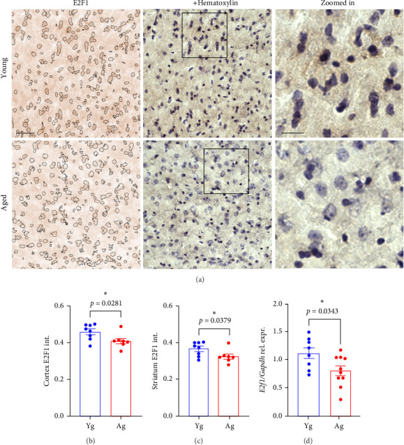
As we age, cerebral endothelial cells (CECs) are less efficient in maintaining genome integrity and accumulate DNA damage. DNA damage in the brain endothelium can lead to the impairment of the blood-brain barrier (BBB), which is a major factor in brain dysfunction and dementia. Thus, identifying factors that regulate DNA repair in the brain endothelium can prevent brain dysfunction associated with aging. E2F1 is a transcription factor that regulates the expression of genes associated with DNA repair, among other functions. We hypothesize that E2F1 is downregulated in the brain vasculature of mice with aging and that E2F1 upregulation can improve cognitive function. We found that in the brain endothelium, E2F1 was significantly less phosphorylated, which is associated with its transcriptional activity, in the brain vasculature of aged mice and cultured CEC derived from aged mice compared with those from young mice. We found that E2f1 overexpression reduced DNA damage in cultured CEC, and targeting the brain vasculature to overexpress E2f1 improved cognition and increased the expression of genes associated with BBB integrity in aged mice. From RNA sequencing data from cultured CEC, we found that E2f1 overexpression significantly upregulated Acod1, which codes for aconitate decarboxylase-1 (ACOD1), an enzyme that produces itaconate. We also found that 4-octyl itaconate (4-OI), a derivative of itaconate, reduced DNA damage, promoted cell proliferation, and restored endothelial barrier function from oxidative stress in cultured CEC. Thus, our study identifies the E2F1-ACOD1 axis as a molecular pathway that can protect the brain endothelium from oxidative stress and aging.

Keywords: DNA damage; E2F1; aging; cognition; endothelial cells; itaconate.


Conflict of interest statement

All authors declare no conflicts of interest.

Figures

Figure 1
Figure 1
E2F1 was downregulated in the brains of aged mice compared with young mice. (A) Representative images of the cortex of 4- (young) and 20-m/o (aged) male mice processed for immunohistochemistry and stained with antibodies against E2F1 (brown) and counterstained with hematoxylin (purple). Hematoxylin-positive particles are identified in the left panel to visualize differences in E2F1 signal between young and aged groups. Scale bar, 100 µm. Zoomed-in scale bar, 50 µm. Quantification of E2F1 intensity from the cortices (B) and striatum (C) tissues from young and aged male and female mice. Data are mean ± SEM from 4 male and 4 female mice pooled together per age. Student's t-test. (D) Bar graph representing gene expression of E2f1 relative to Gapdh in the brains of 4- (Yg) and 20-m/o (Ag) male and female mice. Data are mean ± SEM from 8 to 9 mice/age. Student's t-test.
Figure 2
Figure 2
Phospho-E2F1 and γH2AX were reduced and increased, respectively, in microvessel fractions from aged mice compared with young mice. (A) Representative images of microvessel fractions isolated from 4- (young) and 20-m/o (aged) male mice and immunostained against γH2AX (green) and phosphorylated form E2F1 (pE2F1, red) and the nuclear DAPI dye (blue). Scale bar, 20 µm. Zoomed-in scale bar, 5 µm. (B) Quantification of fluorescence intensity of phospho-E2F1 from (A). (C) Quantification of fluorescence intensity of γH2AX from (A). Note that the cells with higher fluorescence intensity of phospho-E2F1 have lower fluorescence signals from γH2AX. Data are mean ± SEM from 4 to 7 mice/age. Student's t-test.
Figure 3
Figure 3
Cultured aged mouse-derived CECs showed reduced phospho-E2F1 and enhanced DNA damage compared with young mouse-derived CECs. (A) Representative images of cultured CECs isolated from 4- (young) and 20-m/o (aged) male mice stained with antibodies against phospho-E2F1 (pE2F1, green) and the nuclear DAPI dye (blue). Scale bar, 50 µm. (B) Quantification of fluorescence intensity of phospho-E2F1 from (A). (C) Representative images of CECs stained with antibodies against γH2AX (green) and the nuclear DAPI dye (blue). Scale bar, 50 µm. (D) Quantification of fluorescence intensity of γH2AX from (C). Data are mean ± SEM from independent cultures of CECs from four mice/age. Student's t-test.
Figure 4
Figure 4
E2f1 overexpression reduced DNA damage in cultured aged-mouse-derived CECs. (A) Cultured CECs from 20-m/o male mice were transfected either with an empty plasmid and pCAG-GFP (control), or with pCMV-HA-E2F1 and pCAG-GFP (E2F1). Then, cells were fixed and stained with antibodies against γH2AX (red) and the nuclear DAPI dye (blue). Scale bar, 50 µm. (B) Quantification of fluorescence intensity of γH2AX from (A). (C) Quantification of the fluorescence intensity CellTiter-Blue (resorufin) in cultured CECs transfected either with an empty plasmid and pCAG-GFP (cont.), or either with pCMV-HA-E2F1 and pCAG-GFP (E2F1). Measurement was performed 72 h after transfection. Data are mean ± SEM from independent cultures of CECs from four mice. Student's t-test.
Figure 5
Figure 5
E2f1 overexpression in the brain vasculature improved spatial memory and contextual learning and reduced DNA damage in aged mice. 20-m/o male mice were injected (i.v.) with AAV (BR1)-CAG-mE2F1-T2A-GFP (E2F1) or AAV (BR1)-CAG-T2A-GFP, as control. 4-m/o mice were injected with AAV (BR1)-CAG-T2A-GFP as control young mice. Two months after injection, mice were tested for open field (A–C), Y-maze (D), novel object recognition (E), and fear conditioning (F). (A) Distance moved, (B) velocity, (C) percentage of time spent in borders of the arena, (D) preference index, (E) percentage of alternation, and (F) percentage of time inactive. Data are mean ± SEM from 6 to 12 mice/group. One-way ANOVA test, Tukey's multiple comparisons test. (G) Representative images of the cortex of 20-m/o mice previously injected with the control vector, or with the vector coding for E2F1. Brain slices were stained with antibodies against γH2AX (green) and CD31 (red). Scale bar, 50 µm. Zoomed-in scale bar, 25 µm. Note that the cells in CD31-positive vessels in the zoomed in Image 2 (E2F1 mouse) show reduced γH2AX fluorescence than in cells in the zoomed in Image 1 (control). (H) Quantification of fluorescence intensity of γH2AX from (G). Data are mean ± SEM from 5 to 6 mice per type of vector. Student's t-test.
Figure 6
Figure 6
E2f1 overexpression in the brain endothelium increased the expression of genes associated with the BBB in mice. (A) From an mRNA sequencing analysis from cultured CECs isolated from young and aged mice, the heatmap represents the differential expression of genes involved in BBB and vascular permeability. (B) Representative images of cultured CECs isolated from 4-m/o (young) and 20-m/o (aged) mice and immunostained with claudin-5 (green) and the nuclear DAPI dye (blue). Scale bar, 100 µm. Note that the claudin-5 signal is reduced and discontinuous in the monolayer culture of CECs isolated from aged mice, compared with claudin-5 in cultured CECs from young mice. (C) Transendothelial electrical resistance values per area (cm2) of cultured CECs isolated from young and aged mice 72, 96, and 120 h after plating cells on the inserts. Data are mean ± SEM from independent cultures of CECs isolated from 3 mice per age. Student's t-test per each time point between young and aged CECs. (D) Volcano plot showing fold changes for genes involved in BBB integrity and permeability differentially expressed between control CECs versus E2f1-overexpressing CECs in culture. (E–I) Gene expression of genes of interest relative to β-actin in the brains of mice previously injected with the control vector or the vector coding for E2F1. These genes are E2f1 (B), Plvap (C), Esam (D), Cldn1 (E), Jam2 (F). Data are mean ± SEM from 6 to 8 mice per type of vector. Student's t-test.
Figure 7
Figure 7
Acod1 was significantly upregulated by E2f1 overexpression in cultured aged mouse-derived CECs. (A) Cultured CECs isolated from 20 m/o male mice were transfected with pCMV-HA-E2F1 (E2F1) or pCAG (cont.). Then, RNA from cells was isolated and processed for RNA sequencing analysis. The heatmap shows the 500 most expressed genes in control and E2f1-overexpressing CECs. Representation of the biological processes upregulated (B) and downregulated (C) in E2f1-overexpressing CECs compared with control CECs. Each biological process shows a p value. Representation of the most enriched KEGG pathways containing the most significant upregulated (D) and downregulated (E) genes in E2f1-overexpressing CECs, compared with control CECs. Each KEGG pathway shows a p value. (F) Volcano plot showing fold changes for genes differentially expressed between control CECs versus E2f1-overexpressing CECs in culture. (G) Gene expression of E2f1 relative to β-actin in cultured CECs transfected with a control plasmid (cont.) or with a plasmid coding for E2f1 (E2F1). (H) Gene expression of Acod1 relative to β-actin in cultured CECs transfected with a control plasmid (cont.) or with a plasmid coding for E2f1 (E2F1). Data are mean ± SEM from independent cultures of CECs from three aged mice. Student's t-test.
Figure 8
Figure 8
ACOD1 levels were reduced in the brains of aged mice compared with young mice. (A) Representative image of a Western blot for ACOD1 and the housekeeping protein β-actin from the brain lysates of 4-m/o (young) and 20-m/o (aged) mice and (B) bar graph representing the band intensity of ACOD1 relative to the intensity of the correspondent β-actin. Data are mean ± SEM from seven mice. Student's t-test.
Figure 9
Figure 9
A derivative itaconate protected cultured CECs from oxidative stress. (A) Quantification of the fluorescence intensity CellTiter-Blue (resorufin). Cultured CECs were treated with 10 µM 4-OI (1 h) or a vehicle and then treated with 500 µM H2O2 (2 h) or a vehicle. The medium was replaced with fresh complete endothelial medium, and cells were maintained for 24 h. The CellTiter-Blue reagent was added, and fluorescence was measured. Data are mean ± SEM from cultured CECs isolated from three 20-m/o mice. One-way ANOVA test, Tukey's multiple comparisons test. (B) Representative images of cultured CECs derived from aged mice stained with anti-γH2AX and DAPI. Cultured aged mouse-derived CECs were treated with 10 µM 4-OI (1 h) or a vehicle and then treated with 500 µM H2O2 or a vehicle for 2 h. The medium was replaced, and cells were maintained for 24 h. Then, cells were fixed and stained with antibodies against γH2AX (green) and with the nuclear DAPI dye (blue). Scale bar, 25 µm. (C) Quantification of the fluorescence intensity of γH2AX from (B). Data are mean ± SEM from independent cultures of CECs from three aged mice. One-way ANOVA test, Tukey's multiple comparisons test. (D) Transendothelial electrical resistance values per area (cm2) of cultured CECs isolated from aged mice plated in the inserts of transwell plates. Cells were pretreated with 10 µM 4-OI or a vehicle for 1 h, and then treated with 500 µM H2O2 or a vehicle for 2 h. The medium was replaced with complete endothelial medium, and cells were maintained until 72 h. Data are mean ± SEM from independent cultures of CECs from four aged mice. Student's t-test per each time point between H2O2 and 4-OI + H2O2.

Similar articles

References

    1. Andjelkovic A. V., Situ M., Citalan-Madrid A. F., Stamatovic S. M., Xiang J., Keep R. F. Blood-Brain Barrier Dysfunction in Normal Aging and Neurodegeneration: Mechanisms, Impact, and Treatments. Stroke . 2023;54(3):661–672. - PMC - PubMed
    1. Graves S. I., Baker D. J. Implicating Endothelial Cell Senescence to Dysfunction in the Ageing and Diseased Brain. Basic & Clinical Pharmacology & Toxicology . 2020;127(2):102–110. - PMC - PubMed
    1. López-Otín C., Pietrocola F., Roiz-Valle D., Galluzzi L., Kroemer G. Meta-Hallmarks of Aging and Cancer. Cell Metabolism . 2023;35(1):12–35. - PubMed
    1. Garwood C. J., Simpson J. E., Mashhadi S. Al, et al. DNA Damage Response and Senescence in Endothelial Cells of Human Cerebral Cortex and Relation to Alzheimer’s Neuropathology Progression: A Population-Based Study in the Medical Research Council Cognitive Function and Ageing Study (MRC-CFAS) Cohort. Neuropathology and Applied Neurobiology . 2014;40(7):802–814. - PubMed
    1. Jeong H. W., Dieguez-Hurtado R., Arf H., et al. Single-Cell Transcriptomics Reveals Functionally Specialized Vascular Endothelium in Brain. eLife . 2022;11e57520 - PMC - PubMed

Substances

LinkOut - more resources