

Magnetic resonance thermometry in the target volume versus intraluminal probe thermometry for hyperthermia treatment monitoring
- PMID: 40761767
- PMCID: PMC12319249
- DOI: 10.1016/j.phro.2025.100812
Magnetic resonance thermometry in the target volume versus intraluminal probe thermometry for hyperthermia treatment monitoring
Abstract
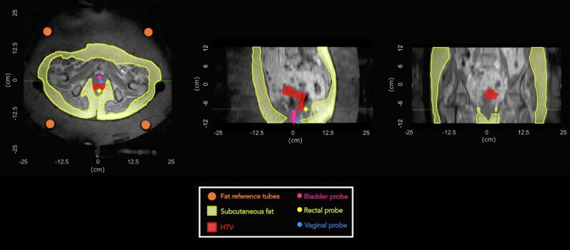
Background and purpose: Hyperthermia, the elevation of target temperature to 39-44 °C, is monitored using temperature probes. However, these provide limited spatial information, sampling only a few discrete locations. Magnetic resonance (MR) thermometry currently offers an option for three-dimensional (3D) temperature monitoring during hyperthermia. This study compares and correlates temperatures measured by intraluminal probes with MR-based temperatures in (1) the anatomical region containing the intraluminal probes and (2) the hyperthermia target volume (HTV), located at a distance from the probes and representing the primary region of clinical interest.
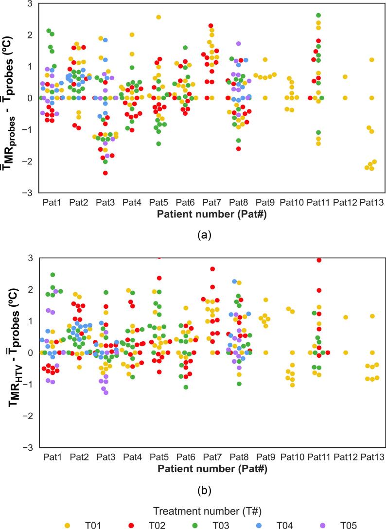
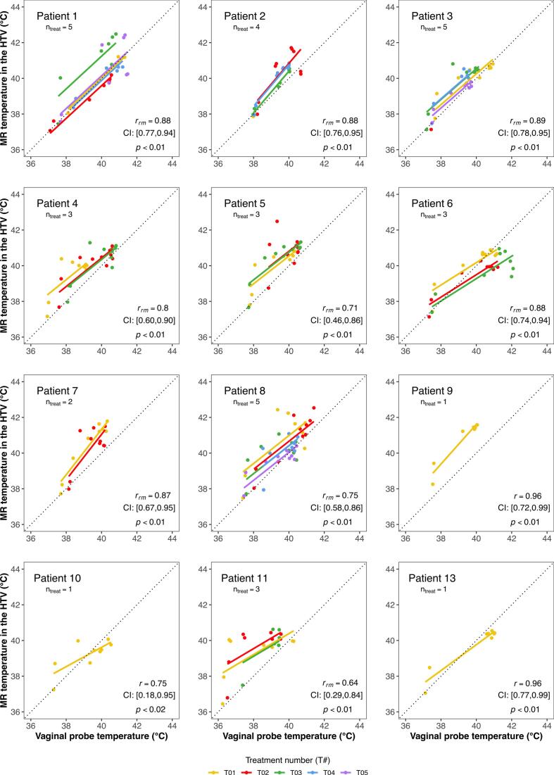
Methods: Thirteen locally advanced cervical cancer (LACC) patients treated with radiotherapy and hyperthermia were included. Hyperthermia was monitored using intraluminal probes and MR thermometry. MR-based temperatures were compared to intraluminal probe temperatures. Repeated measures correlation was applied to correlate probe and MR-based temperatures in the HTV across all data and on a patient-specific basis.
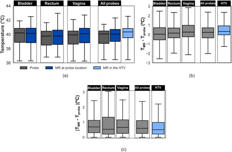
Results: MR-based temperatures at probe locations showed good agreement with probe measurements (median absolute error ≤ 0.7 °C). In the HTV, MR-based temperatures deviated by a median absolute error of 0.5 °C from probe temperatures. Repeated measures correlations (rrm) between MR and probe-based HTV temperatures ranged from 0.74 to 0.79 across all data and 0.64-0.96 on a patient-specific basis.
Conclusions: MR thermometry demonstrated promising performance for retrospective evaluation of temperature distributions in the HTV. While its current reliability for real-time treatment guidance remains limited, our results support further development towards broader clinical implementation in hyperthermia.
Keywords: Hyperthermia; Locally advanced cervical cancer; Magnetic resonance thermometry; Magnetic resonance-guided hyperthermia; Probe thermometry; Repeated measures correlation; Target volume temperature.
© 2025 The Author(s).
Conflict of interest statement
The authors declare the following financial interests/personal relationships which may be considered as potential competing interests: Gerard C. van Rhoon:•Past President of the European Society for Hyperthermic Oncology, retired 2022•Cofounder and shareholder Sensius BV•Holds/submitted several patents on hyperthermia related technology•Member executive committee Editorial Board Int. J. of Hyperthermia Royalties or licenses•E. Majorana Foundation; European School of Antennas; Various EU-Cost Actions•Dr. Sennewald Medizintechnik Gmbh•TU Munich•Japanese STM•SEOR•IT’IS Foundation Consulting fees•WO 2013/028064 Al•WO2020130824A1•WO2022235155A1 Support for attending meetings and/or travel•Received financial support to attend conferences from various companies, societies and charity
Figures
Similar articles
-
Accelerated proton resonance frequency-based magnetic resonance thermometry by optimized deep learning method.Med Phys. 2025 Jul;52(7):e17909. doi: 10.1002/mp.17909. Epub 2025 May 31. Med Phys. 2025. PMID: 40450352
-
Magnetic resonance perfusion for differentiating low-grade from high-grade gliomas at first presentation.Cochrane Database Syst Rev. 2018 Jan 22;1(1):CD011551. doi: 10.1002/14651858.CD011551.pub2. Cochrane Database Syst Rev. 2018. PMID: 29357120 Free PMC article.
-
A T1-based correction method for proton resonance frequency shift thermometry in breast tissue.Med Phys. 2021 Sep;48(9):4719-4729. doi: 10.1002/mp.15085. Epub 2021 Aug 6. Med Phys. 2021. PMID: 34265109 Free PMC article.
-
Comparison of Two Modern Survival Prediction Tools, SORG-MLA and METSSS, in Patients With Symptomatic Long-bone Metastases Who Underwent Local Treatment With Surgery Followed by Radiotherapy and With Radiotherapy Alone.Clin Orthop Relat Res. 2024 Dec 1;482(12):2193-2208. doi: 10.1097/CORR.0000000000003185. Epub 2024 Jul 23. Clin Orthop Relat Res. 2024. PMID: 39051924
-
Systemic pharmacological treatments for chronic plaque psoriasis: a network meta-analysis.Cochrane Database Syst Rev. 2021 Apr 19;4(4):CD011535. doi: 10.1002/14651858.CD011535.pub4. Cochrane Database Syst Rev. 2021. Update in: Cochrane Database Syst Rev. 2022 May 23;5:CD011535. doi: 10.1002/14651858.CD011535.pub5. PMID: 33871055 Free PMC article. Updated.
References
-
- Issels R.D., Lindner L.H., Verweij J., Wessalowski R., Reichardt P., Wust P., et al. Effect of neoadjuvant chemotherapy plus regional hyperthermia on long-term outcomes among patients with localized high-risk soft tissue sarcoma the EORTC 62961-ESHO 95 randomized clinical trial. J Am Med AssocOncol. 2018;4:483–492. doi: 10.1001/jamaoncol.2017.4996. - DOI - PMC - PubMed
-
- Dewey W.C., Hopwood L.E., Sapareto S.A., Gerweck L.E. Cellular responses to combinations of hyperthermia and radiation. Radiology. 1977;123:463–474. - PubMed
LinkOut - more resources
Full Text Sources
