. 2025 Jul 21:16:1593263.
doi: 10.3389/fimmu.2025.1593263. eCollection 2025.

MetE: a promising protective antigen for tuberculosis vaccine development

Affiliations

MetE: a promising protective antigen for tuberculosis vaccine development

Salem Salman Almujri et al. Front Immunol. .

Abstract

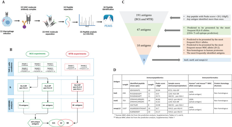
Introduction: Tuberculosis (TB), caused by Mycobacterium tuberculosis (MTB), remains a significant global health concern. The existing vaccine, Bacillus Calmette-Guérin (BCG), provides inconsistent protection, highlighting the pressing need for a more effective vaccine. We aimed to identify novel MTB antigens and assess their protective efficacy as TB vaccine candidates.

Methods: Using immunopeptidomics, we identified 64 and 80 unique mycobacterial antigens derived from BCG and MTB, respectively. We prioritised antigens based on HLA allele coverage through an immunoinformatics approach.

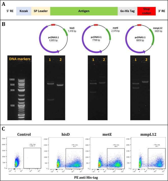
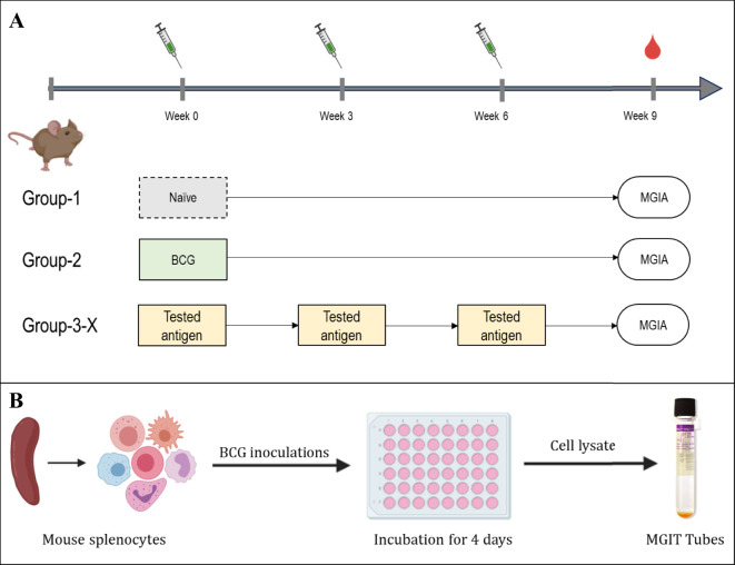
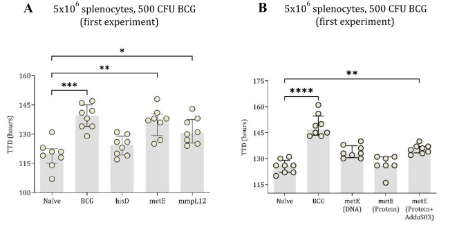
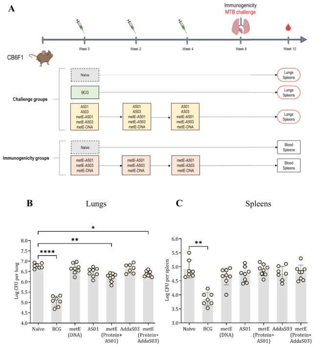
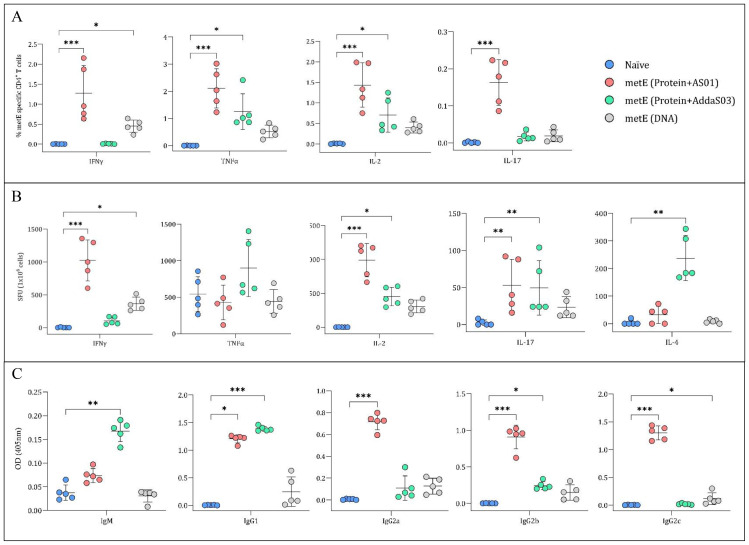
Results: The candidates, hisD, metE, and mmpL12, delivered as DNA vaccines, were evaluated for efficacy in mice using the ex vivo Mycobacterial Growth Inhibition Assay (MGIA) and metE was identified as a promising candidate. In vivo murine MTB challenge experiments confirmed the protective efficacy conferred by metE when formulated as recombinant protein with AS01™ or AddaS03™ adjuvants, compared to the naïve group. The immunogenic profiles of metE formulated in the two different adjuvants differed, with metE-AS01™ inducing antigen-specific IFN-γ, TNF-α, IL-2, IL-17, IgG1 and IgG2a-c, while metE-AddaS03™ induced TNF-α, IL-2, IL-17, IL-4, IgM, IgG1, IgG2b.

Conclusion: Our findings highlight metE as a promising protective antigen for future TB vaccine development.

Keywords: HLA/MHC; antigen discovery; immunoinformatics; immunopeptidomics; mass spectrometry; mycobacterium tuberculosis; tuberculosis; vaccines.


Conflict of interest statement

The authors declare that the research was conducted in the absence of any commercial or financial relationships that could be construed as a potential conflict of interest.

Figures

Figure 1
Figure 1
(A) Step-by-step procedure for antigen identification: THP-1 macrophages are infected with mycobacteria, harvested, lysed for immunoprecipitation, and peptides are eluted from MHC complexes (generated by BioRender). The peptides are separated by HPLC, analyzed via LC-MS/MS, and processed using Peaks software (v10). (B) Sample arrangement for immunopeptidomics analysis: In the BCG experiments, Bettencourt et al. (2020) prepared six samples, which underwent IP with L243 and W6/32 antibodies, identifying 70 antigens. Additional IP with B271 and IVA12 isolated HLA-DP and -DQ, identifying 64 antigens. In the MTB experiments, two samples underwent IP with three antibodies, leading to the identification of 80 antigens, totally 191 unique antigens. (C) Antigen selection criteria: selecting antigens with a Peaks score >20 or identified multiple times yielded 47 antigens out of 191. Antigens with full HLA-II coverage were narrowed down to 10. Finally, non-homologous antigens presented by common HLA-I alleles resulted in three candidates: hisD, metE, and mmpL12. (D) Characterization of candidate antigens using immunopeptidomics and immunoinformatics.
Figure 2
Figure 2
The design and validation of antigen inserts (hisD, metE, and mmpL12). (A) Schematic representation of the plasmid constructs showing insertion of Kozak sequences and signal peptides at the N-terminus, and a 6x-His tag followed by a stop codon at the C-terminus of each insert. (B) DNA agarose gels confirming successful plasmid assembly for hisD, metE, and mmpL12 constructs; lane 1 shows restriction enzyme-digested plasmids with bands at expected insert sizes, lane 2 shows uncut plasmids, and DNA markers are provided as size references. (C) Flow cytometry analysis confirming protein expression of each antigen (hisD, metE, mmpL12) following transfection of HEK293T cells, detected using an anti-6x-His antibody.
Figure 3
Figure 3
Experimental layout of MGIA experiments. (A) immunisation layout. CB6F1 mice were randomly divided into six groups, each containing eight mice. A single group was given a one-time intradermal BCG vaccination, while four groups designated for antigen testing received plasmid DNA vaccinations three times, with two-week intervals. One group of animals remained unvaccinated. (B) Schematic of the MGIA assay procedure; Splenocytes are co-cultured with approximately 300 or 500 CFU of BCG. After a 96-hour incubation period, cells were lysed, and both extracellular and intracellular bacteria were introduced to the BACTEC MGIT system and monitored until the time to detection (TTD) registered as positive.
Figure 4
Figure 4
TTD values of mycobacterial growth as measured by MGIA. In the first experiment, mouse splenocytes, counted at 5x10⁶ (A), were co-cultured with 500 CFU of BCG. These splenocytes were taken from both unvaccinated (naïve) mice and those vaccinated with BCG or DNA vaccines expressing hisD, metE or mmpL12. In the second experimental setup, TTD values were derived from 5x10⁶ mouse splenocytes co-cultured with BCG, inoculated at ~500 CFU (B). Splenocytes originated from naïve mice and those vaccinated with BCG, metE, metE-AddaS03™, or metE-DNA. Plotted circles represent individual mice, lines representing median data, and whiskers indicating interquartile ranges (IQR). For identifying significant deviations compared to the naïve control, a combination of the Kruskal-Wallis and Dunn’s multiple comparisons tests was applied. Significance levels are denoted as * for P values <0.05, ** for P values <0.01, and *** indicating P values <0.001.
Figure 5
Figure 5
Aerosol MTB-Challenge and Immunogenicity Experiments. (A) MTB challenge, immunisation schedule and experimental groups. (B) lung and (C) spleen CFU counts. Plotted circles represent individual mice, lines representing median data, and whiskers indicating interquartile ranges (IQR). For identifying significant deviations compared to the naïve control, a combination of the Kruskal-Wallis and Dunn’s multiple comparisons tests was applied. Significance levels are denoted as * for P values <0.05, ** for P values <0.01, and **** indicating P values <0.0001.
Figure 6
Figure 6
Analysis of Cytokine Secretion and metE-specific Antibody Titres. (A) Percentage of CD4+ T cells producing different cytokines. Mouse splenocytes were stained for CD4+ surface markers and cytokines (IFNγ, TNFα, IL-2, and IL-17) after overnight stimulation with metE protein. Cytokine staining was analysed by flow cytometry. (B) Cytokine secretion measured by ELISpot. Splenocytes were stimulated overnight with metE protein to measure the release of IFNγ, TNFα, IL-2, IL-4, and IL-17 cytokines. (C) MetE-specific antibody titers. Antibody titers (IgM, IgG1, IgG2a, IgG2b, and IgG2c) were analysed from mouse sera collected after the final vaccination by ELISA. Each plotted circle represents individual mice, with lines denoting median values. Significant differences compared to the naïve control were identified using the Kruskal-Wallis test with Dunn’s multiple comparisons test. Significance markers: * for P values <0.05, ** for P values <0.01, and *** for P values <0.001.

Similar articles

References

    1. Global tuberculosis report 2024. Geneva: World Health Organization; (2024). Available online at: https://www.who.int/publications/i/item/9789240101531.
    1. Mangtani P, Abubakar I, Ariti C, Beynon R, Pimpin L, Fine PE, et al. Protection by BCG vaccine against tuberculosis: a systematic review of randomized controlled trials. Clin Infect Dis. (2014) 58:470–80. doi: 10.1093/cid/cit790, PMID: - DOI - PubMed
    1. Rieder HL. International Union against Tuberculosis and Lung Disease, IUATLD. @ in Interventions for tuberculosis control and elimination. Paris: International Union Against Tuberculosis and Lung Disease Paris; (2002).
    1. Bell LC, Noursadeghi M. Pathogenesis of HIV-1 and Mycobacterium tuberculosis co-infection. Nat Rev Microbiol. (2018) 16:80–90. doi: 10.1038/nrmicro.2017.128, PMID: - DOI - PubMed
    1. Lin Y, Slight SR, Khader SA. Th17 cytokines and vaccine-induced immunity. In: Seminars in immunopathology ser. Berlin: Springer; (2010). p. 32. - PMC - PubMed