. 2025 Aug 18;8(8):7188-7200.
doi: 10.1021/acsabm.5c00907. Epub 2025 Aug 5.

Design and Assembly of a Cargo-Agnostic Hollow Two-Lidded DNA Origami Box

Affiliations

Design and Assembly of a Cargo-Agnostic Hollow Two-Lidded DNA Origami Box

Abigail Koep et al. ACS Appl Bio Mater. .

Abstract

DNA origami, a method of folding DNA into precise nanostructures, has emerged as a powerful tool for the design of complex nanoscale shapes. It has great potential as a technology to encapsulate and release cargos spanning small molecules through large proteins, while remaining stable in a variety of ex vivo processing conditions and in vivo environments. While DNA origami has been utilized for drug delivery applications, the vast majority of these structures have been flexible, flat 2D or solid 3D nanostructures. There is a crucial need for a hollow and completely enclosed design capable of holding and eventually releasing a variety of cargos. In this paper, we present the design and assembly of a hollow DNA origami box with two lids. We characterize the isothermal conditions for structural assembly within minutes. We demonstrate that passive loading of small molecules is charge dependent. We also outline an approach to design staple extensions pointing into the cavity or outside of the hollow DNA origami, allowing for the active loading of protein or the potential for decoration with passivating or targeting molecules.

Keywords: Smart nanomaterials; controlled release; helical twist; isothermal assembly; stability.


Figures

1
1
Characterization of hollow DNA origami boxes with two openable lids. (A) CanDo renderings of cadnano files based on the design of the DNA origami box with no lids open, one-lid-open, and two-lids-open. (B) DLS graphs of the size distribution after gradient-temperature assembly and filter purification. The average diameter distribution from five independent assemblies is noted as the black curve and the peak diameter is denoted on the graph (purified at 5000 g for 20 min, 3 μM DNA origami). (C) AFM images of each gradient-temperature assembly. Scale bar represents 200 nm. (D) Shape quantification of the AFM images with sample diagrams and images for each expected assembly shape. Counting was performed on four AFM images from each of two independent assembly experiments per design. The graph shows the fraction of the shapes within an assembly.
2
2
Temperature-dependent assembly of hollow DNA origami boxes under isothermal conditions. (A) Agarose gel of different assemblies over a range of temperatures after heating the assembly mixture for 10 min at 95 °C. (B) Apparent size in equivalent dsDNA base pairs and band intensity of the DNA origami assembly across various assembly temperatures. Red is the closed box assembly. Light blue is one-lid-open assembly. Dark blue is two-lids-open box assembly. (C) Apparent size in equivalent dsDNA base pairs and band intensity of the secondary DNA band that runs more slowly than the DNA origami assembly. Red is the closed box assembly. Light blue is one-lid-open assembly. Dark blue is two-lids-open box assembly. The gray shaded regions are the apparent size/intensity ranges calculated using 95% confidence intervals when assembly is done using a thermal gradient of −1 °C/min starting from 95 °C. Solid lines are melting temperature isotherm of complementary DNA binding fit to the data (c = 5.00 nM, ΔH = −200 kJ/mol, ΔS = −605 J/mol, C p = −214 J/mol K). The thin line shows the distribution of melting temperatures of the staple strands.
3
3
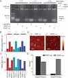
Hollow DNA origami box separation is sensitive to centrifugal force. (A) Agarose gel showing the lack of staples when the assemblies are purified at 5000 g for 20 min compared to 20,000 g for 5 min. (B) DNA origami and (C) normalized staple band fluorescence intensities for closed box assemblies (red), one-lid-open box assemblies (light blue), and two-lids-open box assemblies (dark blue) shown in (A). DNA origami and staple intensities were normalized by dividing the intensity of a particular band by the sum of all band intensities for a particular sample in one lane. (D) In solution AFM images of the origami structures obtained after purification at each of the two centrifugal forces (3 μM DNA origami). Solid white boxes show DNA origami structures within the expected size. Dashed white boxes show particles too small to be DNA origami structures. The scale bar represents 200 nm. (E) Quantification of number of DNA origami boxes vs small box fragments. Counting was performed on one AFM image from a single gradient-temperature assembly experiment per condition.
4
4
Hollow DNA origami boxes are temperature, pH and solvent stable. Electrophoretic resolution through agarose gels of hollow closed box DNA origami isothermal assemblies held for various times (A) at 4 °C, pH 7.5 and 12.5 mM Mg­(OAc)2, (B) at 4 °C, pH 7.5 and 12.5 mM Mg­(OAc)2 (purified), (C) in 4 °C, pH 7.5 and 12.5 mM Mg­(OAc)2 10% DMSO, and (D) assembled in 4 °C, pH 7.5, 12.5 mM Mg­(OAc)2 and incubated in 10% FBS. Quantification of the DNA origami band intensity for DNA origami assemblies kept at different (E) temperatures, (F) pH (purified), and (G) solvent or serum conditions. Each dot represents the average of at least three independent assembly experiments. (H) Quantification of the apparent DNA origami band size in equivalent dsDNA base pairs normalized to the no serum exposure DNA origami band size as measured in equivalent dsDNA base pairs. DNA origami assemblies kept in 10% DMSO, assembled in 12.5 mM Mg­(OAc)2 and incubated in 10% FBS, and assembled in 100 mM NaCl and incubated in 10% FBS. Each bar represents the average of three independent assembly experiments. All error bars represent 95% confidence intervals.
5
5
Hollow DNA origami loading is dependent on the charge of the cargo. Fluorescent molecules with (A) negative, (B) no, and (C) positive charge were loaded in hollow DNA origami boxes, assembled isothermally, and then separated use PEG purification. Chemical structure and charge in parentheses (left) and agarose gels of 10 mg/mL fluorescent molecule loaded in 3 μM hollow DNA origami boxes (right) are shown. The dotted green box indicates the location of loaded DNA origami boxes. The dotted white box indicates the expected location of the unloaded DNA origami boxes. (D) Quantification of the band intensity referring to the DNA origami box loaded with fluorophore. Dark green refers to the signal when fluorophore was added to preassembled boxes and light green refers to the signal when fluorophore was added during DNA origami assembly. Each bar represents the average of three independent assembly experiments. (E) Quantification of the shift of the band induced by the DNA origami box loaded with fluorophore compared to DNA origami box with no fluorophore. Each square represents the average of three independent assembly experiments. All error bars represent 95% confidence intervals.
6
6
Staple extensions from the hollow DNA origami box can be designed for capture of cargo inside or for decoration with targeting molecules outside, using 5 nt extensions. (A) Schematic of 5nt staples extending inside and outside of the hollow DNA origami box (left). Zoomed view of a single DNA helical twist, showing the direction of extension of different staples. (B) Schematic of the experimental process of attaching hollow DNA origami boxes with biotinylated extended staples to streptavidin coated beads to determine if the extension was directed toward the inside or outside. (C) Agarose gel showing the binding capabilities of hollow DNA origami boxes with staple extensions at different positions. Light blue refers to staples oriented outside and purple refers to staples oriented inside. The no bead binding wells are samples taken from the supernatant after beads were spun down. The bead binding wells are samples taken from the solution after beads were exposed to NaOH, removing the biotinylated hollow DNA origami boxes from the streptavidin coated beads and subsequently neutralized with HCl. (D) Quantification of the scaffold band intensity of the hollow DNA origami box. Each bar represents the average of three independent isothermal assembly experiments. All error bars represent 95% confidence intervals.
7
7
Hollow DNA origami loading requires inside staple extensions of a minimum length. (A) Schematic showing that short biotinylated staples on DNA origami boxes, assembled isothermally, do not attach to streptavidin, while long biotinylated staples on DNA origami boxes can attach to streptavidin. Components not shown to scale. (B) Agarose gels of DNA origami boxes with short, outside (left) and long, inside (right) extensions. Green dashed box refers to the hollow DNA origami box with attached streptavidin bound to fluorescent biotin. Schematics of other species of bound streptavidin are shown to the left.

Similar articles

References

    1. Lucas C. R., Halley P. D., Chowdury A. A., Harrington B. K., Beaver L., Lapalombella R., Johnson A. J., Hertlein E. K., Phelps M. A., Byrd J. C., Castro C. E.. DNA Origami Nanostructures Elicit Dose-Dependent Immunogenicity and Are Nontoxic up to High Doses In Vivo. Small. 2022;18(26):2108063. doi: 10.1002/smll.202108063. - DOI - PMC - PubMed
    1. Wamhoff E.-C., Knappe G. A., Burds A. A., Du R. R., Neun B. W., Difilippantonio S., Sanders C., Edmondson E. F., Matta J. L., Dobrovolskaia M. A., Bathe M.. Evaluation of Nonmodified Wireframe DNA Origami for Acute Toxicity and Biodistribution in Mice. ACS Appl. Bio Mater. 2023;6(5):1960–1969. doi: 10.1021/acsabm.3c00155. - DOI - PMC - PubMed
    1. Du R. R., Cedrone E., Romanov A., Falkovich R., Dobrovolskaia M. A., Bathe M.. Innate Immune Stimulation Using 3D Wireframe DNA Origami. ACS Nano. 2022;16(12):20340–20352. doi: 10.1021/acsnano.2c06275. - DOI - PMC - PubMed
    1. Mitchell M. J., Billingsley M. M., Haley R. M., Wechsler M. E., Peppas N. A., Langer R.. Engineering Precision Nanoparticles for Drug Delivery. Nat. Rev. Drug Discovery. 2021;20(2):101–124. doi: 10.1038/s41573-020-0090-8. - DOI - PMC - PubMed
    1. Dutta P. K., Zhang Y., Blanchard A. T., Ge C., Rushdi M., Weiss K., Zhu C., Ke Y., Salaita K.. Programmable Multivalent DNA-Origami Tension Probes for Reporting Cellular Traction Forces. Nano Lett. 2018;18(8):4803–4811. doi: 10.1021/acs.nanolett.8b01374. - DOI - PMC - PubMed

LinkOut - more resources