. 2025 Dec;40(1):2538673.
doi: 10.1080/14756366.2025.2538673. Epub 2025 Aug 5.

Discovery of a novel RSK2 inhibitor for the treatment of metastatic pancreatic cancer

Affiliations

Discovery of a novel RSK2 inhibitor for the treatment of metastatic pancreatic cancer

Chi-Hsiu Chung et al. J Enzyme Inhib Med Chem. 2025 Dec.

Abstract

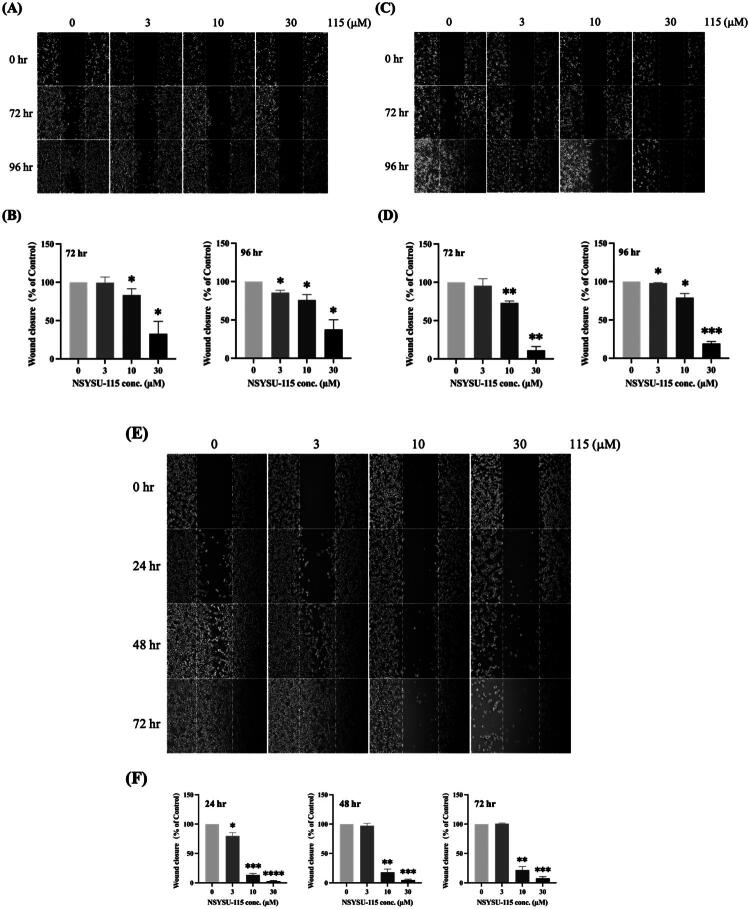
Pancreatic cancer is among the most lethal malignancies, with a five-year survival rate of only 6%. For patients with metastatic disease, current treatments extend median survival by merely four months. This study addresses the urgent need for targeted therapies, as no specific drugs are currently available. Clinical analyses revealed significantly elevated RSK2 expression in pancreatic cancer tissues, associated with shorter survival. We aimed to identify a novel RSK2 inhibitor for metastatic pancreatic cancer. Through structure-based virtual screening, we identified NSYSU-115 as a promising candidate with an IC50 of 45.5 nM. At low concentrations, NSYSU-115 significantly suppressed colony formation, while higher concentrations reduced cell viability and proliferation. It also inhibited phosphorylation of IκBα, a known RSK2 substrate, in a dose- and time-dependent manner. Furthermore, NSYSU-115 impaired cell migration and altered epithelial-mesenchymal transition (EMT) markers. These findings highlight NSYSU-115 as a potent kinase inhibitor with promising therapeutic potential for pancreatic cancer treatment.

Keywords: RSK2 inhibitor; metastasis; pancreatic cancer; structure-based virtual screening.


Conflict of interest statement

The authors report no conflicts of interest.

Figures

Figure 1.
Figure 1.
Overview of study. (A) NCI (National Cancer Institute) chemical library was used for a virtual screening of RSK2 inhibitors. (B) The top-ranked compounds were selected for enzyme-based assays to identify their activity against RSK2. (C) The potent inhibitor was further validated for in vitro cell growth and migration function.
Figure 2.
Figure 2.
An analysis of the selectivity profile across the kinome. (A) NSYSU-115 was tested at 1 μM across a panel of 41 kinases. (B) NSYSU-115 is selective towards RSK2. Red dots indicate kinases where the compounds exhibit an inhibition effect greater than 50%.
Figure 3.
Figure 3.
Kinase inhibition of NSYSU-115. The kinase inhibition assay was used to test the efficacy of compounds. The IC50 (half maximal inhibitory concentration) of NSYSU-115 on RSK2 kinase activity is 45.5 nM.
Figure 4.
Figure 4.
Interactions of NSYSU-115 in RSK2. (A) Groups of compound structures in two sites. Quinoline (purple) and triazine (red) groups were filling in two sites of RSK2. (B) Docking pose of NSYSU-115. The compound (yellow) was docked into the RSK2 binding site (blue). The binding site residues are shown as stick representations, and hydrogen bonds are illustrated with dashed green lines.
Figure 5.
Figure 5.
Effects of NSYSU-115 on cell viability and cell proliferation in pancreatic cancer cell lines. Cell viability was assessed using MTT assays in three pancreatic cancer cell lines: AsPC-1 (A), PANC-1 (B), and MIA PaCa-2 (C). Cell proliferation was measured using SRB assays in AsPC-1 (D), PANC-1 (E), and MIA PaCa-2 (F). Cells were treated with either vehicle control (0.1% DMSO) or various concentrations of NSYSU-115 (0.3, 1, 3, 10, and 30 µM) for 72 hours. The data presented, expressed as mean ± SD, are based on a minimum of three independent experiments. Statistical significance is indicated by asterisks: *, p < 0.05; **, p < 0.01; ***, p < 0.001; ****, p < 0.0001, compared to the control group.
Figure 6.
Figure 6.
Effects of NSYSU-115 on colony-forming ability of pancreatic cancer cell lines. Cells were treated with either vehicle control (0.1% DMSO) or various concentrations of NSYSU-115 (0.3, 1, 3, 10, and 30 µM) for 7 days. Crystal violet was used to stain colonies for enhanced visibility, and the number of colonies was quantified using ImageJ software. The results in (A) and (D) were obtained from AsPC-1, whereas the results in (B) and (E) were obtained from PANC-1. Additionally, the results in (C) and (F) were generated from MIA PaCa-2. The presented data, representing the mean ± SD, are derived from a minimum of three independent experiments. Statistical significance is denoted by asterisks: *, p < 0.05; **, p < 0.01; ***, p < 0.001; ****, p < 0.0001 compared to the control group.
Figure 7.
Figure 7.
Inhibitory effects of NSYSU-115 on RSK2 downstream phosphorylation and degradation. AsPC-1 (A) and PANC-1 (D) cells were treated with either vehicle control (0.1% DMSO) or NSYSU-115 at concentrations of 3, 10, and 30 μM for 48 and 72 hours. Following treatment, cell lysates were subjected to Western blot analysis using specific antibodies. Band intensities were quantified using ImageJ software and normalised to the loading control for AsPC-1 (B) and PANC-1 (E). The ratios of phosphorylated IκBα (p-IκBα) to total IκBα (t-IκBα) were calculated based on the quantitative results and shown for AsPC-1 (C) and PANC-1 (F). The presented data, reflecting the mean ± SD, were obtained from a minimum of three independent experiments. Statistical significance is indicated by asterisks: *, p < 0.05; **, p < 0.01; ***, p < 0.001; ****, p < 0.0001, in comparison to the control group.
Figure 8.
Figure 8.
Inhibitory effects of NSYSU-115 on cell migration in pancreatic cancer cell lines. Cells, including AsPC-1 (A), PANC-1 (C), and MIA PaCa-2 (E), were treated with either vehicle control (0.1% DMSO) or various concentrations of NSYSU-115 (3, 10, and 30 µM) for the indicated time points. A wound distance of 500 µm was marked with white dashed lines, and images were captured using an inverted phase-contrast microscope. For each condition, at least 5 images were randomly selected , and the wound distance was measured using ImageJ software. The quantification results for AsPC-1 (B) at 72 hours are shown on the left, and at 96 hours on the right. Similarly, the quantification results for PANC-1 (D) at 72 hours are displayed on the left, and at 96 hours on the right. For MIA PaCa-2 (F), the quantification results at 24, 48, and 72 hours are displayed at the bottom. Data are presented as the mean ± SD, based on a minimum of three independent experiments. Statistical significance is denoted by asterisks: *, p < 0.05; **, p < 0.01; ***, p < 0.001; ****, p < 0.0001, compared to the control group.
Figure 9.
Figure 9.
Inhibitory effects of NSYSU-115 on vimentin expression in pancreatic cancer cell lines. Cells were treated with either vehicle control (0.1% DMSO) or various concentrations of NSYSU-115 (3, 10, and 30 µM). RT-qPCR analysis was performed to quantify the expression levels of the mesenchymal marker vimentin, using 18S and GAPDH as internal controls. AsPC-1 cells were treated for 48 hours (A) and 72 hours (C), with 18S as the internal control. PANC-1 cells were treated for 48 hours (B) and 72 hours (D), with GAPDH as the internal control. Data are presented as the mean ± SD, based on a minimum of three independent experiments. Statistical significance is indicated by asterisks: *, p < 0.05; **, p < 0.01, compared to the control group.

Similar articles

References

    1. Oberstein PE, Olive KP.. Pancreatic cancer: why is it so hard to treat? Therap Adv Gastroenterol. 2013;6(4):321–337. - PMC - PubMed
    1. Gillen S, Schuster T, Meyer Zum Büschenfelde C, Friess H, Kleeff J.. Preoperative/neoadjuvant therapy in pancreatic cancer: a systematic review and meta-analysis of response and resection percentages. PLoS Med. 2010;7(4):e1000267. - PMC - PubMed
    1. Chang JS, Chen L-T, Shan Y-S, Chu P-Y, Tsai C-R, Tsai H-J.. The incidence and survival of pancreatic cancer by histology, including rare subtypes: a nation-wide cancer registry-based study from Taiwan. Cancer Med. 2018;7(11):5775–5788. - PMC - PubMed
    1. Oettle H, Neuhaus P, Hochhaus A, Hartmann JT, Gellert K, Ridwelski K, Niedergethmann M, Zülke C, Fahlke J, Arning MB, et al. Adjuvant chemotherapy with gemcitabine and long-term outcomes among patients with resected pancreatic cancer: the CONKO-001 randomized trial. JAMA. 2013;310(14):1473–1481. - PubMed
    1. Neoptolemos JP, Moore MJ, Cox TF, Valle JW, Palmer DH, McDonald AC, Carter R, Tebbutt NC, Dervenis C, Smith D, et al. Effect of adjuvant chemotherapy with fluorouracil plus folinic acid or gemcitabine vs observation on survival in patients with resected periampullary adenocarcinoma: the ESPAC-3 periampullary cancer randomized trial. JAMA. 2012;308(2):147–156. - PubMed

MeSH terms

Substances

LinkOut - more resources