. 2025 Oct 15;152(20):dev204718.
doi: 10.1242/dev.204718. Epub 2025 Aug 5.

KLF7 orchestrates hippocampal development through neurogenesis and Draxin-mediated neuronal migration

Affiliations

KLF7 orchestrates hippocampal development through neurogenesis and Draxin-mediated neuronal migration

Yitong Liu et al. Development. .

Abstract

The hippocampus, a brain region that is crucial for cognitive learning, memory and emotional regulation, undergoes its primary development during embryonic and early postnatal stages. Krüppel-like factor 7 (KLF7), a transcription factor associated with autism spectrum disorder and intellectual developmental disorders, plays a pivotal role in brain development. In this study, we investigated the role of KLF7 in hippocampal development using conditional knockout mice [Emx1-Cre;Klf7Flox(F)/F]. We found that KLF7 deletion in hippocampal progenitors resulted in significant hippocampal shrinkage, disrupting neurogenesis, neuronal differentiation and migration. KLF7 mutant mice exhibited abnormal neuronal projections, anxiety- and depression-like behaviors, and memory impairments. Transcriptomic profiling identified Draxin, a neural chemorepellent, as a key downstream target of KLF7. Remarkably, overexpression of Draxin rescued dentate gyrus granule cell migration defects in KLF7 mutant mice. These findings demonstrate that KLF7 is essential for proper hippocampal development and function, regulating neuronal migration through Draxin. This study provides mechanistic insights into the neurological deficits associated with KLF7 pathogenic variants and highlights potential therapeutic targets for neurodevelopmental disorders.

Keywords: Draxin; Hippocampus; Krüppel-like factor 7; Mouse; Neurogenesis; Neuronal migration; Neuronal projections.


Conflict of interest statement

Competing interests The authors declare no competing or financial interests.

Figures

Fig. 1.
Fig. 1.
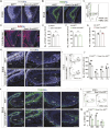
KLF7 deletion disrupts hippocampal development. (A) Nissl staining of hippocampus at different stages. Dashed lines indicate CA-DG interface. (B-D) Quantification of hippocampal, CA, and DG areas in Klf7F/F and Emx1-Cre;Klf7F/F mice (n=4-6). (E) Immunofluorescence for Ctip2 (green) and Math2 (red) in hippocampus at E18.5, P0 and P7. (F-H) Quantification of Ctip2+ (CA1-CA2, DG) and Math2+ (CA3) cells (n=3-4). (I,J) NeuroD immunofluorescence and quantification in DG at P7 (n=3-4). ns, not significant; *P<0.05; ***P<0.001 (unpaired two-tailed t-test). Data are mean±s.e.m. CA, cornu ammonis; DG, dentate gyrus. Scale bars: 500 µm (A); 200 µm (E); 50 µm (I).
Fig. 2.
Fig. 2.
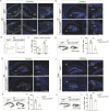
KLF7 cKO reduces hippocampal NPCs and IPCs. (A,B) PAX6 and TBR2 immunostaining at E14.5. Dashed lines indicate hippocampal region. (C) Schematic distribution of PAX6+ and TBR2+ cells at E14.5. (D) EdU staining at E14.5. Dashed lines indicate hippocampus. (E-G) Quantification of PAX6+, TBR2+ and EdU+ cells at E14.5 (n=3-4). (H-M) PAX6 and TBR2 immunostaining, schematic distribution and quantification at E18.5 (n=4). Dashed lines indicate the distinct developing regions of the hippocampus. ns, not significant; **P<0.01; ***P<0.001 (unpaired two-tailed t-test). Data are mean±s.e.m. CH, cortical hem; DMS, dentate migratory stream; DNE, dentate neuroepithelia; HNE, hippocampal neuroepithelium; IZ, intermediate zone; LV, lateral ventricles; VZ, ventricular zone; 1ry, primary germinative matrix; 2ry, secondary germinative matrix; 3ry, tertiary germinative matrix. Scale bars: 500 µm (A,B,D); 200 µm (H,K).
Fig. 3.
Fig. 3.
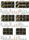
KLF7 cKO reduces postnatal DG neural precursor cells. (A-L) TBR2 immunostaining (A,D,G,J), schematic distribution (B,E,H,K) and quantification (C,F,I,L) in hippocampus at P0, P3, P7 and P14 (n=3-4). ns, not significant; *P<0.05; **P<0.01; ***P<0.001 (unpaired two-tailed t-test). Data are mean±s.e.m. DG, dentate gyrus; DMS, dentate migratory stream; GCL, granule cell layer; ML, molecular layer; SGZ, subventricular zone; 3ry, tertiary germinative matrix. Scale bars: 100 µm.
Fig. 4.
Fig. 4.
KLF7 regulates neural progenitor cell cycle and differentiation. (A,B) EdU and PCNA, PAX6, TBR2 immunostaining from E15.5 to P0. Dashed lines indicate distinct regions. (C-E) Quantification of EdU+PCNA, EdU+PAX6+ and EdU+TBR2+ cells (n=3-4). (F-O) Similar analysis at different hippocampal subregions and time points. ns, not significant; *P<0.05; **P<0.01; ***P<0.001 (unpaired two-tailed t-test). Data are mean±s.e.m. DNE, dentate neuroepithelia; HNE, hippocampal neuroepithelium; IZ, intermediate zone; LV, lateral ventricles; VZ, ventricular zone; 1ry, primary germinative matrix; 2ry, secondary germinative matrix; 3ry, tertiary germinative matrix. Scale bars: 50 µm.
Fig. 5.
Fig. 5.
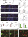
KLF7 cKO disrupts neuronal migration. (A) EdU injection timeline at E12.5/E14.5; analysis at P7. (B-I) EdU immunostaining and quantification in CA, DG and hypothalamus (n=3-4). Dashed lines show the interface between the CA and DG. (J-O) Schematic, immunostaining and quantification of in utero electroporation (IUE) at E14.5/P0 and neuronal migration analysis at P7/P21 (n=3). ns, not significant; **P<0.01; ***P<0.001 (unpaired two-tailed t-test). Data are mean±s.e.m. 3V, third ventricle. Scale bars: 100 µm.
Fig. 6.
Fig. 6.
KLF7 cKO reduces dendritic spines. (A-D) Vglut1 and synaptoporin immunostaining and quantification at P7 (n=4). (E-I) Golgi staining and dendritic spine quantification in CA and DG (n=4). **P<0.01; ***P<0.001 (unpaired two-tailed t-test). Data are mean±s.e.m. Scale bars: 100 µm (A,C); 200 µm (E), 5 µm (F,G).
Fig. 7.
Fig. 7.
KLF7 cKO disrupts dCA1 projections. (A-H) Coronal sections showing monosynaptic input labeling to ipsilateral and contralateral dCA1 PNs along the RC axis in Klf7F/F and Emx1-Cre;Klf7F/F mice. (I-L) Monosynaptic cortical input labeling to dCA1 in both groups. Dashed lines indicate distinct regions of the hippocampus and cortex. Arrows show various hippocampal subregions, including the boundary between the SP and SR layers, the sub region, and the DG. CA, cornu ammonis; DG, dentate gyrus; ECT, ectorhinal cortex; ENT, entorhinal cortex; ML, molecular layer; PRh, perirhinal cortex; SL, stratum lucidum; SLM, stratum lacunosum-moleculare; SO, stratum oriens; SP, stratum pyramidale; SR, stratum radiatum; Sub, subiculum. Scale bars: 200 µm (A-H); 500 µm (I-L).
Fig. 8.
Fig. 8.
KLF7 cKO affects adult behavior. (A-D) Open field test (OFT) movement analysis (n=10). (E,F) Tail suspension test (TST) and forced swim tests (FST) (n=8-11). (G,H) Elevated plus maze (EPM) test (n=10-12). (I,J) Marble burying test (MBT) (n=10-12). (K,L) Y-maze test (n=8). (M-Q) Morris Water Maze (MWM) learning and memory test (n=10-12). ns, not significant; *P<0.05; **P<0.01; ***P<0.001 [unpaired two-tailed t-test (A-M,O-Q), two-way ANOVA followed by Sidak's multiple comparisons test (N)]. Data are mean±s.e.m.
Fig. 9.
Fig. 9.
KLF7 regulates neuronal migration via Draxin. (A) Volcano plot of differentially expressed genes (DEGs) in Emx1-Cre;Klf7F/F mice. (B) GO analysis of DEGs. (C,D) RT-qPCR and luciferase assay validating candidate genes. (E-H) Draxin in situ hybridization, immunofluorescence and quantification at P7 (n=3). (I-L) In utero electroporation of Draxin and neuronal migration analysis at P7/P21 (n=3). ns, not significant; *P<0.05; **P<0.01; ***P<0.001 [unpaired two-tailed t-test (A-I,L), two-way ANOVA followed by Sidak's multiple comparisons test (J)]. Data are mean±s.e.m. Scale bars: 100 µm (E,G); 50 µm (I,K).

Similar articles

References

    1. Ahmed, G., Shinmyo, Y., Ohta, K., Islam, S. M., Hossain, M., Naser, I. B., Riyadh, M. A., Su, Y., Zhang, S., Tessier-Lavigne, M.et al. (2011). Draxin inhibits axonal outgrowth through the netrin receptor DCC. J. Neurosci. 31, 14018-14023. 10.1523/JNEUROSCI.0943-11.2011 - DOI - PMC - PubMed
    1. Alexandre, V., Walz, R., Bianchin, M. M., Velasco, T. R., Terra-Bustamante, V. C., Wichert-Ana, L., Araújo, D., Machado, H. R., Assirati, J. A., Carlotti, C. G.et al. (2006). Seizure outcome after surgery for epilepsy due to focal cortical dysplastic lesions. Seizure-Eur. J. Epilep. 15, 420-427. 10.1016/j.seizure.2006.05.005 - DOI - PubMed
    1. Angevine, J. B.Jr. (1965). Time of neuron origin in the hippocampal region. An autoradiographic study in the mouse. Exp. Neurol. Suppl. Suppl. 2, 1-70. - PubMed
    1. Bátiz, L. F., Castro, M. A., Burgos, P. V., Velásquez, Z. D., Muñoz, R. I., Lafourcade, C. A., Troncoso-Escudero, P. and Wyneken, U. (2016). Exosomes as novel regulators of adult neurogenic niches. Front. Cell. Neurosci. 9, 501. 10.3389/fncel.2015.00501 - DOI - PMC - PubMed
    1. Bayer, S. A. (1980). Development of the hippocampal region in the rat I. Neurogenesis examined with 3H-thymidine autoradiography. J. Comp. Neurol. 190, 87-114. 10.1002/cne.901900107 - DOI - PubMed

Substances

LinkOut - more resources