

A novel endovascular approach using transcalvarial emissary vein access for scalp and skull base AVMs: Case report
- PMID: 40763497
- PMCID: PMC12374205
- DOI: 10.1016/j.ijscr.2025.111760
A novel endovascular approach using transcalvarial emissary vein access for scalp and skull base AVMs: Case report
Abstract
Introduction and importance: Scalp arteriovenous malformations (SAVMs) are rare high-flow vascular lesions with complex angioarchitecture, often involving both superficial and deep components. When the malformation extends toward the skull base or includes deep venous drainage, traditional transarterial or percutaneous access may be insufficient. While direct puncture techniques have gained traction, transosseous access via emissary veins has not previously been described.
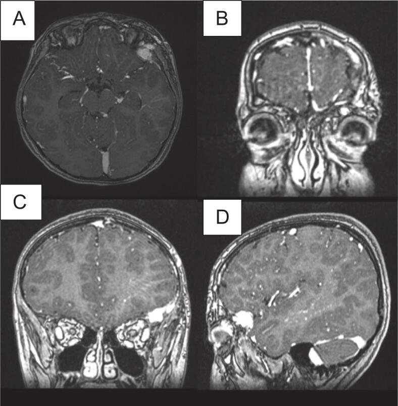
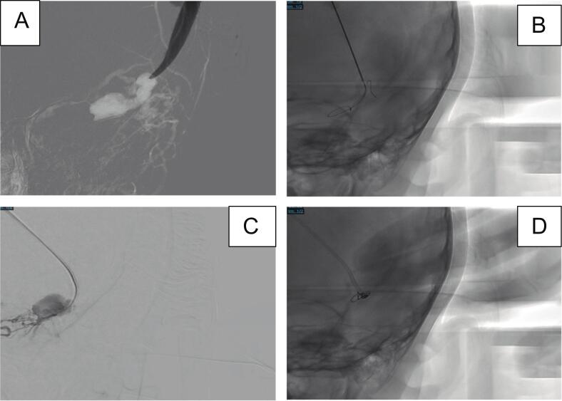
Case presentation: A 12-year-old girl presented with a pulsatile, fluctuant swelling over the left forehead and localized headache. Imaging revealed a mixed-type SAVM involving the left sphenoid ridge, frontal bone, and scalp, supplied by the ophthalmic artery and cortical branches of the middle cerebral artery, with multiple draining veins. Due to tortuous arterial feeders and the risk of non-target embolization, conventional transarterial access was deemed unsafe. A transcalvarial micropuncture technique was employed through a burr hole targeting an emissary vein, granting direct access to a deep venous pouch. Under fluoroscopic guidance, detachable coils were deployed, achieving complete angiographic obliteration. The patient remained neurologically intact, with no complications and an excellent cosmetic outcome. Follow-up imaging at 1, 3, and 6 months showed no recurrence.
Clinical discussion: This case demonstrates the feasibility of using a transosseous emissary vein approach for embolization of complex SAVMs. When conventional access is limited, this technique offers precise, direct venous access with reduced morbidity.
Conclusion: This is the first reported case of SAVM embolization using transcalvarial micropuncture via an emissary vein. The technique presents a novel, safe, and effective adjunct in managing complex scalp AVMs involving the skull base.
Keywords: Aneurysms; Endovascular; SAVM; Transcalvarial access.
Copyright © 2025. Published by Elsevier Ltd.
Conflict of interest statement
Conflict of interest statement The authors declare no commercial or financial conflicts of interest.
Figures


Similar articles
-
Prescription of Controlled Substances: Benefits and Risks.2025 Jul 6. In: StatPearls [Internet]. Treasure Island (FL): StatPearls Publishing; 2025 Jan–. 2025 Jul 6. In: StatPearls [Internet]. Treasure Island (FL): StatPearls Publishing; 2025 Jan–. PMID: 30726003 Free Books & Documents.
-
Dural arteriovenous fistulas of the hypoglossal canal: systematic review on imaging anatomy, clinical findings, and endovascular management.J Neurosurg. 2015 Apr;122(4):883-903. doi: 10.3171/2014.10.JNS14377. Epub 2014 Nov 21. J Neurosurg. 2015. PMID: 25415064
-
Injection sclerotherapy for varicose veins.Cochrane Database Syst Rev. 2021 Dec 10;12(12):CD001732. doi: 10.1002/14651858.CD001732.pub3. Cochrane Database Syst Rev. 2021. PMID: 34883526 Free PMC article.
-
Anterior Approach Total Ankle Arthroplasty with Patient-Specific Cut Guides.JBJS Essent Surg Tech. 2025 Aug 15;15(3):e23.00027. doi: 10.2106/JBJS.ST.23.00027. eCollection 2025 Jul-Sep. JBJS Essent Surg Tech. 2025. PMID: 40821726 Free PMC article.
-
Angiographic predictors of spontaneous obliteration of transarterial partially embolized brain arteriovenous malformations.Interv Neuroradiol. 2023 Aug;29(4):371-378. doi: 10.1177/15910199221092579. Epub 2022 Mar 31. Interv Neuroradiol. 2023. PMID: 35360965 Free PMC article.
References
-
- Wali S., Alsolmi A., Babgi M., Alqurashi A., Alghamdi D., Bajunaid K., Baeesa S. Scalp arteriovenous malformation with dual bilateral arterial feeders: case report and review of literature. Interdiscip. Neurosurg.: Adv. Techniq. Case Manag. 2022;27 doi: 10.1016/j.inat.2021.101431. - DOI
-
- Lee K.M., Kim E.J., Park B.J., Kim K.H. Direct puncture embolization of scalp arteriovenous malformation in a patient with severe hemophilia a: a case report. J. Korean Soc. Radiol. 2011;65(3):229–233. doi: 10.3348/jksr.2011.65.3.229. - DOI
-
- Nishijima I., Ikemura R., Gushiken M., Miyagi K., Kiyoshi Iha K. Nonsurgical treatment of scalp arteriovenous malformation using a combination of ultrasound-guided thrombin injection and transarterial coil embolization. J. Vasc. Surg. 2012;55:833–836. - PubMed
Publication types
LinkOut - more resources
Full Text Sources
