. 2025 Aug 5;15(1):28562.
doi: 10.1038/s41598-025-13218-6.

Utilizing native nanodiscs to isolate active TRPC3 channels and expand structural analysis capabilities

Affiliations

Utilizing native nanodiscs to isolate active TRPC3 channels and expand structural analysis capabilities

Jasmin Baron et al. Sci Rep. .

Abstract

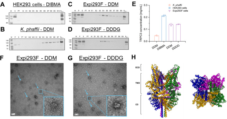
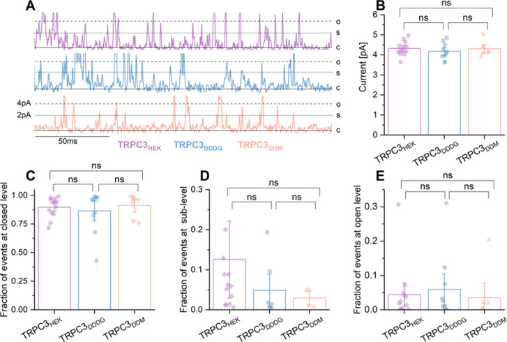
Recent advances in structural biology have provided insights into TRPC3, a TRP family member involved in various (patho)physiological processes. However, the lack of structural information on the channel's open pore hampers understanding of its function and therapeutic potential. Cryogenic electron microscopy holds promise for elucidating TRPC3's open-pore conformation, but challenges remain in isolating it without compromising function. Our study evaluated novel extraction agents in comparison to conventional detergents for isolating functional TRPC3 complexes from HEK293, Komagataella phaffii, and Expi293F cells, identifying Expi293F as optimal for TRPC3 expression. Among the extraction agents screened, dodecyl diglucoside (DDDG) and n-dodecyl-β-D-maltoside (DDM) were the most effective for extracting TRPC3. We successfully purified TRPC3 under native conditions, preserving its tetrameric structure and activity, as confirmed by electron microscopy, mass spectrometry and patch-clamp analysis. This study highlights the importance of extraction agents in advancing TRPC3 research and therapeutic development.

Keywords: Electrophysiology; Nanodiscs; Protein purification; Reconstitution; TRPC3.


Conflict of interest statement

Declarations. Competing interests: The authors declare no competing interests.

Figures

Fig. 1
Fig. 1
Overexpression of TRPC3 in adherent HEK293 (A), Expi293F (B), and K. phaffii cells (C), and electrophysiology of TRPC3-YFP-3xFLAG in adherent HEK293 cells by whole-cell analysis (D–F). A–C) In all cell types, TRPC3 is prominently expressed in the PM. Overexpression of TRPC3 in HEK293 (A) and Expi293F cells (B) was examined by fluorescence microscopy, where the YFP tag was excited at 495 nm, and emission was detected at 530 nm. TRPC3 is highlighted in yellow localizing in the PM. C) TRPC3 is enriched in microsomal fractions of K. phaffii. A CBS7435 his4Δ strain and an otherwise isogenic strain expressing TRPC3-3F6H driven by the strong AOX1 promoter were induced with methanol. Different fractions (total cell lysates (TCL)), as well as cytosolic and microsomal fractions) were prepared, separated by SDS–PAGE, and analyzed by immunoblotting with anti-FLAG antibody (TLCWT = total cell lysate of K. phaffi without TRPC3 (negative controle), TLC = total cell lysate of K. phaffi expressing TRPC3). D-F) Whole-cell patch clamp analysis was conducted in adherent HEK293 cells activated with 10 µM GSK. The TRPC3–YFP–3 x FLAG construct (orange, n = 9), TRPC3–YFP construct (blue, n = 11), and TRPC3–3xFLAG construct (green, n = 10) were compared. No significant variances in TRPC3 channel activity were observed across the constructs. D) Statistical evaluation of maximum current densities of the TRPC3 channels at − 90/70 mV with mean ± SEM are displayed. Statistical analysis was performed using a two-tailed Student’s t-test for normally distributed data and a Wilcoxon test for non-normally distributed data, with significance denoted as follows: ns = non-significant, * p < 0.05, ** p < 0.01, *** p < 0.001. E) Representative time courses of current activation by 10 µM GSK in TRPC3 channels are shown. F) Representative IV curves of activated TRPC3 channels with 10 µM GSK are displayed. Please note: Uncropped fluorescent images of TRPC3-YFP-3xFLAG over expression in adherent HEK293 and Expi293F cells are provided in the Supplementary Information (see Figure S1).
Fig. 2
Fig. 2
BCA assay and Western Blot analysis was conducted on supernatant fractions containing all membrane proteins extracted with DIBMA, Carboxy-DIBMA, DDDG, DDM, l LMNG or FOS-Choline (FOSC) from adherent HEK293 (blue), K. phaffii (orange), and Expi293F suspension cells (purple). A-E) Yields of extracted membrane proteins for DIBMA and Carboxy-DIBMA at different polymer/membrane mass ratios: R = 0.25 (A), R = 0.5 (B), R = 0.75 (C), and R = 1.0 (D). E) Yields of extracted membrane proteins for DDDG, DDM, LMNG and FOS-Choline (FOSC). Except FOS-choline, which resulted in higher protein yields, and membrane-protein extraction from K. phaffii, all extraction agents yielded similar protein yields (50–60%) from all three expression systems. The membrane-protein concentration was normalized to the total membrane-protein concentration before solubilization and expressed as a percentage yield (%). F-G) Western Blot analysis was conducted on the supernatant fractions extracted from adherent K. phaffii (F), adherent HEK293 cells (G) and Expi293F (H) cell membranes using DIBMA and Carboxy-DIBMA at various polymer-to-membrane ratios ranging from R = 0.25 to 1.0, as well as DDM, DDDG, FOS-Choline (FOSC), and LMNG. A specific TRPC3 antibody was used to target TRPC3. F) DIBMA at R = 1.0 and DDM extract the highest quantity of TRPC3. G) DIBMA extracts the highest quantity of TRPC3. H) DDM and DDDG extract the highest amount of TRPC3. Note: Please note that the Western blots on the right side of Panels F and H were cropped from the same blot. For the full, uncropped Western blot, refer to Supplementary Information, Figure S6.
Fig. 3
Fig. 3
Western blot analysis and negative-stain electron microscopy of purified TRPC3. TRPC3 was purified from adherent HEK293 cell membranes using DIBMA (A), K. phaffi cell membranes using DDM (n = 3, B), and Expi293F cell membranes using DDDG (n = 6, C) or DDM (n = 6, D). The resulting concentrations of purified TRPC3 are shown in (E). TRPC3 protein concentration (Vtotal = 150 µL) was lower from K. phaffi cell membranes compared to adherent HEK293 or Expi293F cells. Specifically, TRPC3 concentration was 0.05 mg/mL from K. phaffii cell membranes using DDM, 0.2 mg/mL from HEK293 cell membranes using DIBMA, and 0.15 mg/mL from Expi293F cells using either DDM or DDDG. Negative-stain electron microscopy images of TRPC3 extracted with DDM (F) or DDDG (G) from Expi293F cells reveal the tetrameric structure of TRPC3 channels (blue arrows). The blue box highlights the magnification of a single TRPC3 tetramer as observed in the electron microscopy images. H) General cryo-EM structure of the closed homomeric TRPC3 channel from the side (left) and top (right) view (PDB: 7DXB). Note: Please note that the Western blots in Panel A-D were cropped. For the full, uncropped Western blot, refer to Supplementary Information, Figure S7.
Fig. 4
Fig. 4
Single-channel patch-clamp experiments were conducted on TRPC3 in HEK293 cells (purple) and on reconstituted TRPC3 extracted from Expi293F cells using DDDG (blue) or DDM (orange). (A) Representative single-channel currents showing 200 ms of GSK-induced TRPC3 activity. TRPC3 was either expressed in HEK293 cells (purple) or purified from Exppi293F cells using DDDG (blue) or DDM (orange) and reconstituted into DPhPC bilayers. GSK was applied at 10 µM for adherent HEK293 cells and 20 µM for reconstituted TRPC3. Currents were recorded at + 80 mV. Channel states are indicated as closed (c), sublevels (s), or open (o). (B) Unitary currents at the open (o) level in response to repeated GSK applications of (10 µM for adherent HEK293 cells and 20 µM for reconstituted TRPC3) at a membrane potential of + 80 mV. Data are presented as means ± SEM. Statistical significance was determined using a two-tailed multiple t-test for normally distributed data and the Wilcoxon test for non-normally distributed data. Non-significant differences are denoted as “ns.” C–E) Fraction of events corresponding to the closed (panel C), sublevel (panel D), and open (Panel E) states, derived from Gaussian fitting of single-channel recordings from HEK293 cells expressing TRPC3 (purple), reconstituted TRPC3 purified with DDDG (blue) or with DDM (orange). GSK was applied at 10 µM for HEK293 cells and 20 µM for reconstituted TRPC3 at a membrane potential of + 80 mV. Data are presented as means ± SEM. Statistical significance was assessed using a two-tailed multiple t-test or the Wilcoxon test, with differences considered significant at * p < 0.05, ** p < 0.01, and *** p < 0.001. Non-significant differences are indicated as “ns”. We demonstrated that purified TRPC3 remains functional and its activity is comparable to TRPC3 overexpressed in HEK293 cells. Please note: Uncropped representative traces from Fig. 4A are shown in the Supplementary Information including the statistical analysis of the fraction of events at multiple levels (SI Figure S12).

References

    1. Zhu, X. et al. trp, a novel mammalian gene family essential for Agonist-Activated capacitative Ca2 + Entry. Cell85, 661–671. 10.1016/S0092-8674(00)81233-7 (1996). - PubMed
    1. Chen, X., Sooch, G., Demaree, I. S., White, F. A. & Obukhov, A. G. Transient Receptor Potential Canonical (TRPC) Channels: Then and Now. Cells 9, 1983. (2020). 10.3390/cells9091983 - PMC - PubMed
    1. Gees, M., Colsoul, B. & Nilius, B. The role of transient receptor potential cation channels in Ca2 + Signaling. Cold Spring Harb. Perspect. Biol.2, a003962–a003962. 10.1101/cshperspect.a003962 (2010). - PMC - PubMed
    1. Ong, H. L., de Souza, L. B. & Ambudkar, I. S. Role of TRPC channels in Store-Operated calcium entry. In Calcium Entry Pathways in Non-excitable Cells Advances in Experimental Medicine and Biology., (ed Rosado, J. A.) (Springer International Publishing), 87–109. 10.1007/978-3-319-26974-0_5. (2016). - PubMed
    1. Smyth, J. T. et al. Activation and regulation of store-operated calcium entry. J. Cell. Mol. Med.14, 2337–2349. 10.1111/j.1582-4934.2010.01168.x (2010). - PMC - PubMed

MeSH terms

LinkOut - more resources