. 2025 Aug 5;16(1):7227.
doi: 10.1038/s41467-025-62513-3.

Three positively charged binding sites on the eastern equine encephalitis virus E2 glycoprotein coordinate heparan sulfate- and protein receptor-dependent infection

Affiliations

Three positively charged binding sites on the eastern equine encephalitis virus E2 glycoprotein coordinate heparan sulfate- and protein receptor-dependent infection

Maria D H Alcorn et al. Nat Commun. .

Abstract

Naturally circulating strains of eastern equine encephalitis virus (EEEV) bind heparan sulfate (HS) receptors and this interaction has been linked to neurovirulence. Previous studies associated EEEV-HS interactions with three positively charged amino acid clusters on the E2 glycoprotein. One of these sites has recently been reported to be critical for binding EEEV to the very-low-density lipoprotein receptor (VLDLR), an EEEV receptor protein. The proteins apolipoprotein E receptor 2 (ApoER2) isoforms 1 and 2, and LDLR have also been shown to function as EEEV receptors. Herein, we investigate the individual contribution of each HS interaction site to EEEV HS- and protein receptor-dependent infection in vitro and EEEV replication in animals. We show that each site contributes to both EEEV-HS and EEEV-protein receptor interactions, providing evidence that altering these interactions can affect disease in mice and eliminate mosquito infectivity. Thus, multiple HS-binding sites exist in EEEV E2, and these sites overlap functionally with protein receptor interaction sites, with each type of interaction contributing to tissue infectivity and disease phenotypes.


Conflict of interest statement

Competing interests: W.B.K. is a co-founder of Advanced Virology. M.S.D. is a consultant or advisor for Inbios, Vir Biotechnology, IntegerBio, Moderna, Merck Sharp & Dohme Corporation, and GlaxoSmithKline. The Diamond laboratory has received additional unrelated funding support in sponsored research agreements from Vir Biotechnology, Emergent BioSolutions, and IntegerBio. S.C.W. is a consultant for Valneva. The remaining authors declare no competing interests.

Figures
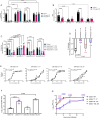
Fig. 1
Fig. 1. E2 mutations independently and uniquely alter EEEV-HS interactions in vitro.
A Relative infectivity of WT or mutant SINV/EEEV for CHO-K1, GAG-deficient CHO-pgsA-745, and HS-deficient CHO-pgsD-677 cells (n = 9 from 3 independent experiments). B Binding of chimeric WT and mutant viruses to CHO-K1 and CHO-pgsA-745 cells (n = 6 from 2 independent experiments, except for SINV/EEEV WT where n = 9 from 3 independent experiments). Viruses were allowed to incubate with cells on ice for 75 min, then cells were washed, and the bound virus was quantified by ddPCR as a ratio of genomes bound to cells versus the host Mmadhc gene. C Relative infectivity of WT or mutant SINV/EEEV on BHK-21 cells treated with heparinases (n = 6 from 2 independent experiments). D Relative indirect binding to heparin-agarose beads by WT and mutant SINV/EEEV. Viruses were incubated with collagen-agarose or heparin-agarose beads; unbound viruses were titered on BHK-21 cells (n = 6 from 2 independent experiments). E Apparent binding affinity showing the nonlinear fit of WT or mutant SINV/EEEV for heparin, with KD apparent and Hill slope (h) as a measure of cooperativity, where h > 1 shows increased cooperative binding and h < 1 shows decreased cooperative binding (n = 6 for mutants and n = 5 for WT from 2 independent experiments). F Genome-to-BHK PFU ratios for EEEV WT and mutant viruses (n = 3 independent experiments). G BHK-21 cells were infected with equal genomes of EEEV WT and mutant EEEVs, corresponding to a multiplicity of infection of 1 for WT (n = 6 from 2 independent experiments). Limit of detection indicated with dashed line. Means ± SD are shown. Significance determined by (A–C) two-way ANOVA with Tukey’s post-hoc tests, D Brown-Forsythe and Welch ANOVA with Dunnett’s post-hoc tests, F one-way ANOVA with Tukey’s post-hoc tests on log-transformed data, or G two-way repeated measures ANOVA with Tukey’s post-hoc tests. Exact p-values are indicated. Source data provided as a Source Data file.
Fig. 2
Fig. 2. E2 mutations that disrupt E2 HS-binding sites also interfere with EEEV-protein receptor interactions.
A Infectivity of K562 cells expressing empty vector (EV), VLDLR, ApoER2 isoform1, or ApoER2 isoform2 (n = 9 from 3 independent experiments, except for EV where n = 12 from 4 independent experiments). B Infectivity of THP-1 cells expressing EV or LDLR (n = 9 from 3 independent experiments). C Binding of chimeric WT and mutant viruses to K562-EV or K562-VLDLR cells (n = 12 from 4 independent experiments for WT, n = 9 from 3 independent experiments for 84–119 and 156–157, and n = 6 from 2 independent experiments for 71–77). Viruses were incubated with cells on ice for 75 min, then cells were washed, and the bound virus was quantified by qPCR, then expressed as a ratio to GAPDH and normalized to EV. D Apparent binding affinity showing the nonlinear fit of WT or mutant SINV/EEEV for VLDLR LA(1-2)-Fc, with KD apparent and Hill slope (h) (n = 6 from 2 independent experiments). E Neutralization of chimeric eGFP reporter WT and mutant viruses by VLDLR LA(1-2)-Fc (100–0.1 µg/mL in tenfold dilutions and 0 µg control) in Vero cells (n = 4 from 2 independent experiments, except for controls where n = 8 for WT and n = 12 for mutants from 2 independent experiments). F Neutralization of chimeric eGFP reporter WT and mutant viruses by VLDLR LA(1-2)-Fc in CHO-K1 and CHO-pgsA-756 cells (n = 4 from 2 independent experiments, except for WT where n = 9 from 3 independent experiments). G Neutralization of chimeric eGFP reporter WT by heparin or BSA control (2000–2 µg/mL in tenfold dilutions and control) in cells overexpressing protein receptors (n = 6 from 2 independent experiments, except for N2a ΔB4galt7-LDLR cells where controls are n = 6 and samples are n = 4 from 2 independent experiments) quantified by flow cytometry. Means ± SD are shown. Significance was determined by two-way ANOVA with Tukey’s post-hoc tests. Exact p-values are indicated. Source data provided as a Source Data file.
Fig. 3
Fig. 3. Passage of HS/protein receptor binding site mutants on cultured cells selects for mutations that impact binding of both receptors.
A Infectivity of double SINV/EEEV mutants found in passaging experiment on CHO-K1, CHO-pgsA-745, or CHO-pgsD-677 cells. Relative infectivity compared to CHO-K1 was determined for all viruses (n = 9 from 3 independent experiments). B Binding of chimeric passaging mutant viruses to CHO-K1 and CHO-pgsA-745 cells(n = 6 from 2 independent experiments). (Data for single mutant parent viruses are the same as in Fig. 1B.) C Apparent binding affinity showing nonlinear fit of chimeric passaging mutants for heparin, with KD apparent and Hill slope (h) (n = 6 from 2 independent experiments). D, E Infectivity of passaging mutants in (D) K562 cells expressing EV, VLDLR, ApoER2 isoform 1, or ApoER2 isoform 2 (n = 9 from 3 independent experiments, except for the 71–77 and 156–157 mutants on K562-EV cells where n = 12 from 4 independent experiments) or E THP-1 EV or LDLR cells (n = 9 from 3 independent experiments) quantified by flow cytometry. (Data for single mutant parent viruses are the same as in Fig. 2A, B). F Binding of chimeric WT and mutant viruses to K562-EV or K562-VLDLR cells, performed as described in Fig. 2C (n = 6 from 2 independent experiments, except for 1561-57 where n = 9 from 3 independent experiments). Binding is expressed as a ratio to GAPDH and normalized to EV. (Data for single mutant parent viruses are the same as in Fig. 2C.) G Apparent binding affinity showing nonlinear fit of chimeric passaging mutants for VLDLR LA(1-2)-Fc, with KD apparent and h (n = 6 from 2 independent experiments). H Neutralization of chimeric passaging mutant viruses by VLDLR LA(1-2)-Fc (100–0.1 µg in tenfold dilutions and 0 µg control) in CHO-K1 and CHO-pgsA-745 cells (n = 4 from 2 independent experiments). Means ± SD are shown. Significance was determined by (A) one-way ANOVA with Bonferroni’s post-hoc tests and (B, D–F, and H) two-way ANOVA with Tukey’s post-hoc tests. Exact p-values are indicated. Source data provided as a Source Data file.
Fig. 4
Fig. 4. Mutation of HS/protein receptor binding sites attenuates EEEV disease.
A, B CD-1 mice were infected with equal genomes of WT and mutant EEEV strains equivalent to 103 WT PFU in fp. (n = 15 from 3 independent experiments). A Survival. B Weight change. C, D CD-1 mice were inoculated ic. with equal genomes of WT and mutant SINV/EEEV virus equivalent to 104 WT PFU (n = 15 from 3 independent experiments: SINV/EEEV WT and SINV/EEEV 84–119, and n = 10 from 2 independent experiments: SINV/EEEV 71–77 and SINV/EEEV 156–157). C Survival. D Weight change. E, F CD-1 mice were inoculated in the fp. with equal genome copies of WT and mutant EEEV strains equivalent to 103 WT PFU (n = 10 from 2 independent experiments). At 2 dpi serum was collected and analyzed. E IFNα levels in serum. F Viral genomes present in the serum measured by qRT-PCR. G CD-1 mice were inoculated in both rear fp with equal genome copies of WT and mutant EEEV or VEEV nLuc expressing virus equivalent to 103 PFU of WT. At 8 hpi, PLNs were collected and replication was quantified (n = 12 for VEEV WT and n = 14 for mock from 2 independent experiments, and n = 26 from 3 independent experiments for EEEV WT, 71–77, 84–119, and 156–157). H–J CD-1 mice (n = 10 from 2 independent experiments) were inoculated in the fp. with equal genome copies of nLuc expressing virus equivalent to 103 PFU of WT. Virus dissemination was monitored daily using an in vivo imaging system (IVIS), and total flux (photons/second) was quantified. H Foot, where animals were infected. I PLNs; area roughly behind the knee on ventral view. J Head; dorsal view. Data shows (B, D) means ± SEM, (E–G) means ± SD, and (H–J) means ± 95% confidence interval (CI). Significance determined by (A, C) log-rank test, (E, G) Brown-Forsythe and Welch one-way ANOVA tests with Dunnett’s post-hoc tests, (F) one-way ANOVA with Bonferroni’s post-hoc test, and (H–J) two-way repeated measures ANOVA with Dunnett’s post-hoc tests. Exact p-values are indicated. Source data provided as a Source Data file.
Fig. 5
Fig. 5. Mutation of HS/protein receptor binding sites diminishes protection conferred by a receptor decoy inhibitor.
Survival data for mice treated intraperitoneally with 100 µg of VLDLR LA(1-2)-Fc (dashed line) or LDLRAD3 LA1-Fc (solid line) as a negative control. Six hours after treatment, mice were infected fp. with equal genome copies of WT and mutant EEEV viruses equivalent to 103 WT PFU (n = 6 mice from 2 independent experiments for LDLRAD3 LA1-Fc and n = 8 mice from 2 independent experiments for VLDLR LA(1-2)-Fc). A EEEV WT (FL93-939). B EEEV 71–77. C EEEV 84–119. D EEEV 156–157. Significance of survival for mice treated with control versus VLDLR LA(1-2)-Fc was determined by log-rank test. Exact p-values are indicated. Source data provided as a Source Data file.
Fig. 6
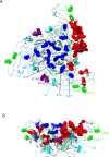
Fig. 6. Location of protein receptor- and HS-binding residues on the EEEV E2 trimer.
A ribbon model of the structure of EEEV E2 proteins as they appear in the E1/E2 trimeric spike. Side chains are displayed for residues involved in HS-binding (blue), residues identified in structural analysis as being involved in binding to one VLDLR molecule (red), and residues that are involved in both HS-binding and are direct contacts for VLDLR-binding (purple). Side chains for residues mutated during in vitro passage are lime green (K562-VLDLR) or cyan (BHK-21). A Top view. B Side view. Figures were made using UCSF ChimeraX,.

Update of

Similar articles

References

    1. Griffin, D. E. & Weaver, S. C. Alphaviruses in Fields Virology Volume 1 Emerging Viruses Vol. 1 (eds. Howley, P. M. and Knipe, D. M.) 194–244 (Philadelphia, PA Lippincott Williams & Wilkins, 2021).
    1. Gardner, C. L. et al. Eastern and Venezuelan equine encephalitis viruses differ in their ability to infect dendritic cells and macrophages: impact of altered cell tropism on pathogenesis. J. Virol.82, 10634–10646 (2008). - PMC - PubMed
    1. Gardner, C. L., Ebel, G. D., Ryman, K. D. & Klimstra, W. B. Heparan sulfate binding by natural eastern equine encephalitis viruses promotes neurovirulence. Proc. Natl. Acad. Sci. USA108, 16026–16031 (2011). - PMC - PubMed
    1. Gardner, C. L. et al. Natural variation in the heparan sulfate binding domain of the eastern equine encephalitis virus E2 glycoprotein alters interactions with cell surfaces and virulence in mice. J. Virol.87, 8582–8590 (2013). - PMC - PubMed
    1. Trobaugh, D. W. et al. RNA viruses can hijack vertebrate microRNAs to suppress innate immunity. Nature506, 245–248 (2014). - PMC - PubMed

MeSH terms

LinkOut - more resources