. 2025 Aug 5;16(1):7226.
doi: 10.1038/s41467-025-62337-1.

14-3-3 binding maintains the Parkinson's associated kinase LRRK2 in an inactive state

Affiliations

14-3-3 binding maintains the Parkinson's associated kinase LRRK2 in an inactive state

Juliana A Martinez Fiesco et al. Nat Commun. .

Abstract

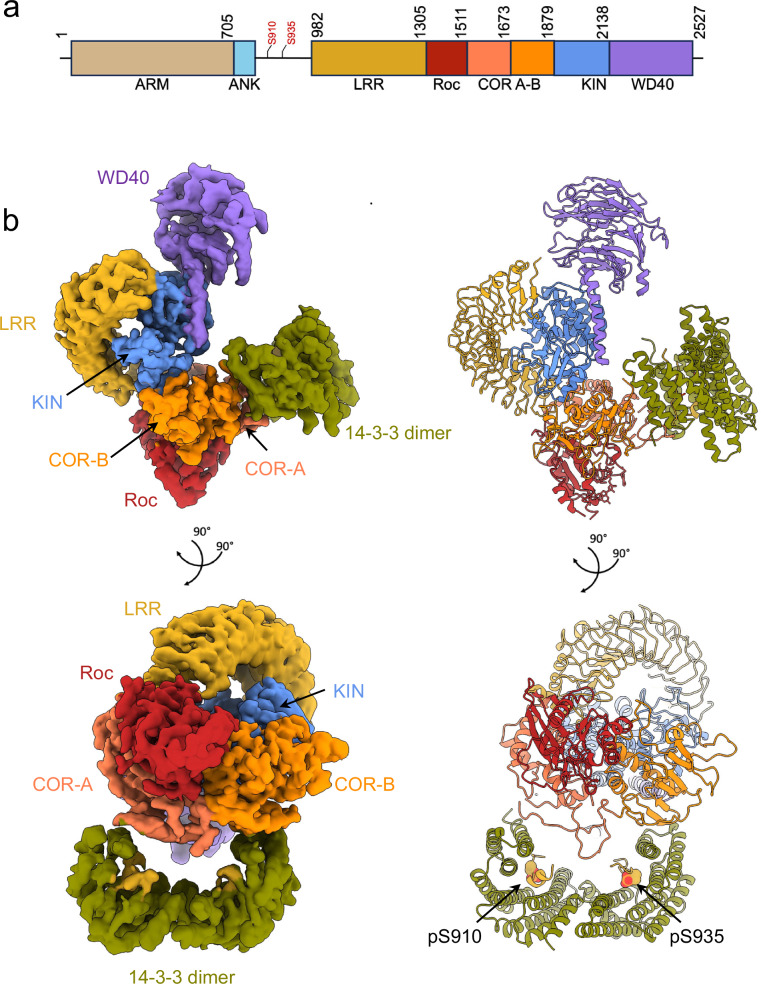
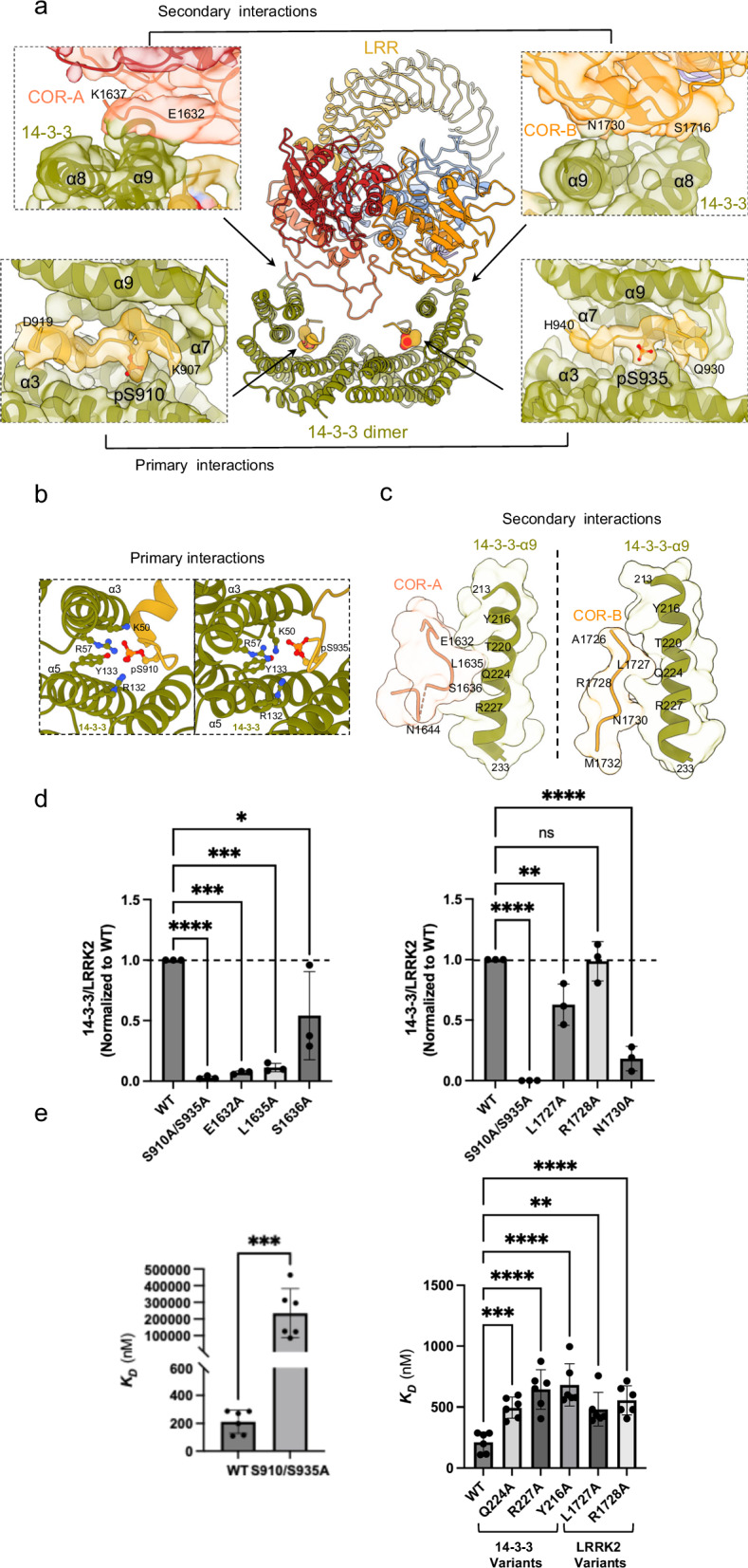
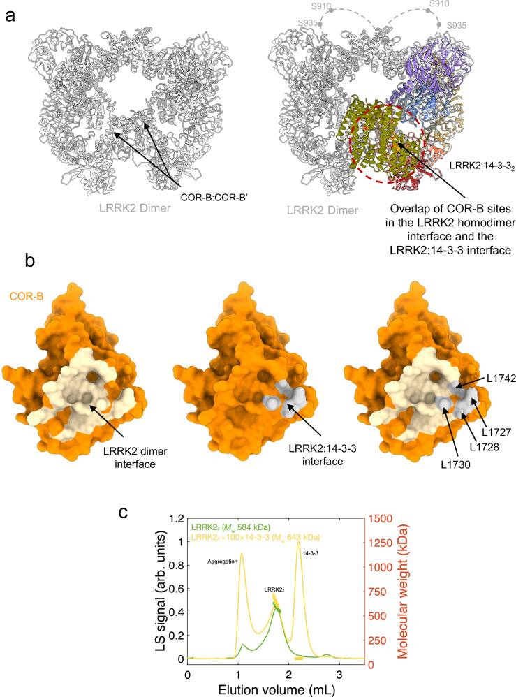
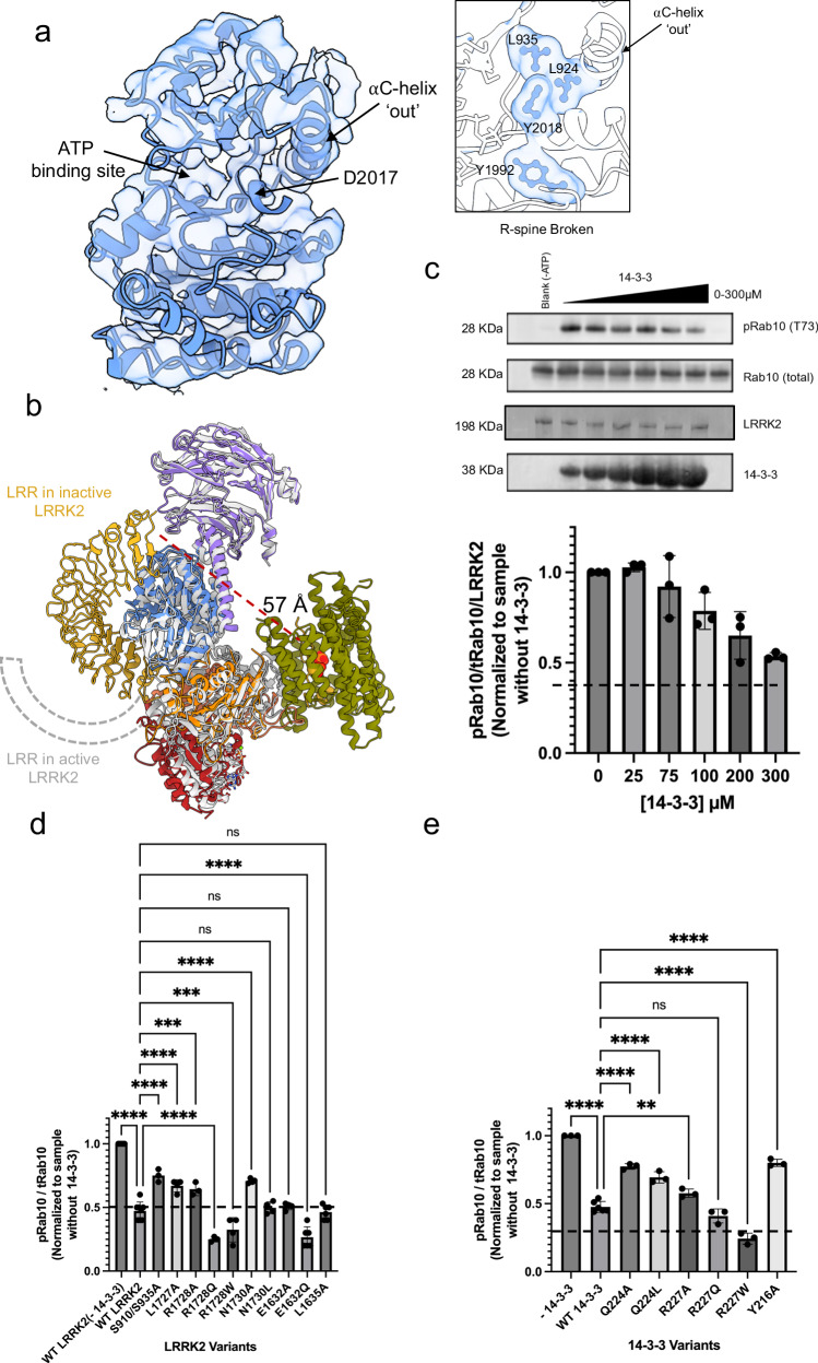
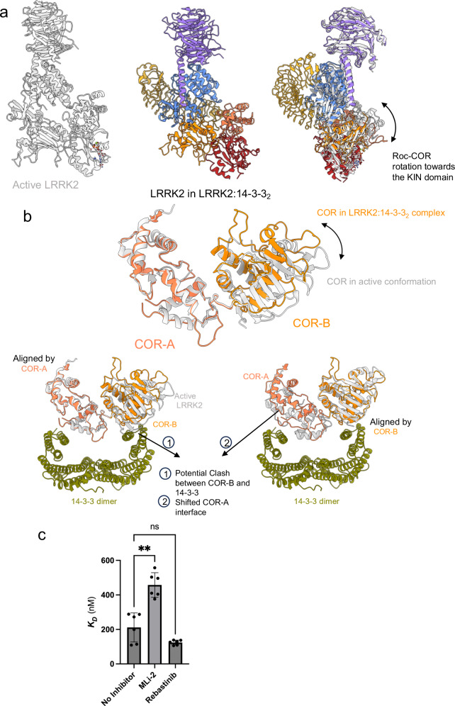
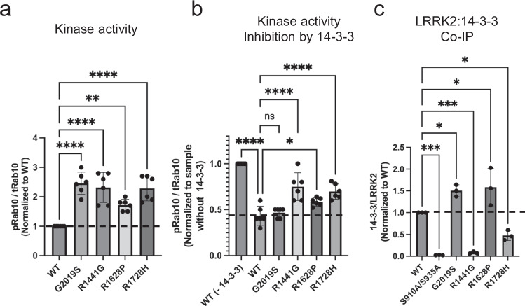
Leucine-rich repeat kinase 2 (LRRK2) is an essential regulator in cellular signaling and a major contributor to Parkinson's disease (PD) pathogenesis. 14-3-3 proteins are critical modulators of LRRK2 activity, yet the structural basis of their interaction has remained unclear. Here, we present the cryo-electron microscopy structure of the LRRK2:14-3-32 autoinhibitory complex, revealing how a 14-3-3 dimer stabilizes an autoinhibited LRRK2 monomer through dual-site anchoring. The dimer engages both phosphorylated S910/S935 sites and the COR-A/B subdomains within the Roc-COR GTPase region. This spatial configuration constrains LRR domain mobility, reinforces the inactive conformation, and likely impedes LRRK2 dimerization and oligomer formation. Structure-guided mutagenesis studies show that PD-associated mutations at the COR:14-3-32 interface and within the GTPase domain weaken 14-3-3 binding and impair its inhibitory effect on LRRK2 kinase activity. Furthermore, we demonstrate that type I LRRK2 kinase inhibitor, which stabilizes the kinase domain in its active conformation, reduces 14-3-3 binding and promotes dephosphorylation at pS910 and pS935. Together, these findings provide a structural basis for understanding how LRRK2 is maintained in an inactive state, elucidate the mechanistic role of 14-3-3 in LRRK2 regulation, inform the interpretation of PD biomarkers, and suggest therapeutic strategies aimed at enhancing LRRK2-14-3-3 interactions to treat PD and related disorders.


Conflict of interest statement

Competing interests: The authors declare no competing interests.

Figures

Fig. 1
Fig. 1. Structure of the LRRK2:14-3-32 complex.
a Schematic representation of LRRK2 domain organization. Residues S910 and S935, which serve as 14-3-3 binding sites upon phosphorylation, are highlighted in red. b Cryo-EM density map at a resolution of 3.96 Å (left), with the corresponding structural model shown on the right. The model is colored according to the domain color code in (a) and shown in two different orientations for clarity.
Fig. 2
Fig. 2. Detailed interactions and mutational effects at the LRRK2:14-3-32 binding interfaces.
a Overview of the LRRK2:14-3-32 contact regions in LRRK2:14-3-32 complex. Insets detail the primary and secondary interaction sites, supported by the corresponding cryo-EM densities. b Close-up view of the primary interactions, where LRRK2 phosphorylation sites pS910 and pS935 engage with the 14-3-3 substrate binding grooves. Assignment of this interaction is supported by integration of structural fitting, mass spectrometry, and biochemical validation. c Close-up view of the secondary interactions, showing LRRK2 COR-A and COR-B subdomain residues contacting the α−9 helices of the 14-3-3 dimer. d Quantitative analysis of LRRK2/14-3-3 interactions through Co-IP experiments of LRRK2 with endogenous 14-3-3, comparing WT LRRK2 with mutants at the secondary interface, as well as primary interface mutants (S910A/S935A). Data illustrate the impact of mutations on the interaction strength. Refer to Supplementary Fig. 16 for representative membrane images and source data for complete membrane images. Data are mean ± SEM (n  =  3 independent experiments), significance of difference was quantified using one-way Brown–Forsythe and Welch ANOVA test and reported with the exact p values in the source data file. e Binding affinity between (WT or mutant) LRRK2 and (WT or mutant) 14-3-3 proteins was determined by MST. Mutations at the primary (left) and secondary (right) binding sites were analyzed. Data are mean ± SEM (n  =  3 independent experiments), significance of difference was quantified using one-way Brown–Forsythe and Welch ANOVA test and reported with the exact p values in the source data file. Refer to Supplementary Fig. 11 for full binding curves.
Fig. 3
Fig. 3. Overlap of the LRRK2:14-3-32 and LRRK2 homodimer interfaces.
a Structural comparison of the inactive LRRK2 homodimer (PDB: 7LHW, in gray, left) and the LRRK2:14-3-32 complex (colored as in Fig. 1), showing the overlapping interfaces mediated by the COR-B domain (right). Loops containing the S910 and S935 sites, flexible and unresolved in the dimer structure, are illustrated with dashed lines. b Surface representations of the COR-B domain. Left: residues involved in the LRRK2 dimer interface shaded in tan. Middle: residues involved in the LRRK2:14-3-32 interface shaded in gray. Right: Overlay of the two interfaces showing steric clash, suggesting mutual exclusivity between LRRK2 dimerization and 14-3-3 binding. c SEC-MALS chromatogram of the pre-formed LRRK2 dimer in the presence and absence of 14-3-3, indicating changes in molecular weight distribution.
Fig. 4
Fig. 4. 14-3-3 binding maintains LRRK2 in an inactive conformation and inhibits its kinase activity.
a Structural representation of the kinase domain in the LRRK2:14-3-32 complex, showing an inactive conformation characterized by an outward αC helix and a broken regulatory (R-) spine (inset). b Overlay of the LRRK2:14-3-32 complex (colored as in Fig. 1) with the active LRRK2 monomer from a LRRK2 tetramer (PDB: 8FO9, in gray), highlighting the conformational differences in the LRR domain, depicted as a cartoon. c In vitro kinase assay showing inhibition of LRRK2 activity by increasing concentrations of 14-3-3, measured by Rab10 phosphorylation levels (pRab10) in western blots, normalized to LRRK2 protein levels. Effects of interface mutations on kinase activity inhibition by 14-3-3, with results for LRRK2 mutations shown in (d) and 14-3-3 mutations in (e). Data in (ce) are mean ± SEM (n  =  3 independent experiments), with representative blots for inhibition assays shown in (c). See source data for membrane images and for significance of difference with the one-way Brown–Forsythe and Welch ANOVA test with the exact p values when applicable.
Fig. 5
Fig. 5. Structural comparison of the LRRK2:14-3-32 complex with active LRRK2.
a Side by side comparison of the LRRK2:14-3-32 complex (colored as in Fig. 1) and the active conformation of LRRK2 derived from a tetrameric structure (PDB: 8FO9, in gray), highlighting the Roc-COR domain rotation (indicated by an arrow). 14-3-3 proteins are omitted for clarity. b Superimposition of the COR domain from the LRRK2:14-3-32 complex (shown in coral) with the COR domain from the active LRRK2 (shown in gray). Alignments were performed by the COR-A domain (top and bottom left) and by the COR-B domain (bottom right), to illustrate the structural shifts and effects. c Binding affinity measurements of LRRK2 and 14−3-3γ in the presence of kinase inhibitors, as determined by MST in vitro. Data in (c) are mean ± SEM (n  =  3 independent experiments), significance of difference was quantified using one-way Brown–Forsythe and Welch ANOVA test and reported with the exact p values in the source data file. Refer to Supplementary Fig. 17 for full binding curves.
Fig. 6
Fig. 6. Impact of PD-associated mutations in Kinase and GTPase domains on LRRK2:14-3-32 interaction.
a In vitro LRRK2 kinase activity assay comparing wild-type (WT) and various PD-related hyperactive mutants in the kinase and GTPase domains, showing increased Rab10 phosphorylation levels indicative of enhanced kinase activity. b Comparative analysis of 14-3-3 inhibition of kinase activity across WT and PD mutants, illustrating that mutations differentially impair LRRK2 regulation by 14-3-3. c Co-IP assays quantifying cellular interaction between LRRK2 WT/ PD-related mutations and endogenous 14-3-3 in cells, revealing altered binding affinities caused by specific mutations. LRRK2 kinase activity and co-IP data (ac) are mean ± SEM (n = 3 independent experiments). Refer to Supplementary Fig. 16 and source data for membrane images and for significance of difference with the one-way Brown–Forsythe and Welch ANOVA test with the exact p values when applicable.

Update of

Similar articles

Cited by

References

    1. Gasser, T. Molecular pathogenesis of Parkinson disease: insights from genetic studies. Expert Rev. Mol. Med.10.1017/S1462399409001148 (2009). - PubMed
    1. Lill, C. M. et al. Comprehensive research synopsis and systematic meta-analyses in Parkinson’s disease genetics: The PDGene Database. PLoS Genet.8, e1002548 (2012). - PMC - PubMed
    1. Ross, O. A. et al. Association of LRRK2 exonic variants with susceptibility to Parkinson’s disease: a case–control study. Lancet Neurol.10, 898–908 (2011). - PMC - PubMed
    1. Satake, W. et al. Genome-wide association study identifies common variants at four loci as genetic risk factors for Parkinson’s disease. Nat. Genet.41, 1303–1307 (2009). - PubMed
    1. Simón-Sánchez, J. et al. Genome-wide association study reveals genetic risk underlying Parkinson’s disease. Nat. Genet.41, 1308–1312 (2009). - PMC - PubMed

MeSH terms

Substances

LinkOut - more resources