. 2025 Aug 5;16(1):423.
doi: 10.1186/s13287-025-04542-9.

Chorion tissue- and plasma-derived extracellular vesicles exhibit superior anti-inflammatory and chondroprotective effects

Affiliations

Chorion tissue- and plasma-derived extracellular vesicles exhibit superior anti-inflammatory and chondroprotective effects

Livia K Fecskeova et al. Stem Cell Res Ther. .

Abstract

Background: Extracellular vesicles (EVs) are the foundation of modern regenerative medicine using a cell-free approach. While current research mainly explores EVs from biological fluids and cell culture supernatants, tissue-derived EVs hold great promise, but remain largely underexplored. Since healthy placental tissues such as the chorion are widely available after full-term delivery, ethically unobjectionable, and possess exceptional regenerative potential, we sought to compare the biological effects of EVs derived directly from chorion tissue with those from chorion-derived mesenchymal stromal cell EVs and plasma EVs.

Method: We compared the biological impact of EVs from various sources (chorion tissue CHO-Ti, MSCs from chorion CHO-MSC and platelet-poor plasma PPP) and isolated by various techniques on the gene expression of osteoarthritic chondrocytes. Additionally, we assessed the effect of enriched soluble proteins of CHO-MSC and CHO-Ti secretome vs. their EVs. EVs were characterized by particle number and size (NTA), protein content (BCA assay) and immunophenotype (flow cytometry). Changes in gene expression of chondrocytes were quantified by RT-qPCR.

Results: CHO-Ti-EVs and PPP-EVs showed particularly beneficial effect on the inflammatory process, with their biological impact surpassing that of CHO-MSC-EVs. Chondroprotective markers COL2A and ACAN were robustly upregulated by CHO-Ti-EVs and PPP-EVs but showed only modest or variable increases with CHO-MSC-EVs. COMP expression, however, was specifically enhanced by CHO-MSC-derived components. Furthermore, our results also indicate that the therapeutic properties of the CHO-Ti secretome are exclusively linked to EVs. Among CHO-MSC-EVs, purification combined with UC resulted in the highest purity, however EVs purified by SEC presented a more favourable surface marker profile and better biological effects. The observed variability suggests that different EV preparations harbour distinct subpopulations that influence regulatory pathways differently and highlight the importance of EV source and isolation methodology in determining biological activity.

Conclusion: CHO-Ti-EVs showed promising effects on cartilage regeneration and inflammation modulation, suggesting they may represent a viable alternative to plasma- and CHO-MSC-EVs. Moreover, the chorion represents a readily accessible and abundant source of perinatal tissue obtainable non-invasively after full-term delivery, further supporting the translational potential of CHO-Ti-EVs.

Keywords: Cell-free therapy; Chondrocyte; Chorion tissue; Extracellular vesicles; MSC-EVs; Platelet-poor plasma; Tissue-derived EVs.


Conflict of interest statement

Declarations. Ethics approval and consent to participate: This study was performed in line with the principles of the Declaration of Helsinki. Approval was granted by the Ethics Committee of the P. J. Safarik University and L. Pasteur University Hospital in Kosice, Slovakia. Title of the approved projects: ID 2023/EK/06026: Extracellular vesicles from mesenchymal stem cells — a new therapeutic strategy in the treatment of inflammatory joint diseases. Date of approval: 23rd May, 2023. ID 2023/EK/06028: Potential use of circulating extracellular vesicles in the diagnosis of osteoarthritis. Date of approval: 19st June, 2023. Informed consent was obtained from all individual participants included in the study. Consent for publication: Not applicable. Competing interests: The authors declare that they have no competing interests.

Figures

Fig. 1
Fig. 1
Workflow of the experiments. Isolation and characterization of EVs/proteins of various sources and isolated by various methods, and their application to chondrocytes
Fig. 2
Fig. 2
Analysis of chondrocyte’s phenotype (a) by immunofluorescence: The percentage of chondrocytes expressing aggrecan (i), collagen type II (ii), or collagen type I (iii) over the total chondrocytes was calculated using ImageJ software, within 10 different fields (500 × 500 μm) for each marker. Details of labeled cells (squares) show expression of aggrecan (i´), type II collagen (ii´) or type I collagen (iii). Scale bars (i, ii, iii) = 200 μm; (i´, ii´, iii´) = 50 μm; (b) by RT-qPCR shown as the log2-value of the relative quantity (RQ) of gene expression of collagen type I, type II and aggrecan (ACAN), normalized to GAPDH, in three replicates of chondrocytes. Statistical significance calculated by t-test. *p < 0.05, ** p < 0.01, *** p < 0.001
Fig. 3
Fig. 3
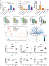
Characterization of EVs. (a) Protein concentrations by BCA, (b) particle number/mL by NTA and (c) relative purity of EV samples isolated by various techniques and from various sources. Mean of 3 biological replicates ± SD. *p < 0.05; **p < 0.01; ***p < 0.001. (d) Expression of the top 5 EV surface markers, including the EV-specific surface markers CD81, CD63 and CD9, by MACSPlex analysis. Data were normalized to mean fluorescence intensity (MFI) of CD9/CD63/CD81 of EVs. (e) PCA analysis of 37 EV surface markers of EVs isolated from various sources (PPP n = 4, CHO-MSCs n = 6, CHO-Ti n = 4). Ellipses represent the 95% confidence intervals with centred symbols representing the mean value. (f) Different expression of EV surface markers of CHO-MSC-EVs based on the isolation technique. Mean ± SD. *p < 0.05; **p < 0.01; ***p < 0.001
Fig. 4
Fig. 4
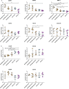
Changes in the expression of pro-inflammatory/degradation (a) and chondroprotective (b) genes of chondrocytes co-cultivated with EVs isolated by different methods (P precipitation, UC precipitation + ultracentrifugation, SEC size exclusion chromatography) and from various biological sources (CHO-MSCs, CHO-Ti, PPP). Expression is shown as the log2 value of fold change, compared to expression of the untreated control. Symbols represent individual replicates, the mean ± SD is shown. *p < 0.05; **p < 0.01; ***p < 0.001
Fig. 5
Fig. 5
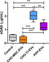
sGAG production in chondrocytes treated with CHO-MSC-EVs-SEC, CHO-Ti-EVs and PPP-EVs, all isolated using SEC. Chondrocytes were treated with EVs for 10 days. Results show 3 biological replicates of OA- chondrocytes. *p < 0.05; **p < 0.01; ***p < 0.001
Fig. 6
Fig. 6
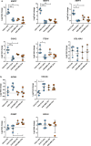
Changes in the expression of pro-inflammatory/degradation (a) and chondroprotective (b) genes of chondrocytes co-cultivated with the enriched soluble protein (PR) or EVs fraction of CHO-Ti (blue) or CHO-MSCs (orange). Expression is shown as the log2 value of fold change, compared to the expression of the untreated control. Symbols represent individual replicates, the mean ± SD is shown. * p < 0.05; ** p < 0.01; *** p < 0.001
Fig. 7
Fig. 7
Heatmap of the relative expression of ECM-degrading and ECM-building (chondroprotective) genes of 4 biological replicates (A, B, C, D) of OA chondrocytes co-cultivated with the enriched soluble protein (CHO-Ti-PR and CHO-MSC-PR) or EVs fraction from different EV sources (chorion tissue CHO-Ti, platelet-poor plasma PPP and CHO-MSCs isolated by different methods, such as precipitation CHO-MSC-EVs-P, precipitation + ultracentrifugation CHO-MSC-EVs-UC, size exclusion chromatography CHO-MSC-EVs-SEC). Values are scaled by row and treatment groups are clustered by Euclidean distances

Similar articles

References

    1. Wu X, Jiang J, Gu Z, Zhang J, Chen Y, Liu X. Mesenchymal stromal cell therapies: Immunomodulatory properties and clinical progress. Stem Cell Res Ther. 2020;11:345. 10.1186/s13287-020-01855-9. - PMC - PubMed
    1. Yin K, Wang S, Zhao RC. Exosomes from mesenchymal stem/stromal cells: a new therapeutic paradigm. Biomark Res. 2019;7:8. 10.1186/s40364-019-0159-x. - PMC - PubMed
    1. Hunter DJ, Bierma-Zeinstra S, Osteoarthritis. Lancet. 2019;393:10182. 10.1016/S0140-6736(19)30417-9. - PubMed
    1. Chen D, Shen J, Zhao W, Wang T, Han L, Hamilton JL, Im HJ. Osteoarthritis: toward a comprehensive Understanding of pathological mechanism. Bone Res. 2017;5:16044. 10.1038/boneres.2016.44. - PMC - PubMed
    1. Wang Y, Yu D, Liu Z, Zhou F, Dai J, Wu B, Zhou J, Heng BC, Zou XH, Ouyang H, Liu H. Exosomes from embryonic mesenchymal stem cells alleviate osteoarthritis through balancing synthesis and degradation of cartilage extracellular matrix. Stem Cell Res Ther. 2017;8:189. 10.1186/s13287-017-0632-0. - PMC - PubMed

Substances

LinkOut - more resources