. 2025 Aug 6;21(1):506.
doi: 10.1186/s12917-025-04914-6.

Preparation and immunogenicity studies of NvIBDV VP2-ferritin nanoparticles

Affiliations

Preparation and immunogenicity studies of NvIBDV VP2-ferritin nanoparticles

Shimin Gao et al. BMC Vet Res. .

Abstract

Background: Infectious bursal disease (IBD), caused by infectious bursal disease virus (IBDV), is a highly contagious disease that is prevalent worldwide and poses a significant threat to the poultry industry. While commercially available vaccines are used for prevention, IBD outbreaks remain frequent.

Objective: The continuous mutation of virulent strains and their ability to evade traditional vaccine protection complicate IBD control, which necessitates the development of novel vaccines and a deeper understanding of viral mutation mechanisms.

Method: Utilizing the self-assembly capability of ferritin (Fe), the hypervariable region (HVR) protein of a novel variant IBDV (NvIBDV) VP2 was displayed on the ferritin shell, forming regular nanoparticles. The full-length NvIBDV VP2 protein and the NvIBDV VP2-HVR-Fe fusion protein were prokaryotically expressed in E. coli and purified to prepare a VP2 protein vaccine and a VP2-Fe nanoparticle vaccine. An inactivated NvIBDV vaccine served as a control for evaluating immunogenicity and protection.

Results: Recombinant prokaryotic expression vectors pET-VP2-Fe (encoding VP2-HVR-Fe) and pET-VP2 (encoding full-length VP2) were successfully constructed. Soluble VP2-Fe and VP2 proteins were expressed and purified. Electron microscopy confirmed the formation of a cage-like nanoparticle structure for VP2-Fe. Immunization of SPF chickens with NvIBDV VP2-Fe nanoparticles induced a robust immune response characterized by high antibody titers and a significantly high protection rate against viral challenge.

Conclusion: The successfully constructed recombinant subunit nanoparticle vaccine, which displays the NvIBDV VP2 HVR on ferritin, effectively increased the antibody titer and provided superior immune protection. This approach offers a feasible strategy for developing novel IBDV subunit vaccines.

Keywords: Ferritin; Infectious bursal disease virus; Nanoparticles; Novel mutant strain; Subunit vaccine.


Conflict of interest statement

Declarations. Ethics approval and consent to participate: All animal experiments were performed in accordance with the Guidelines for Care and Use of Laboratory Animals of Shanxi Agricultural University, China, and the animal study was reviewed and approved by the Animal Ethical Committee of Shanxi Agricultural University, China. Approval No. SXAU-EAW-2024 C.NR.012026285. Consent for publication: Not applicable. Competing interests: The authors declare no competing interests.

Figures

Fig. 1
Fig. 1
Construction of VP2-Fe, VP2 recombinant plasmid. (Figures A-C show the cropped images.) (A) Linearization of the target gene VP2 and the prokaryotic expression vector pET-32a M: DL 5000 DNA Marker; 1: indicates the full length of VP2. 2: VP2-pET-32a carrier; 3: VP2 high variable region; 4: VP2-Fe-pET-32a carrier. (B) pET - VP2, pET - VP2 - Fe bacteria liquid PCR M: DL 5000 DNA Marker; 1–5: pET-VP2 single colony; 6–10: pET-VP2-Fe single colony. (C) Recombinant plasmid pET - VP2 - Fe, pET - VP2 enzyme identification M: DL 5000 DNA Marker; 1: uncut pET-VP2-Fe; 2: pET-VP2-Fe after double digestion; 3: uncut pET-VP2; 4: pET-VP2 after double digestion
Fig. 2
Fig. 2
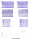
Expression of VP2-Fe, VP2 recombinant protein. (Figures A-F show the cropped images.) (A)/(B) VP2-Fe and VP2 under different temperatures and precipitation on the expression of recombinant proteins A: VP2-Fe recombinant protein; B: VP2 recombinant proteinM: Protein molecular quality standard; 1: no induced supernatant; 2: no induced precipitation; 3:37 °C supernatant; 4:37 °C precipitation; 5:30 °C supernatant; 6:30 °C precipitation; 7:25 °C supernatant; 8:25 °C precipitation; 9:20 °C supernatant; 10:20 °C precipitation; 11:16 °C supernatant; 12:16 °C precipitation. (C) Different induction time of VP2-Fe and VP2 clear expression on the results M: Protein molecular quality standard; 1: VP2-Fe not induce supernatant; 2–6: VP2-Fe induced, respectively 6 h, 12 h, 18 h, 24 h, 30 h; 7: VP2 did not induce supernatant; 8–12: VP2, respectively induced 6 h, 12 h, 18 h, 24 h, 30 h. (D) Different IPTG concentration of VP2-Fe and VP2 clear expression on the results M: Protein molecular quality standard; 1: Supernatant induced by pET-32a empty carrier; 2–7: The final concentration of IPTG was 0.2, 0.4, 0.6, 0.8, 1, 1.2 mmol/L to induce 24 h VP2-Fe recombinant protein supernatant; 8–13: The final concentration of IPTG was 0.2, 0.4, 0.6, 0.8, 1, 1.2 mmol/L to induce 18 h of VP2 recombinant protein supernatant (E)/(F) Purification results of VP2-Fe and VP2 recombinant proteins E: VP2-Fe recombinant protein; F: VP2 recombinant protein M: Protein molecular quality standard; 1: flow through liquid; 2: The first wash; 3: The second wash; 4: First elution; 5: Second elution; 6: third elution; 7: Fourth elution; 8: Fifth elution; 9: Dialysis concentration (G) BCA protein standard curve
Fig. 3
Fig. 3
Western blotting assay and electron microscopy of VP2-Fe, VP2 recombinant protein. (Figures A show the cropped images.). (A) VP2 - Fe, VP2 recombinant protein of Western blotting test results M: Protein molecular quality standard; 1: VP2-Fe recombinant protein; 2: VP2 recombinant protein. (B)The structure of VP2-Fe recombinant protein was observed under electron microscopy
Fig. 4
Fig. 4
Vaccine quality test results. (A) Centrifuge after emulsification. (B) Coating plate after emulsification. (C) Emulsify and drop into the water
Fig. 5
Fig. 5
Detection of IBDV antibody levels in serum and proliferation of peripheral blood lymphocytes. (A) The antibody titers of chickens in each group were detected by ELISA(*** P < 0.001, ns P > 0.05). (B) Peripheral blood lymphocyte proliferation results(*** P < 0.001,** P < 0.01,* P < 0.05)
Fig. 6
Fig. 6
Attack Protection Experiment. (A) Comparison of bursa of Fabris and spleen in each group 7 days after challenge. (B) Groups of spleen index, the index of bursa results figure(** P < 0.01, ns P > 0.05). (C) The standard curve of real-time fluorescent quantitative q-PCR for PMD18-T-VP2

Similar articles

References

    1. Liao Jiedan C, Yuming Z. Research progress on molecular pathogenesis of infectious bursal disease virus [in chinese]. China Poult. 2018;40(05):1–5.
    1. Fan L, Wu T, Wang Y, et al. Novel variants of infectious bursal disease virus can severely damage the bursa of Fabricius of immunized chickens. Vet Microbiol. 2020;240:108507. 10.1016/j.vetmic.2019.108507. - PubMed
    1. Myint O, Suwanruengsri M, Araki K, et al. Bursa atrophy at 28 days old caused by variant infectious bursal disease virus has a negative economic impact on broiler farms in Japan. Avian Pathol. 2021;50(1):6–17. 10.1080/03079457.2020.1822989. - PubMed
    1. Xu A, Pei Y, Zhang K, et al. Phylogenetic analyses and pathogenicity of a variant infectious bursal disease virus strain isolated in China. Virus Res. 2020;276:197833. 10.1016/j.virusres.2019.197833. - PubMed
    1. Castón JR, Martínez-Torrecuadrada JL, Maraver A, et al. C terminus of infectious bursal disease virus major capsid protein VP2 is involved in definition of the T number for capsid assembly. J Virol. 2001;75(22):10815–28. 10.1128/JVI.75.22.10815-10828.2001. - PMC - PubMed

MeSH terms

LinkOut - more resources