Review
. 2025 Aug 5;16(1):424.
doi: 10.1186/s13287-025-04531-y.

Melatonin and angiogenesis potential in stem cells

Affiliations
Review

Melatonin and angiogenesis potential in stem cells

Somayyeh Rashidi et al. Stem Cell Res Ther. .

Abstract

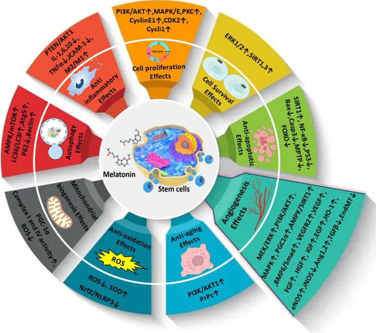
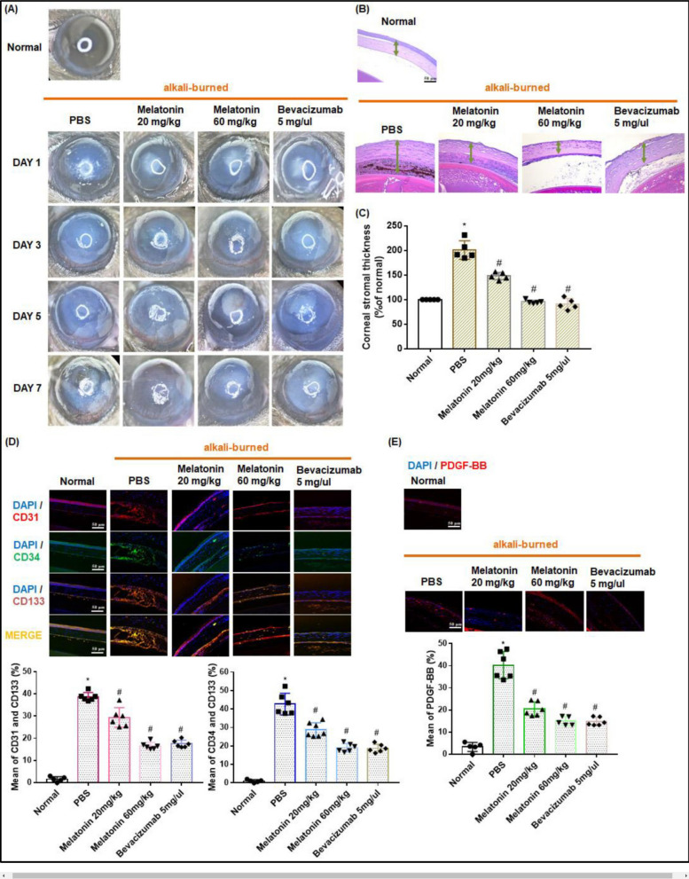
Ischemic diseases, especially coronary artery diseases and myocardial infarction, are the leading cause of human death in the clinical setting. Unfortunately, most of the available clinical interventions can partially restore the function of ischemic myocardium, resulting in the progression of chronic heart failure.The induction of vascular tissue formation, hereafter known as angiogenesis, can provide blood perfusion and prevent the expansion of ischemia-related pathologies. In recent years, the discovery and advent of multiple stem cells into human regenerative medicine have led to the alleviation of certain end-stage pathological conditions via direct differentiation into the mature and functional cells or secretion of various cytokines and angiogenesis factors in a paracrine manner. Melatonin (mel) is a natural molecule with direct and indirect pleiotropic actions on different biological phenomena. This neurohormone is primarily known for its antioxidant, tumoricidal, and anti-inflammatory actions in several pathological conditions. Whether and how mel regulates the angiogenesis behavior of stem cells is currently under debate. Here, we collected and evaluated recent data related to the angiogenic properties of mel on stem cells. Data from the present article may help us in the development of new therapeutic regimes in patients with ischemic conditions.

Keywords: Cell differentiation; Melatonin; Neovascularization; Paracrine communication; Stem cells.


Conflict of interest statement

Declarations. Ethics approval and consent to participate: No human and/or animal samples were used in the current manuscript. The study was registered as titled “pro-angiogenesis/anti-angiogenesis capacity of melatonin on stem cells under ischemic conditions.” to the research ethics committees of the vice-chancellor in research affairs, tabriz university of medical sciences. Consent for publication: Not applicable. Competing interests: None declared.

Figures

Fig. 1
Fig. 1
Schematic illustration related to pleotropic effects of mel in stem cells. Designed by powerpoint software
Fig. 2
Fig. 2
The schematic illustration of mel biogenesis in pinealocytes within the brain pineal gland. Created by biorender's web-based software
Fig. 3
Fig. 3
Mechanism of angiogenesis versus vasculogenesis. In response to hypoxic/ischemic conditions, the release of proangiogenesis factors activates the mature ecs in the close blood vessels (angiogenesis). The pioneer ecs, also known as tip cells, migrate in a cytokine gradient density toward the ischemic/hypoxic zone. The lumen formation and proliferation of stalk and phalanx ecs can extend the developing blood vessels toward the target zone. The addition of pericytes to the abluminal surface of de novo vessels stabilizes the integrity of the vascular wall. It is also possible that the activated bone marrow epcs migrate toward the ischemic/hypoxic zone and are involved in the development of new vascular structure in a paracrine and/or direct orientation toward mature ecs (vasculogenesis) [7]. Reproduced with the permission of the publisher. 2024. Cell proliferation
Fig. 4
Fig. 4
Mechanisms of action of mel. This hormone exerts its effects by engaging membrane receptors mt1 and mt2. These receptors belong to the g-protein-coupled receptor superfamily. Along with these receptors, mel can activate cytosolic enzyme, namely quinone reductase 2 (qr2; also known as mt3), and nuclear receptors rzr/ror. The attachment of mel to mt1 and mt2 recruits several intracellular effectors such as pkc, plcβ, and pka. In the presence of mel, gαi activation of mt1 can contribute to the stimulation of plcβ, while the promotion of gβ/γ is followed by the stimulation of pkc and erk pathways. The mt1 receptor also functions via membrane adenyl cyclase, which inhibits creb phosphorylation by activating camp and pkc. Mel promotes conformational changes in mt2 and thereby activates the αi subunit. Along with these changes, pkg is triggered via guanylate cyclase. Also, mt2 can engage pkc and erk1/2 complexes. Created by biorender's web-based software
Fig. 5
Fig. 5
Study of tissue regeneration in mice with hindlimb ischemia (a-l). Measuring blood perfusion using laser doppler imaging in mice with hindlimb ischemia that received phosphate-buffered saline (pbs), mscs, and mel-pretreated mscs (a, b). Data indicated that blood perfusion was significantly stimulated in mice that received mel-treated mscs compared to mscs and pbs groups after 28 days (b: **p < 0.01 vs. pbs; #p < 0.05, ##p < 0.01 vs. mscs). Gross view of ischemic hindlimb in terms of toe loss, foot necrosis, and limb salvage 28 days after injection of mscs, and/or mel-treated mscs (c). The foot loss is reduced in ischemic mice treated with mscs and mel-treated mscs. Distribution of different parameters after 28 days (d). Immunofluorescence staining indicated enhanced microvascular intensity in the ischemic area with green-color cd31 capillaries and red-colored α-sma arterioles (e–g; scale bar: 50 μm). Panels f and h indicated capillaries and arterioles in different experimental groups after 28 days post-transplantation (**p < 0.01 vs. pbs; ##p < 0.01 vs. mscs). Measuring the fibrotic changes and collagen fibers using sirius red staining in the ischemic area after 28 days (i; scale bar: 100 μm). Data showed that the levels and density of collagen fibers were reduced in the presence of mscs, especially mel-treated mscs. fibrosis was quantified as % of sirius red-stained collagen area (j; **p < 0.01 vs. pbs; ##p < 0.01 vs. mscs). h & e staining was performed to monitor tissue necrotic changes in the ischemic limb after 28 days (k; scale bar: 100 μm). the necrotic area was quantified as the % of necrosis (l; **p < 0.01 vs. pbs; ##p < 0.01 vs. mscs). along with the reduction of fibrotic changes, the necrosis was also diminished in the ischemic mice after receiving mel-mscs. One-way anova and tukey’s post hoc test (mean ± sem). phosphate-buffered saline pbs; [85]. Reproduced with the permission of the publisher. 2019. Biomolecules & therapeutics
Fig. 6
Fig. 6
Monitoring the angiogenesis properties of mel in lethally irradiated diabetic wild-type mice with ischemic hindlimb that received bone marrow transplantation from transgenic egfp+ mice (a, b). Simultaneous expression of egfp and cd31 at the site of ischemia indicated the successful migration and maturation of donor bone marrow epcs in the presence of mel (a; scale bar: 50 μm). Data revealed a statistically significant difference in the number of recruited egfp+/cd31+ cells/hpf in diabetic mice with hindlimb ischemia that received mel compared to matched control mice (b; #p < 0.05 vs. diabetes). It was suggested that mel can reduce the abnormal epc function and stimulate blood perfusion via the up-regulation of enos, ampk, and ho-1, and reduction of oxidative stress (c). student’s t-test. Wild-type mice = 21, diabetes-control, n = 27, and diabetes + melatonin treatment = 24). egfp enhanced green fluorescent protein. [99]. Reproduced with the permission of the publisher. 2022. International journal of molecular sciences
Fig. 7
Fig. 7
Monitoring the cytoprotective impact of mel on mouse epcs (ah). Pre-treated epcs with 50 μm mel for 2 h were incubated with 400 μg/ml ages for 24 h. Ages can affect the mitochondrial integrity via the alteration of mitochondrial permeability transition pore (mptp) opening, indicated by the reduction of green-fluorescent calcein-am and cobalt (a, b; scale bar, 50 μm). Pre-treatment with mel can reduce mptp opening in epcs and close it to near control levels. Mel can prevent the apoptotic changes in age-treated epcs via the reduction of bax, cytochrome c, and caspase-9, and the increase of bcl-2 (c and d). Monitoring the autophagy response in age-incubated mouse epcs (eh). Western blotting showed that mel can reverse the detrimental effects of ages on autophagy effectors (lc3↑, p62↓, and lamp2↑) in mouse epcs (e and g). Immunofluorescence images indicated the reduction of red-colored p62, and an increase of green-colored lc3 punctata inside epcs in mel-treated epcs incubated with ages, indicating the activation and completion of autophagy response (f and h: scale bar: 25 μm). One-way anova followed by tukey’s test (mean ± sem). n = 3. **p < 0.01 or *p < 0.05. [101]. Reproduced with the permission of the publisher. 2018. Experimental & molecular medicine
Fig. 8
Fig. 8
Monitoring the healing process of cutaneous tissue wounds in diabetic mice on days 7 and 21 using h & e staining (a: scale bar: 1000 μm). Images revealed the reduction of wound area in diabetic mice that received mel. High-magnified images and immunofluorescence staining confirmed the increase of α-sma+ arterioles in the wound area after 7 days (b, c; scale bar: 1000 μm). Measuring the wound length in different experimental groups after 7 and 21 days (d). The number of vessels and α-sma+ arterioles were counted in h & e and immunofluorescent images, respectively, 7 days after diabetes mellitus induction (e and f; scale bar: 100 μm). Masson’s trichrome staining was done to monitor the distribution of collagen fibers at the site of cutaneous tissue injury on days 7 and 21 (f; scale bar: 1000 μm). The levels of collagen fibers were higher in diabetic mice that received mel compared to matched diabetic controls. One-way anova followed by tukey’s test (mean ± sem). n = 24. **p < 0.01. [101]. Reproduced with the permission of the publisher. 2018. Experimental & molecular medicine
Fig. 9
Fig. 9
The inhibitory effect of mel on angiogenesis in a mouse model of corneal alkali burn (ae). The angiogenesis levels in normal and alkali-burned corneas (a). stereomicroscopy indicated that the angiogenesis intensity was diminished in mel-treated mice in a dose-dependent manner (20 mg/kg or 60 mg/kg) compared to other groups that received pbs or 5 mg/μl bevacizumab (n = 6). Corneal stromal thickness was evaluated by h & e staining 7 days after alkali burn injury (b and c). Mel can diminish the swelling of the cornea in a dose-dependent manner. Immunofluorescence staining revealed that mel blunted the angiogenesis behavior by reduction of mature cd31+ ecs, and cd34+, or cd133 + epcs at the site of injury (d and e). n = 6; one-way anova. *p < 0.05 vs. uninjured corneas; and #p < 0.05 vs. damaged corneas. [102]. Reproduced with the permission of the publisher. 2023. Cells

Similar articles

  • Systemic treatments for metastatic cutaneous melanoma.
    Pasquali S, Hadjinicolaou AV, Chiarion Sileni V, Rossi CR, Mocellin S. Pasquali S, et al. Cochrane Database Syst Rev. 2018 Feb 6;2(2):CD011123. doi: 10.1002/14651858.CD011123.pub2. Cochrane Database Syst Rev. 2018. PMID: 29405038 Free PMC article.
  • The Black Book of Psychotropic Dosing and Monitoring.
    DeBattista C, Schatzberg AF. DeBattista C, et al. Psychopharmacol Bull. 2024 Jul 8;54(3):8-59. Psychopharmacol Bull. 2024. PMID: 38993656 Free PMC article. Review.
  • Systemic Inflammatory Response Syndrome.
    Baddam S, Burns B. Baddam S, et al. 2025 Jun 20. In: StatPearls [Internet]. Treasure Island (FL): StatPearls Publishing; 2025 Jan–. 2025 Jun 20. In: StatPearls [Internet]. Treasure Island (FL): StatPearls Publishing; 2025 Jan–. PMID: 31613449 Free Books & Documents.
  • Short-Term Memory Impairment.
    Cascella M, Al Khalili Y. Cascella M, et al. 2024 Jun 8. In: StatPearls [Internet]. Treasure Island (FL): StatPearls Publishing; 2025 Jan–. 2024 Jun 8. In: StatPearls [Internet]. Treasure Island (FL): StatPearls Publishing; 2025 Jan–. PMID: 31424720 Free Books & Documents.
  • Derivation of trophoblast stem cells from naïve human pluripotent stem cells.
    Dong C, Beltcheva M, Gontarz P, Zhang B, Popli P, Fischer LA, Khan SA, Park KM, Yoon EJ, Xing X, Kommagani R, Wang T, Solnica-Krezel L, Theunissen TW. Dong C, et al. Elife. 2020 Feb 12;9:e52504. doi: 10.7554/eLife.52504. Elife. 2020. PMID: 32048992 Free PMC article.

References

    1. Roth GA, Mensah GA, Johnson CO, Addolorato G, Ammirati E, Baddour LM, Barengo NC, Beaton AZ, Benjamin EJ, Benziger CP, Bonny A. Global burden of cardiovascular diseases and risk factors, 1990–2019: update from the GBD 2019 study. J Am Coll Cardiol. 2020;76(25):2982–3021. - PMC - PubMed
    1. Frąk W, Wojtasińska A, Lisińska W, Młynarska E, Franczyk B, Rysz J. Pathophysiology of cardiovascular diseases: new insights into molecular mechanisms of atherosclerosis, arterial hypertension, and coronary artery disease. Biomedicines. 2022;10(8):1938. - PMC - PubMed
    1. Salari N, Morddarvanjoghi F, Abdolmaleki A, Rasoulpoor S, Khaleghi AA, Hezarkhani LA, Shohaimi S, Mohammadi M. The global prevalence of myocardial infarction: a systematic review and meta-analysis. BMC Cardiovasc Disorders. 2023;23(1):206. - PMC - PubMed
    1. Pham VA, et al. Myocardial infarction model induced by isoproterenol in rats and potential cardiovascular protective effect of a nattokinase-containing hard capsule. Phytomedicine Plus. 2023;3(3): 100472.
    1. Goldblum RR, et al. Oxidative stress pathogenically remodels the cardiac myocyte cytoskeleton via structural alterations to the microtubule lattice. Dev Cell. 2021;56(15):2252–66. - PMC - PubMed

LinkOut - more resources