. 2025 Aug 5;16(1):7225.
doi: 10.1038/s41467-025-62565-5.

SERBP1-PCIF1 complex-controlled m6Am modification in glutamatergic neurons of the primary somatosensory cortex is required for neuropathic pain in mice

Affiliations

SERBP1-PCIF1 complex-controlled m6Am modification in glutamatergic neurons of the primary somatosensory cortex is required for neuropathic pain in mice

Yue Huang et al. Nat Commun. .

Abstract

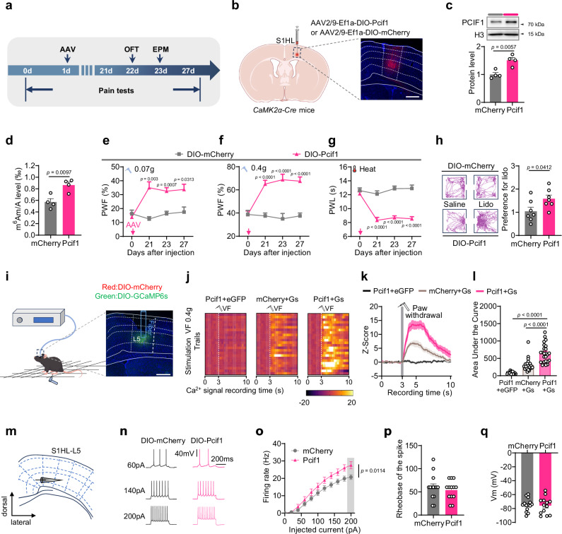
Nerve injury-induced changes in pain-associated genes contribute to genesis of neuropathic pain and comorbid anxiety. Phosphorylated CTD interacting factor-1 (PCIF1)-triggered N6, 2'-O-dimethyladenosine (m6Am) mRNA modification represents an additional layer of gene regulation. However, the role of PCIF1 in these disorders is elusive. Here, we report PCIF1 is increased in glutamatergic neurons of the hindlimb region of the primary somatosensory cortex in mouse with neuropathic pain and anxiety, but not inflammatory pain or anxiety alone. Serpine-1 mRNA-binding protein-1 (SERBP1) is identified as a PCIF1 cofactor, their complex mediates m6Am deposition onto mRNA. Blocking SERBP1-PCIF1 upregulation in glutamatergic neurons of the hindlimb region of the primary somatosensory cortex abolishes m6Am gain on maf1 homolog, negative regulator of RNA polymerase III (Maf1), elevates MAF1 protein, and mitigates neuropathic pain and anxiety. Conversely, mimicking this increase adds m6Am onto Maf1, reduces MAF1, and induces comorbidity symptoms. These findings highlight the significance of m6Am in neuropathic pain-anxiety comorbidity and identify SERBP1-PCIF1 in glutamatergic neurons of the hindlimb region of the primary somatosensory cortex as a potential therapeutic target.


Conflict of interest statement

Competing interests: The authors declare no competing interests.

Figures

Fig. 1
Fig. 1. Neuropathic pain increases the levels of m6Am and PCIF1 protein in S1HL.
a m5C, ac4C, m6Am, m7G, and m6A levels in contralateral primary somatosensory cortex (S1HL) on days 7 and 21 after surgery (n = 8 mice). b Expression level of METTL3, METTL14, PCIF1, ALKBH5, and FTO protein in the contralateral S1HL after surgery (n = 10 mice). c, e Expression of PCIF1 protein in contralateral S1HL at different times after spared nerve injury (SNI; n = 10 mice) or chronic constriction injury (CCI; n = 8 mice) or sham surgery. d, f Quantitative analysis of Pcif1 mRNA expression in contralateral S1HL by qRT-PCR at different times post-SNI or post-CCI or sham surgery (n = 8 mice). gk Expression of PCIF1 protein in contralateral S1HL in a model of cisplatin-induced neuropathic pain (Cisp), diabetic neuropathic pain (DNP) or chronic restraint stress (CRS) (n = 8 mice). i, j Expression of PCIF1 protein in contralateral S1HL at different times after complete Freund’s adjuvant (CFA)injection (Saline group: n = 8 mice, CFA group: n = 10 mice) or formalin (FM) injection (Saline group: n = 12 mice, FM group: n = 10 mice). l, m Co-expression analysis of PCIF1 with neuron (NeuN), astrocyte (S100β) or microglia (Iba1) immunofluorescence staining in S1HL of naïve mice (n = 4 mice). Scale bar, 200 μm. n Schematic showing isolation of GABAergic neurons and glutamatergic neurons from the L2/3, L5, and L6 layers of S1HL of mice subjected to SNI surgery for quantitative analysis of Pcif1 mRNA expression. o, p Expression of Pcif1 mRNA in GABAergic or glutamatergic neurons of S1 layers 2-6. n = 12 single cells form mice. The data are represented as mean ± SEM. For RNA modifications, mRNA and protein, S1 samples from two mice were pooled to create one sample. One-way ANOVA followed with Dunnett’s post hoc test was used for panels (a, b). Two-way ANOVA followed with Fisher’s LSD post hoc test was used for panels (cf, i, j, o and p). Student’s unpaired t-test (two-tailed) was used for panels (g, h and k).
Fig. 2
Fig. 2. Increased S1HLGlu PCIF1 is required for induction and maintenance of neuropathic pain.
a Viral injection schematic and Pcif1-shRNA vector design. b S1HL PCIF1 levels significantly decreased 35 days post AAV-CaMK2α-shPcif1 in SNI mice (n = 16 mice). ce Pre-injection of AAV-CaMK2α-shPcif1 into contralateral S1HL prevented SNI-induced mechanical allodynia (c, d) and thermal hyperalgesia (e) (n = 10 mice). fh Post-injection of shPcif1 reversed established contralateral SNI-induced mechanical allodynia (f, g) and thermal hyperalgesia (h) (n = 8 mice). i Experimental timeline in Pcif1fl/fl mice. j Schematic and images showing reduced PCIF1 in contralateral S1HLGlu neurons after AAV-CaMK2α-Cre injection in Pcif1fl/fl mice. Scale bar, 20 µm. k, l Pre-injection of AAV-CaMK2α-Cre reduced ipsilateral S1HL PCIF1 (k, n = 12 mice) and m6Am (l, n = 8 mice) levels post-SNI. mo Pre-injection of AAV-CaMK2α-Cre prevented SNI-induced mechanical hypersensitivity (mn) and thermal hyperalgesia (o) (n = 10 mice). p Left: Fiber photometry setup for Ca2+ imaging in awake Pcif1fl/fl mice (drawn by figdraw.com). Right: image shows AAV-CaMK2α-GCaMP6s (Green)/AAV-CaMK2α-mCherry (Red). Scale bar, 500 µm. Heatmaps (q), average Ca2+ transients (r) and AUC quantification of the GCaMP6s signal (3-7 seconds, s) from S1HLGlu neurons of Pcif1fl/fl mice receiving 0.4 g von Frey stimulation (n = 24 trails from 8 mice). t Patch-clamp schematic in S1HLGlu neurons. ux Sample traces (u), statistical data (v), and rheobase of the spike (w) for action potential firing and the membrane potential (x) recorded from S1HLGlu neurons (n = 13 cells from 4 mice). e PWF: paw withdrawal frequencies; PWL: Paw withdrawal latencies. OFT: open filed test; EPM: elevated plus maze; Gs: GCaMP6s. The data are represented as mean ± SEM. S1 samples from two mice were pooled to create one sample (b, l, k). One-way ANOVA followed with Tukey’s post hoc test was used for panels (b, k, l, s and vx). Two-way ANOVA followed with Tukey’s post hoc test was used for panels (ch and mo).
Fig. 3
Fig. 3. Upregulating S1HLGlu PCIF1 induces neuropathic pain- like behavior.
a Timeline for virus injection and behavior tests in CaMK2α-Cre mice. b Schematic of the experimental paradigm (left) and a representative image showing mCherry expression in S1HL after injection of AAV-DIO-Pcif1 or AAV-DIO-mCherry into CaMK2α-Cre mice (right). Scale bar, 500 µm (left). PCIF1 (c) and m6Am (d) levels significantly increased 28 days post AAV-DIO-Pcif1 (vs. mCherry) in CaMK2α-Cre mice (n = 8 mice). PCIF1 upregulation in S1HLGlu neurons induced contralateral mechanical allodynia (e, f), thermal hyperalgesia (g), as measured by conditioned place preference (h) (eg: n = 8 mice, h: n = 7 mice). i Schematic (drawn by figdraw.com) and image of GCaMP6s recording setup in awake mice (scale bar: 500 µm). Scale bar, 500 µm. Heatmaps (j), average Ca2+ transients (k), and AUC quantification of the GCaMP6s signal (l) from S1HLGlu neurons of CaMK2α-Cre mice receiving 0.4 g von Frey stimulation (n = 24 trails from 8 mice). m Patch-clamp schematic in S1HLGlu neurons. Sample traces (n), statistical data (o), and rheobase of the spike (p) for action potential firings and the membrane potential (q) recorded from S1HLGlu neurons in mice with upregulated PCIF1 expression, as described for (c, d) above (DIO-mCherry group: n = 14 cells from 4 mice, DIO-Pcif1 group: n = 13 cells from 4 mice). The data are represented as mean ± SEM. S1 samples from two mice were pooled to create one sample (c, d). Student’s unpaired t-test (two-tailed) was used for (c, d, h, p and q). Two-way ANOVA followed with Bonferroni’s post hoc test was used for (eg and o). One-way ANOVA followed with Bonferroni’s post hoc test was used for (l).
Fig. 4
Fig. 4. SERBP1 and PCIF1 interact and co-catalyze m6Am modifications on mRNA.
a The SERBP1 peptide identified by an MS analysis after an anti-PCIF1 co-immunoprecipitation assay in HEK293T cells. b Co-immunoprecipitation (IP) analysis of SERBP1 binding to PCIF1, using anti-PCIF1. Tissue was collected from the contralateral S1HL on day 21 after SNI or sham surgery in wild-type mouse premicroinjected with AAV-CaMK2α-shPcif1 (to knock down Pcif1 in glutamatergic cells) or a scrambled control. Input, the purified protein control. IB: immunoblotting (n = 10 mice). S1 samples from two mice were pooled to create one sample. c Construction schematic for the mammalian two-hybrid system; the luciferase reporter, with full-length Serbp1 and Gal4, and the VP16 transcription factor with a PCIF1 domain. The binding activity of SERBP1 and different PCIF1 domains (N-WW, d; helical, e; MTase, f; C-terminal, g) at 48 h after their co-transfection into HEK293T cells (n = 3 dishes of cells). h m6Am levels in HT22 cells after 48-hour transfection using: Vehicle (RNase-free water), SERBP1 plasmid with Control (empty vector), SERBP1 with Δ-MTase, SERBP1 with Δ-Helix, or SERBP1 with PCIF1 (n = 4 dishes of cells). i Schematic of CRY2/CIBN optogenetic regulation of the m6Am level through control of PCIF1 and SERBP1 binding activity. Serbp1 and Pcif1 were fused to the Cry2 and Cibn vectors, respectively. j, k Diagram showing Serbp1-Cry2 and Pcif1-Cibn co-transfection and activation of their interaction by blue light (j), and the quantitation of m6Am levels (k) (n = 3 dishes of cells). PBS, transfection of PBS control. The data are represented as mean ± SEM. One-way ANOVA followed with Tukey’s post hoc test was used for (b). One-way ANOVA followed with Dunnett’s post hoc test was used for (dh and k).
Fig. 5
Fig. 5. The interaction of SERBP1 and PCIF1 contributes to neuropathic pain.
a Schematic of virus injections and optical fiber implantation for activation of the optogenetic CRY2/CIBN system. b m6Am levels at 10 min after 1-h administration of blue light in wild-type mice injected 5 days previously in S1HL with Lenti-Pcif1-Cibn (Pci-Cibn) and Lenti-Serbp1-Cry2 (Ser-Cry2) or their controls Lenti-Cibn (Ctrl-Cibn) and Lenti-Cry2 (Ctrl-Cry2) (n = 10 mice). Pre, Pci-Cibn/Ser-Cry2-injected mice without blue light; Post, Pci-Cibn/Ser-Cry2-injected mice after 1-h blue light. Paw-withdrawal frequencies (PWF) to 0.07 g (c) and 0.4 g (d) von Frey filaments and paw-withdrawal latencies (PWL) to heat stimuli (e) on the contralateral side of mice injected as described in B. Behavioral tests were conducted 10 min after the end of 1 h blue light administration (n = 11 mice). f, g The level of PCIF1 protein (f) and m6Am (g) on day 7 after microinjection of AAV-DIO-shSerbp1 (shSerbp1) or AAV-DIO-scrambled-shRNA (Scr) in S1HL of CaMK2α-Cre mice preinjected with AAV-DIO-Pcif1 (Pcif1) or AAV-DIO-mCherry (mCherry) (F: n = 12 mice, G: n = 10 mice). Effect of SERBP1 downregulation on the contralateral mechanical (h, i) and heat (j) hypersensitivities induced by PCIF1 upregulation by DIO-Pcif1 (n = 10 mice). Red arrows, DIO-Pcif1 or DIO-mCherry injection. Blue arrows, DIO-shSerbp1 or DIO-mCherry injection. The data are represented as mean ± SEM. S1 samples from two mice were pooled to create one sample (b, f and g). Student’s unpaired t-test (two-tailed) was used for (b). Student’s paired t-test (two-tailed) was used for (ce). One-way ANOVA followed with Tukey’s post hoc test was used for (f and g). Two-way ANOVA followed with Tukey’s post hoc test was used for (hj).
Fig. 6
Fig. 6. Increased PCIF1 is responsible for gain of m6Am at Maf1 mRNA in the S1HL after nerve injury.
a Flowchart of the screening for downstream targets of PCIF1 by m6Am-Seq and RNC-Seq. SNI+shPcif1 indicates PCIF1 downregulation in S1HL of SNI mice via injection of AAV-shPcif1. b Distribution of m6Am peaks across mRNA segments of S1HL from three groups. c Motif analysis of m6Am peaks of mRNAs’ genomic sequences. The minus sign in X axis, upstream genomic nucleotides. d Venn diagram analysis revealed 436 genes consistently regulated by PCIF1 and SERBP1, based on integrated analysis of three complementary datasets: (i) genes with SNI-induced m6Am increases that were reversed by PCIF1 knockdown (m6Am-Exo-Seq), (ii) genes with increased SERBP1 binding post-SNI (SERBP1-RIP-Seq), and (iii) genes that exhibited reduced translation efficiency following SNI (RNC-Seq). e Gene ontology (GO) analysis of biological processes for the 436 genes. f Heatmap visualization of p-values and fold changes for the 436 genes. g Top 10 genes with most significance among 436 genes. h The representative image of m6Am peaks on Maf1 gene in S1HL. i Maf1 m6Am in S1HL on day 14 post-SNI measured via RNA immunoprecipitation (RIP)-PCR with four PCR primer pairs. The first two pairs include the RNA cap site (n = 8 mice). The forward F, and reverse R arrows represent paired PCR primers. j, k Maf1 m6Am (j) and the binding level of PCIF1 to Maf1 mRNA (k) in S1HL of SNI or Sham Pcif1fl/fl mice after preinjection of AAV-CaMK2α-Cre (Cre) or AAV-CaMK2α-mCherry (Ctrl) into S1HL (n = 10 mice). Maf1 m6Am level (l) and PCIF1 binding to Maf1 mRNA (m) on day 21 after injection of AAV-DIO-Pcif1 or AAV-DIO-mCherry into S1HL of naïve CaMK2α-Cre mice (n = 10 mice). For (im), data are represented as mean ± SEM. S1 samples from two mice were pooled to create one sample (jm). Student’s unpaired t-test (two-tailed) was used for panels (i, j, k). One-way ANOVA followed with Tukey’s post hoc test was used for (l and m).
Fig. 7
Fig. 7. Increased PCIF1 leads to SNI-induced downregulation of MAF1 in S1HL.
a MAF1 protein levels on day 21 after injection of AAV-DIO-Pcif1 (AAV-Pcif1; to induce PCIF1 expression) or AAV-DIO-mCherry (DIO-mCherry) into S1HL of naïve CaMK2α-Cre mice (n = 10 mice). b The level of MAF1 protein in SNI or Sham Pcif1fl/fl mice preinjected with AAV-CaMK2α-Cre (CaMK2α-Cre; to knock out PCIF1) or AAV-CaMK2α-mCherry (CaMK2α-mCherry) into S1HL. Tissue was harvested 21 days after injection (n = 12 mice). c, d Co-expression analysis of MAF1 (red) with NeuN (a neuronal marker, cyan), S100β (an astrocyte marker, cyan), or Iba1 (a microglia marker, cyan) immunofluorescence staining in the S1HL of naïve mice. n = 4 mice. Scale bar, 200 μm. e Co-expression analysis of Maf1 with Serbp1 and Pcif1 in CaMK2α neurons by single-cell PCR. Numbers 1-8 represent eight individual neurons. M, DNA marker. f CRISPR-dCasRx/PCIF1 “writing” m6Am to the given site in Maf1 mRNA. gRNA, small guide RNA. g Identification of dCasRx-Pcif1 fusion protein expression on day 5 after microinjection of CRISPR-dCasRx-Pcif1 into S1HL. h, i The Maf1 m6Am (h) and MAF1 protein (i) levels on day 5 after co-microinjection of CRISPR-dCasRx-Pcif1 and gRNA-26 or gRNA-60 into S1HL in naïve mice (n = 10 mice). For (a, b and h, i), data are represented as mean ± SEM. S1 samples from two mice were pooled to create one sample (a, b, h and i). Student’s unpaired t-test (two-tailed) was used for (a). One-way ANOVA followed with Tukey’s post hoc test was used for (b, h and i).
Fig. 8
Fig. 8. MAF1 mediates the regulatory effect of PCIF1 on neuropathic pain and comorbid anxiety.
a Reduced S1HL MAF1 protein 21 days post AAV-DIO-shMaf1 injection in CaMK2α-Cre mice (n = 8 mice). Knockdown of S1HL Maf1 or Co-injection of CRISPR-dCasRx-Pcif1 + gRNA-26/60 induced contralateral mechanical allodynia (b, c, e, f) and thermal hyperalgesia (d, g) (n = 10 mice). h MAF1 levels increased after injection of AAV-DIO-MAF1 into CaMK2α-Cre mice (n = 12 mice). ik Overexpressing MAF1 in contralateral S1HL attenuated SNI-induced mechanical allodynia (i, j) and thermal hyperalgesia (k) (n = 10 mice). l S1HL PCIF1/MAF1 levels after co-injection of AAV-DIO-Pcif1 ± AAV-DIO-Maf1 in naïve CaMK2α-Cre mice (n = 8 mice). mo MAF1 overexpression blocked PCIF1-induced pain-like behaviors (n = 10 mice). p Schematic (left, drawn by figdraw.com) and GCaMP6s expression image (right) for fiber photometry. Heatmaps (q), average Ca2+ transients (r), and AUC quantification (s) of GCaMP6s signals recorded from S1HLGlu neurons as mice received 0.4 g von Frey stimulation (n = 24 trails from 8 mice). Scale bar, 200 µm. t Schematic of patch-clamp recordings from labeled S1HLGlu neurons in slices. Sample traces (u), statistical data (v), and rheobase of the spike (w) for action potential firing and the membrane potential (x) recorded from S1HLGlu neurons (n = 13 cells from 4 mice). Data are represented as mean ± SEM. S1 samples from two mice were pooled to create one sample (a, h and l). Student’s unpaired t-test (two-tailed) was used for (a) and (v). Two-way ANOVA followed with Bonferroni’s post hoc test was used for (bd). Two-way ANOVA followed with Tukey’s post hoc test was used for (eg). One-way ANOVA followed with Tukey’s post hoc test was used for (h, l, s, w and x). Two-way ANOVA followed with Tukey’s post hoc test was used for panels (ik and mo).
Fig. 9
Fig. 9. Cartoon showing the mechanism by which the SERBP1-PCIF1 complex in S1HLGlu neurons contributes to the development and maintenance of neuropathic pain and comorbid anxiety.
Peripheral nerve injury induces an increase in the SERBP1-PCIF1 complex, which in turn regulates m6Am-controlled MAF1 expression in S1HL, leading to reduced inhibition and enhanced neuronal excitability (drawn by figdraw.com).

Similar articles

References

    1. van Hecke, O., Austin, S. K., Khan, R. A., Smith, B. H. & Torrance, N. Neuropathic pain in the general population: a systematic review of epidemiological studies. Pain155, 654–662 (2014). - PubMed
    1. Attal, N., Bouhassira, D. & Colvin, L. Advances and challenges in neuropathic pain: a narrative review and future directions. Br. J. Anaesth.131, 79–92 (2023). - PubMed
    1. Mills, S. E. E., Nicolson, K. P. & Smith, B. H. Chronic pain: a review of its epidemiology and associated factors in population-based studies. Brit J. Anaesth.123, e273–e283 (2019). - PMC - PubMed
    1. Li, X. Y. et al. Alleviating neuropathic pain hypersensitivity by inhibiting PKMzeta in the anterior cingulate cortex. Science330, 1400–1404 (2010). - PubMed
    1. Jin, Y. et al. A somatosensory cortex input to the caudal dorsolateral striatum controls comorbid anxiety in persistent pain. Pain161, 416–428 (2020). - PubMed

LinkOut - more resources