. 2025 Aug 5;8(1):1155.
doi: 10.1038/s42003-025-08594-8.

Prenatal linguistic exposure shapes language brain responses at birth

Affiliations

Prenatal linguistic exposure shapes language brain responses at birth

Andréanne René et al. Commun Biol. .

Abstract

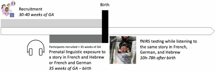
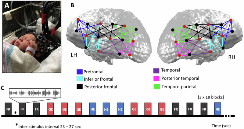
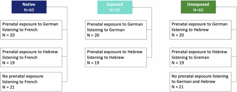
Newborns have an immature brain network responsible for speech processing that resembles the adult language network. However, it remains unclear how prenatal experience modulates this network. To test this, we exposed 39 fetuses to a story in their native language and in a foreign language during the last month of gestation, while another group of 21 fetuses received no experimental prenatal exposure. Within 3 days of life, neonates' brain responses were recorded using functional near-infrared spectroscopy (fNIRS) whilst they listened to the same story in their native language and in two foreign languages, one of which neonates had been prenatally exposed to. Results revealed that brain responses to the native language and the prenatally exposed foreign language were similar, whereas they differed in the left temporal and right prefrontal regions when listening to a prenatally unexposed foreign language. Findings indicate that foetuses' linguistic environment influences speech processing at birth.


Conflict of interest statement

Competing interests: The authors declare no competing interests.

Figures

Fig. 1
Fig. 1. Study design.
Timeline of recruitment, prenatal linguistic exposure according to fetal gestational age (GA) and fNIRS recording during a passive listening story task within 3 days after birth. Participants recruited after 35 weeks of GA underwent no prenatal exposure to German or Hebrew.
Fig. 2
Fig. 2. Experimental procedure for the fNIRS recording at birth.
A A newborn participant with the fNIRS headgear in their crib. B Probe placement and their approximate spatial projection onto a newborn MRI brain template. Coupling of light sources (red dots) and detectors (black dots) yielded a total of 72 channels (colored lines). Colors represent the six regions of interest (ROI) subdivisions identified in the left (LH) and right (RH) hemispheres based on anatomical landmarks and 10-10 coordinates. C Example of the experimental paradigm. The three languages were presented in blocks of three consecutive story segments of the same language (French (FR) in black, German (GE) in red, and Hebrew (HE) in blue). The order of the languages was randomized and counterbalanced across participants.
Fig. 3
Fig. 3. Grand average hemodynamic concentration changes according to language exposure conditions across all participants.
A Topographical representation of mean HbO concentration changes according to language familiarity for the native (n = 60), exposed (n = 39) and unexposed (n = 60) language conditions. Front (F) and back (B) of topographical representation are indicated for left and right hemispheres. B HbO concentration changes for Native in blue, Exposed (German or Hebrew, depending on the prenatal linguistic group) in cyan, Unexposed (Hebrew or German, depending on the prenatal linguistic group) in green. Channels are plotted following the headgear placement shown in Fig. 2 for the left and right hemispheres. The black vertical lines indicate the onset of the stimulation period. An asterisk (*) over a region indicates significant (P < 0.05) differences between native and unexposed conditions.
Fig. 4
Fig. 4. Repeated measures ANOVA results.
Data of hemodynamic HbO concentration changes were plotted in box plots for the three exposure conditions and six ROIs, in the left hemisphere (left panel) and in the right hemisphere (right panel). The white diamonds show the mean of the data. Black arrows indicate significant post hoc differences between native and unexposed conditions. An asterisk (*) over a region indicates significant (P < 0.05) hemispheric differences for the native and the exposed conditions in the posterior temporal, temporo-parietal, prefrontal, and posterior frontal regions. No hemispheric differences were found for the unexposed condition. Blue arrows show significant differences between regions within the same hemisphere for the native condition. Hemodynamic HbO concentration mean and standard error estimates for all exposure conditions, ROIs, and hemispheres are available in Supplementary Table S2.
Fig. 5
Fig. 5. Language familiarity conditions.
Participants’ fNIRS data were split into three language familiarity conditions (Native in blue, Exposed in cyan, Unexposed in green) according to prenatal exposure conditions (Prenatal exposure (German), Prenatal exposure (Hebrew), No prenatal exposure) and language of story.

References

    1. Byers-Heinlein, K. & Fennell, C. T. Perceptual narrowing in the context of increased variation: insights from bilingual infants. Dev. Psychobiol.56, 274–291 (2014). - PubMed
    1. Hervé, P.-Y., Zago, L., Petit, L., Mazoyer, B. & Tzourio-Mazoyer, N. Revisiting human hemispheric specialization with neuroimaging. Trends Cogn. Sci.17, 69–80 (2013). - PubMed
    1. Kuhl, P. K. Brain mechanisms in early language acquisition. Neuron67, 713–727 (2010). - PMC - PubMed
    1. Wang, D., Buckner, R. L. & Liu, H. Functional specialization in the human brain estimated by intrinsic hemispheric interaction. J. Neurosci.34, 12341–12352 (2014). - PMC - PubMed
    1. Gervain, J., Macagno, F., Cogoi, S., PeñA, M. & Mehler, J. The neonate brain detects speech structure. Proc. Natl. Acad. Sci. USA105, 14222–14227 (2008). - PMC - PubMed

LinkOut - more resources