Preoperative MRI and CA19-9 for predicting occult lymph node metastasis in small pancreatic ductal adenocarcinoma (≤ 2 cm)
- PMID: 40764914
- PMCID: PMC12326715
- DOI: 10.1186/s12880-025-01854-3
Preoperative MRI and CA19-9 for predicting occult lymph node metastasis in small pancreatic ductal adenocarcinoma (≤ 2 cm)
Abstract
Aim: Accurate prediction of occult lymph node metastasis (OLNM) in small pancreatic ductal adenocarcinoma (sPDAC) (≤ 2 cm) is crucial for curative management. This study aims to explore clinical and MRI features associated with OLNM in sPDAC and their pathological and prognostic implications.
Materials and methods: This retrospective study included 135 patients with pathologically confirmed sPDAC who underwent surgery between September 2014 and September 2023. Preoperative multi-sequence MRI, clinical data, and pathological features were analyzed. Univariate and multivariate logistic regression models were used to identify risk predictors of OLNM in sPDAC. Receiver operating characteristic (ROC) analysis was performed to assess diagnostic performance and Kaplan-Meier survival analysis was used to evaluate prognostic outcomes.
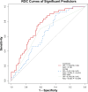
Results: OLNM was present in 43 (31.9%) sPDAC patients. Univariate and multivariate analysis identified elevated CA19-9 (> 100 U/mL) (OR = 2.404, P = 0.040) and low apparent diffusion coefficient (ADC) values (OR = 0.243, P = 0.031) as independent predictors of OLNM. The combined clinical-radiological model demonstrated an AUC of 0.740, significantly higher than CA19-9 (AUC = 0.653, P = 0.021) or ADC alone (AUC = 0.635, P = 0.035). sPDAC patients with OLNM exhibited higher rates of lymphovascular invasion (44.2%, P = 0.013) and pathological fat invasion (86.0%, P = 0.030). OLNM was associated with significantly worse OS and DFS (P = 0.034 and 0.043).
Conclusions: OLNM is associated with adverse pathological features and poorer prognosis. The combination of preoperative MRI assessment of ADC and CA19-9 may aid in identifying sPDAC patients at high risk for OLNM.
Clinical trial number: Not applicable.
Keywords: Magnetic resonance imaging; Occult lymph node metastasis; Pancreatic ductal adenocarcinoma.
© 2025. The Author(s).
Conflict of interest statement
Declarations. Ethics approval and consent to participate: This retrospective study was approved by the ethics committee of Zhongshan Hospital, Fudan university (B2024-250R), which waived the requirement for written informed consent owing to the use of deidentified retrospective data. This study was performed in accordance with the Declaration of Helsinki. Consent for publication: Not applicable. Competing interests: The authors declare no competing interests.
Figures


Similar articles
-
Predictors of occult lymph node metastasis in clinical T1 lung adenocarcinoma: a retrospective dual-center study.BMC Pulm Med. 2025 Mar 1;25(1):99. doi: 10.1186/s12890-025-03559-3. BMC Pulm Med. 2025. PMID: 40025457 Free PMC article.
-
Development and validation of an explainable machine learning model for predicting occult lymph node metastasis in early-stage oral tongue squamous cell carcinoma: A multi-center study.Int J Surg. 2025 Aug 1;111(8):5022-5035. doi: 10.1097/JS9.0000000000002641. Epub 2025 Jun 5. Int J Surg. 2025. PMID: 40479496
-
Evaluation of Occult Liver Metastases in Pancreatic Adenocarcinoma by Diffusion-Weighted Related Magnetic Resonance Imaging.J Magn Reson Imaging. 2025 Aug;62(2):536-548. doi: 10.1002/jmri.29747. Epub 2025 Mar 10. J Magn Reson Imaging. 2025. PMID: 40062724
-
Predictive role of radiomics features extracted from preoperative cross-sectional imaging of pancreatic ductal adenocarcinoma in detecting lymph node metastasis: a systemic review and meta-analysis.Abdom Radiol (NY). 2023 Aug;48(8):2570-2584. doi: 10.1007/s00261-023-03940-y. Epub 2023 May 18. Abdom Radiol (NY). 2023. PMID: 37202642
-
The prognostic impact of para-aortic lymph node metastasis in pancreatic cancer: A systematic review and meta-analysis.Eur J Surg Oncol. 2016 May;42(5):616-24. doi: 10.1016/j.ejso.2016.02.003. Epub 2016 Feb 13. Eur J Surg Oncol. 2016. PMID: 26916137
References
-
- Conroy T, Pfeiffer P, Vilgrain V, Lamarca A, Seufferlein T, O’Reilly EM, Hackert T, Golan T, Prager G, Haustermans K, et al. Pancreatic cancer: ESMO clinical practice guideline for diagnosis, treatment and follow-up. Ann Oncol. 2023;34(11):987–1002. - PubMed
-
- Iacobuzio-Donahue CA. The war on pancreatic cancer: progress and promise. Nat Rev Gastro Hepat. 2023;20(2):75–6. - PubMed
-
- Fyfe I. AI predicts pancreatic cancer risk. Nat Rev Gastro Hepat. 2023;20(7):413. - PubMed
-
- Yurgelun MB. Building on more than 20 years of progress in pancreatic cancer surveillance for High-Risk individuals. J Clin Oncol. 2022;40(28):3230–4. - PubMed
MeSH terms
Substances
Grants and funding
LinkOut - more resources
Full Text Sources
Medical

