. 2025 Aug 5;12(1):46.
doi: 10.1186/s40779-025-00631-1.

Decreased IL-33 in the brain following repetitive mild traumatic brain injury contributes to cognitive impairment by inhibiting microglial phagocytosis

Affiliations

Decreased IL-33 in the brain following repetitive mild traumatic brain injury contributes to cognitive impairment by inhibiting microglial phagocytosis

Ze-Xi Jia et al. Mil Med Res. .

Abstract

Background: Repetitive mild traumatic brain injury (rmTBI) is a significant risk factor for neurodegeneration, characterized by pathological protein deposition and persistent neuroinflammation. Research has observed increased interleukin-33 (IL-33) levels in the peripheral blood of patients with rmTBI, suggesting IL-33 may participate in regulating the pathological development of rmTBI. The study aims to elucidate the impact and mechanism of IL-33 in the progression of neuropathology following rmTBI, and to explore its potential as a therapeutic target to improve the neurological outcome.

Methods: The study employed an rmTBI mouse model using the wild-type (WT) and IL-33 knockout mice. Cognitive function was assessed via the Y-maze and Barnes tests. The main cell type expressing IL-33 and its receptor, suppression of tumorigenicity 2 (ST2), was then investigated in the mouse brain through immunofluorescence colocalization. As the primary neural cell responsible for ST2 expression, microglia were studied in vitro using the BV2 cell line. The effects of lipid droplets (LDs) accumulation and amyloid-beta (Aβ) phagocytosis were measured to elucidate the impact of IL-33 on BV2 cells' phagocytosis. Additionally, HT22 neuronal apoptosis was assessed by flow cytometry. Finally, the cognitive effects of intranasal administration of IL-33 were evaluated in mice.

Results: IL-33KO mice exhibited pronounced cognitive impairment after rmTBI. In the mouse brain, astrocytes were identified as the primary source of IL-33 secretion, while microglia predominantly expressed ST2. Transcriptome sequencing revealed that IL-33 significantly influenced phagocytosis function. IL-33 mitigated LDs accumulation in BV2 cells and enhanced Aβ phagocytosis in vitro. In addition, the culture medium of BV2 cells with activated IL-33/ST2 signaling reduced HT22 neuronal apoptosis and axonal damage. Furthermore, intranasal administration of IL-33 was observed to be effective in alleviating neurodegeneration and cognitive outcome of rmTBI mice.

Conclusions: Dysfunction of the IL-33/ST2 axis following rmTBI leads to cognitive dysfunction via impairing microglial phagocytosis capacity and promoting neuronal damage. IL-33 would be a promising therapeutic target for alleviating neurodegeneration following rmTBI.

Keywords: Cognition; Interleukin-33 (IL-33); Microglia; Repetitive mild traumatic brain injury (rmTBI).


Conflict of interest statement

Declarations. Ethics approval and consent to participate: The collection and processing of clinical samples for this study were approved by the Ethics Committee of the General Hospital of Tianjin Medical University (IRB2021-YX-056–01), and informed consent was obtained from all participants at enrollment. All animal studies were approved by the Animal Care and Use Committee at Tianjin Medical University (IRB2024-DWFL-044) and complied with the National Institutes of Health Guidelines for the Care and Use of Laboratory Animals. Consent for publication: Not applicable. Competing interests: The authors declare that they have no competing interests.

Figures
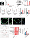
Fig. 1
Fig. 1
The dynamic change of IL-33 levels following rmTBI. a TEM image displaying purified exosomes. Scale bar = 1 μm. b Analysis of size distribution for purified exosomes conducted via NTA. c Western blotting analysis of characteristic exosomal biomarkers, including Alix, CD63, and CD9. d mRNA levels of total IL-33, exosomal IL-33, and vWF in blood from patients with rmTBI and healthy control individuals (n = 6). e ELISA measurements of total circulating IL-33 and exosomal IL-33 in blood at specified time points post-rmTBI (n = 6). f Representative immunofluorescence staining images showing IL-33 (red), CD31 (green), and DAPI (blue) in the aortas of mice at specified time points post-rmTBI. Scale bar = 50 μm. g Array data were quantified using ImageJ to generate a protein profile, which is displayed in a heatmap. h ELISA was performed to measure IL-33 levels in the mouse brain at specified time points post-rmTBI (n = 5). Data are represented as the mean ± SEM. *P < 0.05, **P < 0.01, ***P < 0.001. TEM transmission electron microscope, NTA nanoparticle tracking analysis, DAPI 4,6-diamidino-2-phenylindole dihydrochloride, rmTBI repetitive mild traumatic brain injury, vWF von Willebrand factor, ELISA enzyme-linked immunosorbent assay
Fig. 2
Fig. 2
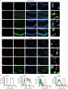
Cellular localization of IL-33 in the hippocampal (dentate gyrus) and cortical regions of WT-rmTBI mice. a Representative immunofluorescence staining images of IL-33 (red), Olig2+ oligodendrocytes (green), GFAP+ astrocytes (green), TMEM119+ microglia (green), NeuN+ neurons, and DAPI (blue) in the hippocampus and cortex of mice on day 42 post-rmTBI (n = 5, 3 slides/mouse. Consistent fields of view were selected across all slides for subsequent quantification). Scale bar = 50 μm (main images) and 25 μm (magnified insets). b Fluorescence intensity plots of IL-33 and various cells. The red curves show the relative intensity of IL-33, and the green curves show Olig2+ oligodendrocytes, GFAP+ astrocytes, TMEM119+ microglia, and NeuN+ neurons. DAPI 4,6-diamidino-2-phenylindol dihydrochloride, IL-33 interleukin-33, Olig2 oligodendrocyte lineage transcription factor 2, GFAP glial fibrillary acidic protein, TMEM119 transmembrane protein 119, NeuN neuron-specific nuclear protein, WT wild-type, rmTBI repetitive mild traumatic brain injury
Fig. 3
Fig. 3
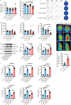
IL-33 deficiency exacerbated cognitive deficits induced by rmTBI. a Total number of entries and percentage of spontaneous alternations in the Y-maze (n = 10). b Average speed, total distance, and crossing times during the exploration phase of the Barnes maze for mice (n = 10). c Training and spatial exploration paths of mice in the Barnes maze experiment from days 42 to 46 post-rmTBI. d Western blotting analysis of APP, p-tau, and tau on day 42 post-rmTBI (n = 6). e Reconstructed S16-tau PET/CT images of mice and average SUV values in the hippocampal region of the mouse brain (n = 3). f ELISA measurements of IL-6, IL-1β, TNF-α, IL-4, IL-10, and TGF-β in the mouse brain on day 42 post-rmTBI (n = 6). Data are represented as the mean ± SEM. *P < 0.05, **P < 0.01, ***P < 0.001, ns non-significant. DAPI 4,6-diamidino-2-phenylindol dihydrochloride, PET/CT positron emission tomography/computed tomography, SUV standardized uptake value, WT wild-type, KO knockout, APP amyloid precursor protein, rmTBI repetitive mild traumatic brain injury, ELISA enzyme-linked immunosorbent assay, IL interleukin, TNF-α tumor necrosis factor-α, TGF-β transforming growth factor-β, SUVbw standardized uptake value based on body weight
Fig. 4
Fig. 4
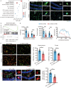
IL-33 deficiency impaired microglial phagocytic function. a KEGG enrichment scatter plot showing representative differential pathways between IL-33KO and WT groups at 42 d post-rmTBI. b GSEA analysis related to the phagocytic process in IL-33KO-rmTBI mice. c Representative immunofluorescence staining images of Iba1+ microglia (green) and DAPI (blue) in the hippocampus and cortex of WT-rmTBI and IL-33KO-rmTBI mice. Scale bar = 50 μm (main images) and 20 μm (magnified insets). d Endpoint counts, process length, and Sholl analysis of Iba1+ cells (n = 6). e Representative immunofluorescence staining images and quantification of CD68+ (red), Iba1+ microglia (green) in the hippocampus and cortex of WT-rmTBI and IL-33KO-rmTBI mice (n = 6). Scale bar = 50 μm. f Representative immunofluorescence staining images and quantification of APP+ (green), Iba1+ microglia (red), and DAPI (blue) in the hippocampus of WT-rmTBI and IL-33KO-rmTBI mice (n = 6). Scale bar = 50 μm (main images) and 20 μm (magnified insets). Data are represented as the mean ± SEM. *P < 0.05, **P < 0.01, ***P < 0.001. DAPI 4,6-diamidino-2-phenylindol dihydrochloride, KEGG Kyoto Encyclopedia of Genes and Genomes, GSEA Gene Set Enrichment Analysis, rmTBI repetitive mild traumatic brain injury, Iba1 ionized calcium-binding adapter molecule 1, APP amyloid precursor protein, IL-33 interleukin-33, TNF tumor necrosis factor, ECM extracellular matrix, Th T helper cell, WT wild-type, KO knockout
Fig. 5
Fig. 5
IL-33 improved phagocytic function by reducing LD accumulation in BV2 microglial cells. a Representative image of siRNA-ST2 (green) transfected BV2 cells. Scale bar = 50 µm. RT-qPCR (b) and Western blotting (c) analysis showing changes in ST2 levels in BV2 cells following transfection. d Representative immunofluorescence image of Iba1+ BV2 cells (red) after 12 h of treatment with Aβ1-42 (green) and uptake index of Aβ1-42 in BV2 cells. Scale bar = 50 μm (main images) and 25 μm (magnified insets). e Representative immunofluorescence staining images of BODIPY+ (green) in BV2 cells. Scale bar = 50 μm (main images) and 25 μm (magnified insets). Quantification was performed by flow cytometry (n = 3). f Representative immunofluorescence image of Iba1+ BV2 cells (red) after 12 h of treatment with Aβ1-42 (green) following IL-33 intervention. Scale bar = 50 μm (main images) and 25 μm (magnified insets). g The uptake of Aβ1-42 by BV2 cells under different concentrations of IL-33 intervention (n = 6). h Representative immunofluorescence staining images of BODIPY+ (green) in BV2 cells after IL-33 intervention. Scale bar = 50 μm (main images) and 25 μm (magnified insets). Quantification was performed by flow cytometry (n = 3). Data are represented as the mean ± SEM. *P < 0.05, **P < 0.01, ***P < 0.001, ns non-significant. DAPI 4,6-diamidino-2-phenylindol dihydrochloride, ST2 suppression of tumorigenicity 2, LPS lipopolysaccharide, Aβ1-42 amyloid-beta 1–42, Iba1 ionized calcium-binding adapter molecule 1, BODIPY boron-dipyrromethene, VEH vehicle, IL-33 interleukin-33, FITC fluorescein isothiocyanate, LDs lipid droplets, RT-qPCR real-time quantitative polymerase chain reaction
Fig. 6
Fig. 6
IL-33/ST2 signaling activation in BV2 cells contributed to their neuroprotective effects in vitro. a Flow cytometric analysis of apoptosis after different treatments (n = 3). b Representative immunofluorescence staining images and quantification of β-tubulin (red) and DAPI (blue) following different treatments (n = 3). Scale bar = 50 μm (main images) and 25 μm (magnified insets). Arrows indicate axons. Data are represented as the mean ± SEM. *P < 0.05, **P < 0.01, ***P < 0.001. DAPI 4,6-diamidino-2-phenylindol dihydrochloride, WT wild-type, LPS lipopolysaccharide, ST2 suppression of tumorigenicity 2, IL-33 interleukin-33, 7-AAD 7-aminoactinomycin D, APC allophycocyanin
Fig. 7
Fig. 7
Exogenous IL-33 improved the cognitive outcome of rmTBI mice. a Total number of entries and percentage of spontaneous alternations in the Y-maze (n = 8). b Average speed, total distance, and crossing times during the exploration phase of the Barnes maze for mice (n = 8). c Training and spatial exploration paths of mice in the Barnes maze experiment from days 42 to 46 post-rmTBI. Data are represented as the mean ± SEM. **P < 0.01, ***P < 0.001. rmTBI repetitive mild traumatic brain injury, PBS phosphate buffer saline, IL-33 interleukin-33
Fig. 8
Fig. 8
Intranasal administration of the recombinant IL-33 protein attenuated the accumulation of microglial lipid droplets, enhanced Aβ phagocytosis, and promoted neuronal survival in the brain, thus contributing to improving the cognitive outcome of rmTBI mice. rmTBI repetitive mild traumatic brain injury, IL-33 interleukin-33, ST2 suppression of tumorigenicity 2, IL-1RAcP interleukin-1 receptor accessory protein, LD lipid droplet, Aβ amyloid-beta

Similar articles

References

    1. Jiang JY, Gao GY, Feng JF, Mao Q, Chen LG, Yang XF, et al. Traumatic brain injury in China. Lancet Neurol. 2019;18(3):286–95. - PubMed
    1. Maas AIR, Menon DK, Adelson PD, Andelic N, Bell MJ, Belli A, et al. Traumatic brain injury: integrated approaches to improve prevention, clinical care, and research. Lancet Neurol. 2017;16(12):987–1048. - PubMed
    1. Zhu Y, Williams J, Beyene K, Trani JF, Babulal GM. Traumatic brain injury, seizures, and cognitive impairment among older adults. JAMA Netw Open. 2024;7(8):e2426590. - PMC - PubMed
    1. Armstrong RC, Sullivan GM, Perl DP, Rosarda JD, Radomski KL. White matter damage and degeneration in traumatic brain injury. Trends Neurosci. 2024;47(9):677–92. - PubMed
    1. Li H, Sun D, Zhao Z, Fang J, Li M, Lv C, et al. Neutrophil membrane-derived nanoparticles protect traumatic brain injury via inhibiting calcium overload and scavenging ROS. J Nanobiotechnology. 2024;22(1):477. - PMC - PubMed