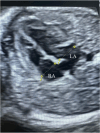
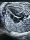
Prognostic value of foramen ovale morphology and hemodynamics in late-onset fetal growth restriction: a 3D ultrasonography-based study
- PMID: 40764963
- PMCID: PMC12326826
- DOI: 10.1186/s12884-025-07985-3
Prognostic value of foramen ovale morphology and hemodynamics in late-onset fetal growth restriction: a 3D ultrasonography-based study
Abstract
Objective: To assess the structural and hemodynamic characteristics of the foramen ovale (FO) in fetuses with late-onset fetal growth restriction (LO-FGR) using three-dimensional (3D) ultrasonography and Doppler imaging, and to examine their associations with Doppler parameters in FGR and composite adverse perinatal outcomes (CAPO).
Methods: This case-control study included 40 fetuses with LO-FGR and 40 matched controls exhibiting appropriate-for-gestational-age (AGA) between 34 and 37 weeks. FO area was measured using 3D spatio-temporal image correlation (STIC) imaging, and FO width and pulsatility index (PI) were evaluated using 2D and Doppler ultrasonography. FO parameters were compared between the groups, and partial correlation analyses adjusted for gestational age to assess their associations with FGR and CAPO. Additionally, Receiver Operating Characteristic (ROC) curve analysis was conducted to evaluate the predictive value of FO parameters for CAPO within the FGR group.
Results: FO area (p < 0.001), FO width (p < 0.001), left atrial (LA) width (p = 0.029), FO/LA ratio (p < 0.001), and FO/RA ratio (p = 0.024) were significantly reduced in the FGR group compared to the controls. Among FGR fetuses, those who developed CAPO had lower FO area (p = 0.009), FO width (p = 0.001), LA width (p = 0.006), FO/LA ratio (p < 0.001), and FO/RA ratio (p = 0.041). In ROC analysis, the FO/LA ratio exhibited the highest predictive value for predicting CAPO (AUC: 0.851, p < 0.001).
Conclusion: Alterations in FO morphology are significantly associated with adverse perinatal outcomes in LO-FGR. The FO/LA ratio may serve as a reliable and noninvasive parameter for risk stratification. Incorporating advanced fetal cardiac morphometry could improve prenatal surveillance in FGR.
Keywords: Composite adverse perinatal outcome; Fetal growth restriction; Fetal hemodynamics; Foramen ovale; Three-dimensional ultrasonography.
© 2025. The Author(s).
Conflict of interest statement
Declarations. Ethics approval and consent to participate: The study protocol was approved by the Ankara Etlik City Hospital Ethics Committee (approval number: AESH-BADEK-2025-0074). and written informed consent was obtained from all participants. The study adhered to the ethical principles outlined in the Declaration of Helsinki. Consent for publication: Not applicable. This study does not contain any individual person’s data in any form (including any individual details, images, or videos) that would require consent for publication. Competing interests: The authors declare no competing interests.
Figures


Similar articles
-
Perinatal risk assessment in pregnancies complicated by early-onset fetal growth restriction: development and internal validation of a prediction model for composite adverse perinatal outcome.Ultrasound Obstet Gynecol. 2025 Aug;66(2):175-185. doi: 10.1002/uog.29265. Epub 2025 Jul 7. Ultrasound Obstet Gynecol. 2025. PMID: 40623699 Free PMC article.
-
The role of umbilical vein blood flow assessment in the prediction of fetal growth velocity and adverse outcome: a prospective observational cohort study.Am J Obstet Gynecol. 2025 Jul;233(1):66.e1-66.e14. doi: 10.1016/j.ajog.2025.01.001. Epub 2025 Jan 4. Am J Obstet Gynecol. 2025. PMID: 39756605
-
Predictive value of fetal Doppler velocimetry, fetal growth trajectory and maternal serum biomarkers for short-term adverse perinatal outcome: secondary analysis of DRIGITAT study.Ultrasound Obstet Gynecol. 2025 Jul 11. doi: 10.1002/uog.29266. Online ahead of print. Ultrasound Obstet Gynecol. 2025. PMID: 40641303
-
Severe smallness as predictor of adverse perinatal outcome in suspected late small-for-gestational-age fetuses: systematic review and meta-analysis.Ultrasound Obstet Gynecol. 2022 Sep;60(3):328-337. doi: 10.1002/uog.24977. Ultrasound Obstet Gynecol. 2022. PMID: 35748873
-
Fetal cerebro-placental ratio and adverse perinatal outcome: systematic review and meta-analysis of the association and diagnostic performance.J Perinat Med. 2016 Mar;44(2):249-56. doi: 10.1515/jpm-2015-0274. J Perinat Med. 2016. PMID: 26756084
References
-
- American College of Obstetricians and Gynecologists’ Committee on Practice Bulletins—Obstetrics and the Society forMaternal-FetalMedicin. ACOG practice bulletin 204: fetal growth restriction. Obstet Gynecol. 2019;133(2):e97–109. 10.1097/AOG.0000000000003070. - PubMed
-
- Burton GJ, Jauniaux E. Pathophysiology of placental-derived fetal growth restriction. Am J Obstet Gynecol. 2018;218(2S):S745–61. 10.1016/j.ajog.2017.11.577. - PubMed
-
- Erbilen EA, Varol FG. Tyrosine Kinase-2, Angiopoietin-2, and thrombomodulin axes in the Late-Onset fetal growth restriction: A prospective cohort study. Gynecol Obstet Reproductive Med. 2024;30(3):159–66. 10.21613/GORM.2023.1500.
MeSH terms
LinkOut - more resources
Full Text Sources
Research Materials
Miscellaneous

