. 2025 Aug 5;23(1):860.
doi: 10.1186/s12967-025-06902-8.

Ivabradine induces RAD51 degradation, potentiating PARP inhibitor efficacy in non-germline BRCA pathogenic variant triple-negative breast cancer

Affiliations

Ivabradine induces RAD51 degradation, potentiating PARP inhibitor efficacy in non-germline BRCA pathogenic variant triple-negative breast cancer

Ho Tsoi et al. J Transl Med. .

Abstract

Background: Triple-negative breast cancer (TNBC) is an aggressive subtype lacking targetable proteins for treatment. PARP inhibitors (PARPi) are effective in BRCA-mutated cancers but have limited utility in non-germline BRCA-mutated (non-gBRCAm) TNBC. We hypothesized that inducing BRCAness by targeting RAD51, a key homologous recombination protein, could sensitize non-gBRCAm TNBC to PARPi.

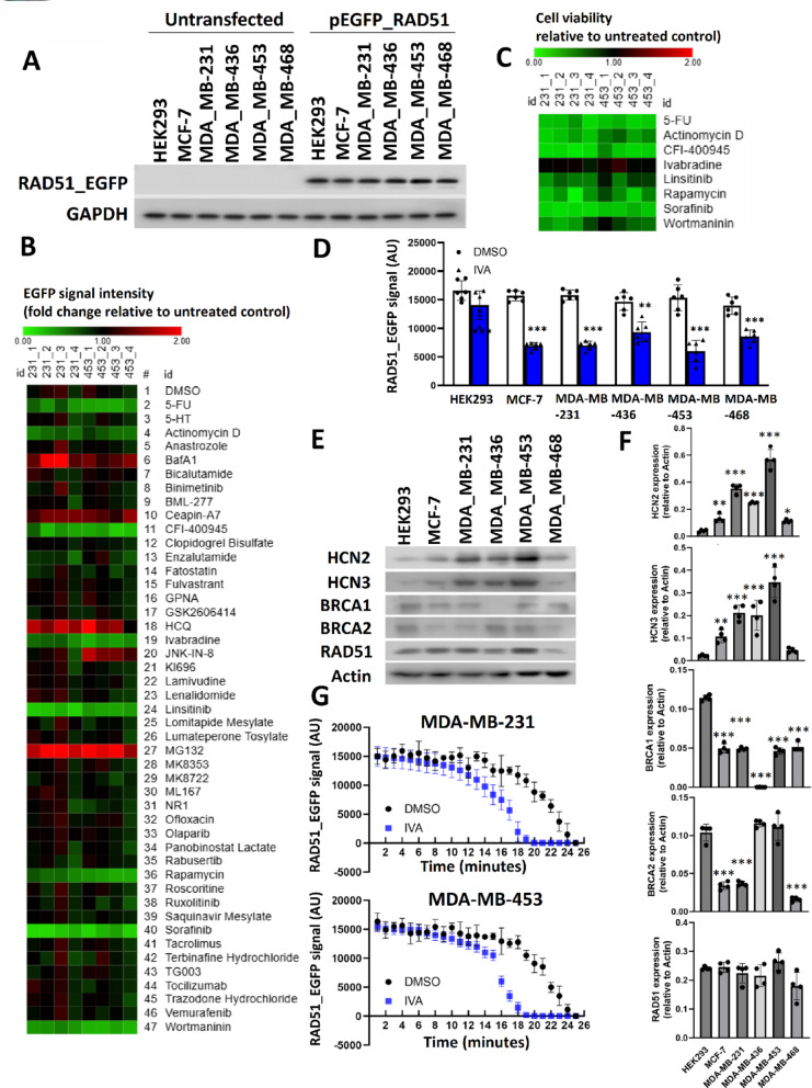
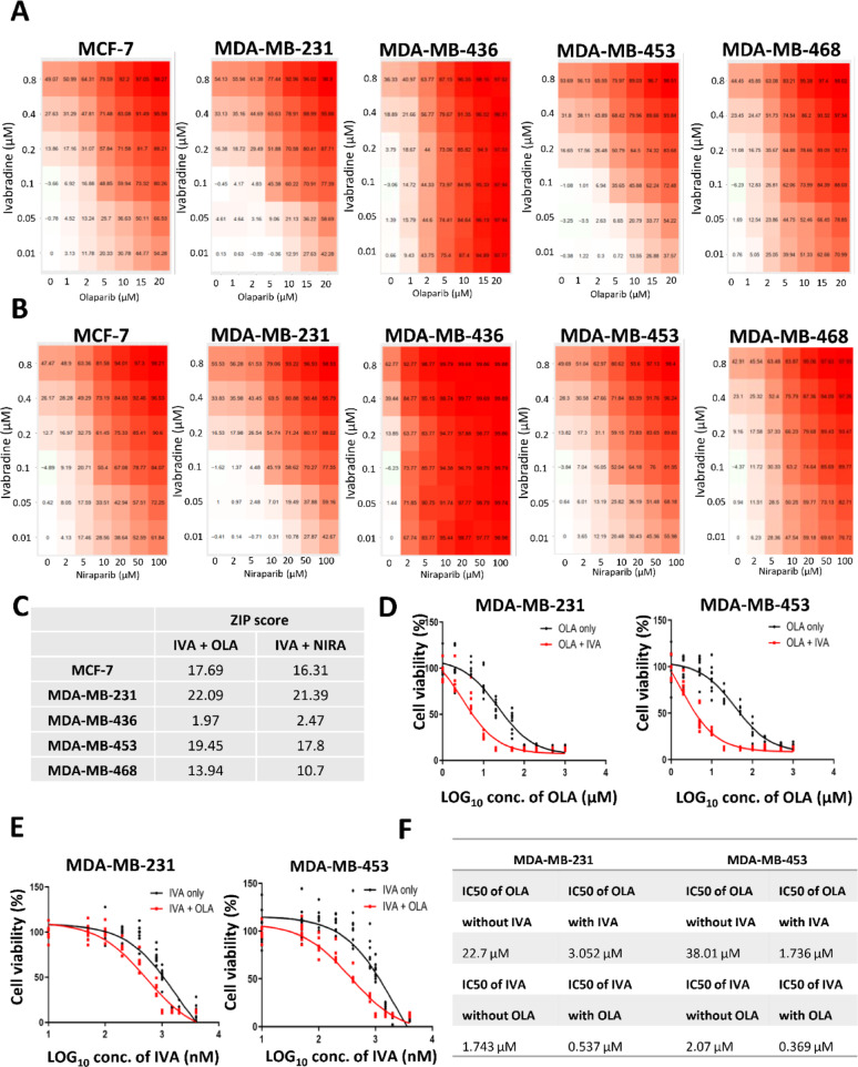
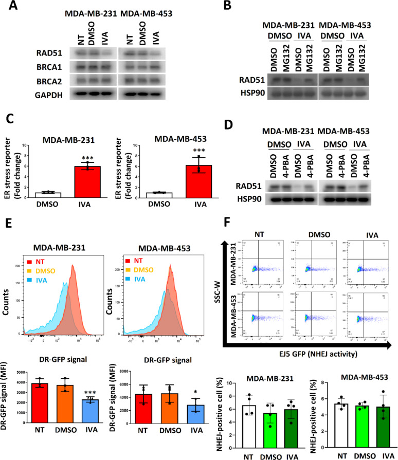
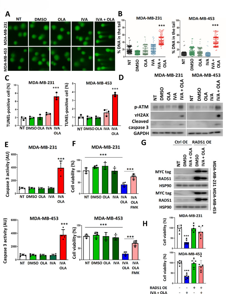
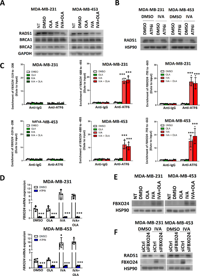
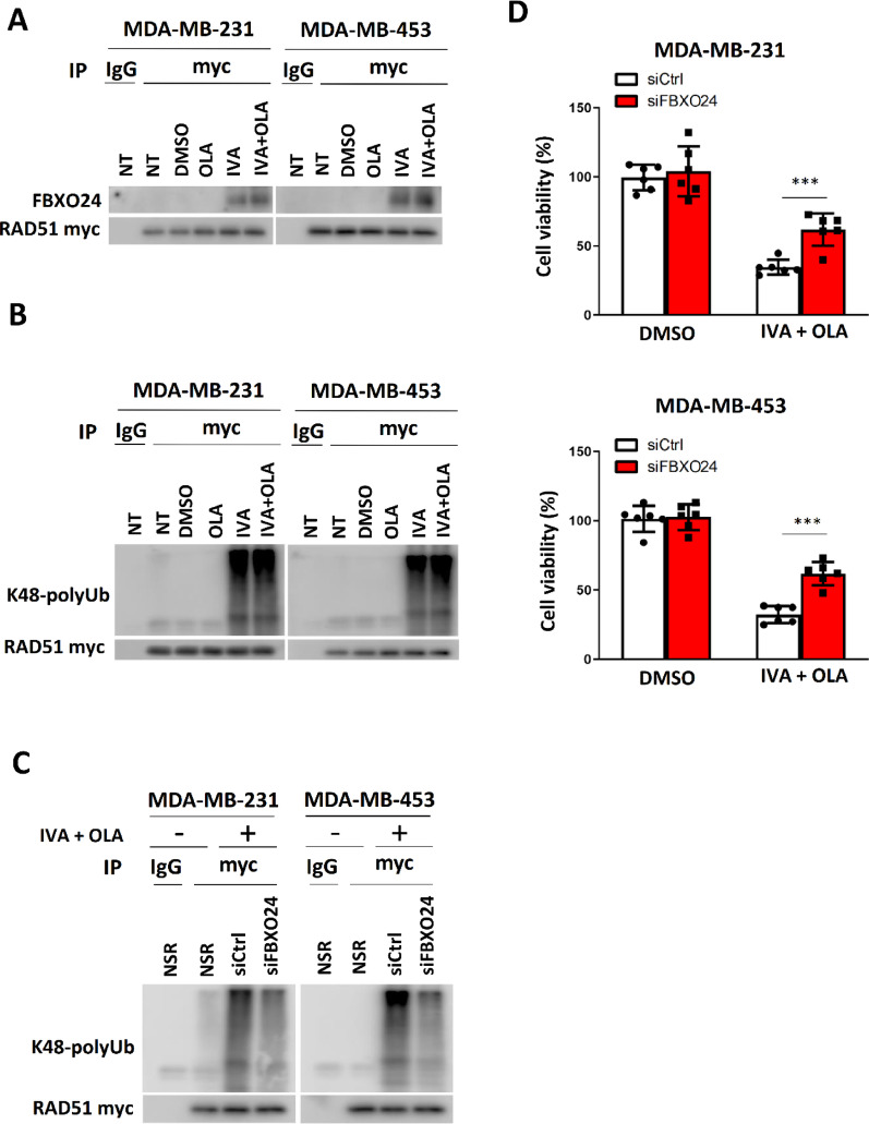
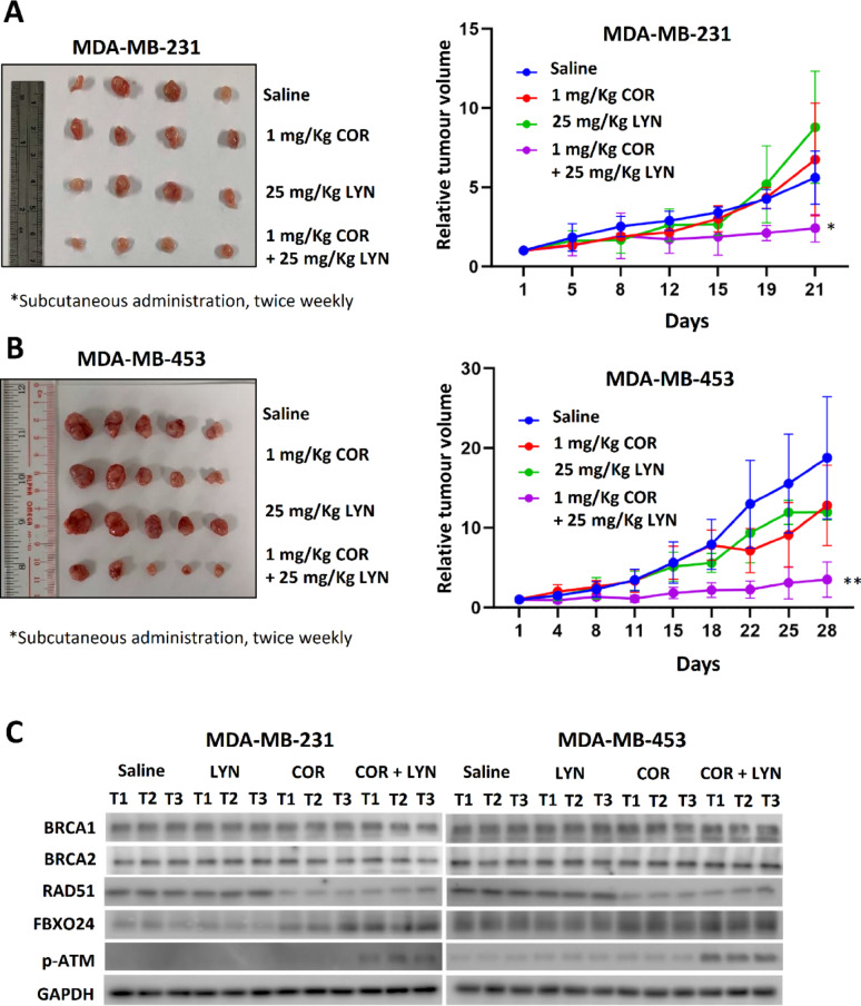
Methods: EGFP-tagged RAD51 was generated and EGFP signal was monitored for identifying agents that affected RAD51 protein expression and stability. Cell viability was assayed using cell counting kit-8. Synergism of ivabradine and olaparib was determined using SynergyFinder 3.0. DR-GFP, EJ5-GFP and comet assays were employed to evaluate the degree of DNA repair and damage, respectively. Protein and mRNA levels were determined by western blot and qPCR, respectively. ChIP was used to determine the binding to ATF6 to the promoter of FBXO24. CoIP was employed to determine the interaction between RAD51 and FBXO24. Xenografts on nude mice and PDTX were in vivo models for validating the combined effect of ivabradine and olaparib.

Results: Using an EGFP-RAD51 reporter, we identified ivabradine as a RAD51-reducing agent. In vitro studies with TNBC cell lines demonstrated that ivabradine synergized with PARPi to reduce cell viability (ZIP score > 10), induce apoptosis, and impair HR-mediated DNA repair. This synergy was confirmed in vivo using xenografts and patient-derived tumor xenografts, where co-treatment with clinical grade ivabradine (Coralan) and PARPi olaparib (Lynparza) led to substantial tumor growth inhibition without notable toxicity. Mechanistically, ivabradine triggered ER stress, activating ATF6 to upregulate FBXO24-dependent ubiquitination, leading to RAD51 degradation, resulting in the condition of BRCAness. Chromatin immunoprecipitation and co-immunoprecipitation confirmed the ATF6-FBXO24-RAD51 cascade. These findings reveal a novel mechanism by which ivabradine, an FDA-approved cardiac drug, induces BRCAness, by degrading RAD51 via the ATF6-FBXO24 axis, thus, by mimicking HR deficiency hypersensitizes BRCA-proficient TNBC to olaparib.

Conclusion: This study highlights the translational potential of repurposing ivabradine as a therapeutic strategy for non-gBRCAm TNBC. By addressing a critical unmet need of this aggressive breast cancer subtype, it can potentially expand the utility of PARPi.

Keywords: BRCAness; Breast cancer; Ivabradine; Olaparib; Synthetic lethality; TNBC; Targeted therapy.


Conflict of interest statement

Declarations. Ethics approval and consent to participate: The procedures on mice were reviewed and approved by Committee on the Use of Live Animals in Teaching and Research, The University of Hong Kong (5236-19). Approval was granted from The Institutional Review Board of the University of Hong Kong/Hospital Authority Hong Kong West Cluster (HKU/HA HKW IRB No. UW 16–391) for collecting human tissue and blood samples. Consent for publication: Not applicable. Competing interests: USK, HT, and CG are the inventors of the patent (Use of HCN Inhibitors for Treatment of Cancer; EP3615052B1). The other authors declare that they have no competing interests.

Figures

Fig. 1
Fig. 1
Identification of IVA as a modifier of ectopic EGFP-tagged RAD51. A The ectopic expression of RAD51_EGFP in the cell lines. Western blot with anti-EGFP was employed to detect the EGFP-tagged RAD51. GAPDH was the loading control. B Screening to identify inhibitors to reduce EGFP-tagged RAD51 in MDA-MB-231 and MDA-MB-453. The cells were treated with 5 µM of each inhibitor for 48 h. The EGFP signal was recorded and compared to the untreated control. A heatmap was plotted to show the effect of the chemicals on the EGFP signal. C Heatmap showed the effect of the selected inhibitors on cell viability. CCK8 assay was used after 48 h of the treatment. D The treatment of IVA significantly reduced the expression of EGFP-tagged RAD51 in MDA-MB-231 and MDA-MB-453 but not in HEK293. Students’ t-test was used for the statistical analysis. The result was shown as mean ± SD from 6 independent experiments. E The expression of BRCA1, BRCA2, RAD51, HCN2 and HCN3. Western blot was employed to examine the expression of the indicated proteins in the cell lines. Actin was used as the loading control. F Quantification of E. Image J was employed to determine the band intensity in E. The band intensity of the candidate protein relative to actin was determined. Results showed mean ± SD from 4 independent experiments. G The effect of IVA on the protein stability of EGFP-tagged RAD51. After 6 h, the EGFP signal was recorded per minute for 25 min. The result was shown as mean ± SD from 6 independent experiments. ** represents P < 0.01. *** represents P < 0.001
Fig. 2
Fig. 2
The synergistic effect of IVA and OLA. A the effect of different concentrations of IVA and OLA on cell viability. 5 breast cancer cell lines were examined. The cells were treated for 72 h. CCK8 was used for cell viability assay. The mean percentage of the inhibition from 5 dependent experiments was shown. B the effect of different concentrations of IVA and niraparib (NIRA) on cell viability. C ZIP scores in the 5 breast cancer cells. ZIP score was calculated using SynergyFinder 3.0. D Comparing the response to OLA in the presence or absence of IVA. The cells were treated with different concentrations of OLA with 0.1 µM of IVA. Cell viability was determined using CCK8 after 72 h of the treatment. Results were shown as mean ± SD from 9 independent experiments. E Comparing the response to IVA in the presence or absence of IVA. The cells were treated with different concentrations of IVA with 5 µM of OLA. Cell viability was determined using CCK8 after 72 h of the treatment. Results were shown as mean ± SD from 9 independent experiments. F The IC50 values of OLA in the presence or absence of IVA and IVA in the presence or absence of OLA were determined
Fig. 3
Fig. 3
IVA compromised DNA repair mediated by HR. A IVA treatment reduced RAD51 expression. The cells were treated with 0.1 µM of IVA for 72 h. Western blot was employed. GAPDH was the loading control. B Proteasome inhibitor MG132 abolished the effect of IVA on RAD51 expression. The cells were treated with 100 nM of IVA and 5 μM of MG132 for 72 h. Western blot was performed. C IVA induced ER stress. The cells were treated with 0.1 µM of IVA. Luciferase reporter assay with ATF4 response element was performed 48 h post-treatment. Untreated control was used as the reference. Results were shown as mean ± SD from 3 independent experiments. D 4-PBA abolished the effect of IVA on RAD51 expression. The cells were treated with 0.1 µM of IVA and 10 µM of 4-PBA for 72 h. Western blot was performed. HSP90 was the loading control. E IVA suppressed HR. DR-GFP assay was performed. The cells were treated with 0.1 µM of IVA for 72 h. Mean Fluorescence Intensity (MFI) was determined. Results were shown as mean ± SD from 3 independent experiments. F IVA did not affect NHEJ. EJ5-GFP assay was performed. MDA-MB-231 and MDA-MB-453 were treated with 0.1 µM of IVA for 72 h. The GFP signal was analyzed using flow cytometry. Representative traces were shown. Results were shown as mean ± SD from 4 independent experiments. One-way ANOVA was used. NT represents no treatment control. *** represents P < 0.001
Fig. 4
Fig. 4
IVA and OLA co-treatment triggered DNA damage and apoptosis. A The co-treatment enhanced DNA damage. Comet assay was performed. B Statistical analysis of the comet assay in (A). The cells were treated with 0.1 µM of IVA and/or 5 μM of OLA for 72 h. 200 nuclei were analysed. Results were shown as mean ± SD. C IVA and OLA co-treatment enhanced the proportion of cells with DNA damage. TUNEL assay was performed after 72 h of treatment. Results were shown as mean ± SD from 3 independent experiments. D The co-treatment enhanced the expression of p-ATM and ɤH2AX, and cleaved caspase 3. Western blot was performed. E The co-treatment significantly enhanced the enzymatic activity of the caspase 3. Results were shown as mean ± SD from 4 independent experiments. F Pan-caspase inhibitor Z-VAD-FMK compromised the effect of the co-treatment on cell viability. 10 μM of Z-VAD-FMK, 0.1 µM of IVA and/or 5 μM of OLA were used for 72 h of treatment. Results were shown as mean ± SD from 4 independent experiments. G The effect of the co-treatment on cells expressing exogenous RAD51. The cells were transfected with pcDNA3.1 or pcDNA3.1_myc_RAD51. Ctrl OE indicated control overexpression, while RAD51 OE indicated RAD51 overexpression. 24-h post-transfection, the cells were treated with 100 nM of IVA and 5 μM of OLA for 48 h. Western blot was performed to detect exogenous MYC-tagged RAD51 and total RAD51 (exogenous and endogenous). H RAD51 overexpression compromised the effect of the co-treatment on cell viability. The cells were transfected with pcDNA3.1_myc_RAD51. 24 h post-transfection, the cells were treated with 100 nM of IVA and 5 μM of OLA for 72 h. Results were shown as mean ± SD from 6 independent experiments. NT represents no treatment control. *** represent P < 0.001
Fig. 5
Fig. 5
IVA employed ATF6 to induce the expression of FBXO24 to mediate RAD51 down-regulation. A IVA treatment and co-treatment of IVA and OLA reduced the expression of RAD51 only. The cells were treated with 0.1 µM of IVA and/or 5 μM of OLA for 72 h. Western blot was performed. GAPDH was the loading control. B ATF6 inhibition abolished the effect of RAD51 down-regulation mediated by IVA. 5 μM of ATF6 inhibitor Ceapin-A7, 0.1 µM of IVA and 5 μM of OLA were used. The cells were treated for 72 h. Western blot was performed. HSP90 was the loading control. C IVA enhanced ATF6 binding to the promoter region of FBXO24. The cells were treated with 0.1 µM of IVA and/or 5 μM of OLA for 72 h. ChIP was performed with anti-ATF6. qPCR was employed to determine the enrichment of 3 different regions of FBXO24 promoter in the elute. Results were shown as mean ± SD from 4 independent experiments. One-way ANOVA was employed. D ATF6 inhibition abolished the effect of IVA on FBXO24 induction. 5 μM of ATF6 inhibitor Ceapin-A7 and 0.1 µM of IVA and/or 5 μM of OLA were used for treating the cells for 48 h. qPCR was performed. Results were shown as mean ± SD from 4 independent experiments. E IVA treatment enhanced FBXO24 protein expression. The cells were treated with 0.1 µM of IVA and/or 5 μM of OLA for 72 h. Western blot was used. HSP90 was the loading control. F Knockdown of FBXO24 compromised the effect of IVA on RAD51 down-regulation. The cells were treated with 0.1 µM of IVA and 15 μM of siFBXO24 for 72 h. Western blot was used. HSP90 was the loading control. NT represents no treatment control. *** represent P < 0.001
Fig. 6
Fig. 6
The ATF6-FBXO24 axis was essential for the efficacy of IVA and OLA co-treatment. A RAD51 interacted with FBXO24. The cells were transfected with pcDNA3.1_myc_RAD51. 24 h post-transfection, the cells were treated with 0.1 µM of IVA and/or 5 μM of OLA for 48 h. CoIP was performed with either anti-mouse IgG or anti-myc antibodies. Western blot was employed to evaluate the level of indicated protein candidates in the immunoprecipitant. B IVA treatment enhanced the degree of ubiquitination on RAD51. The cells were transfected with pcDNA3.1_myc_RAD51. 24 h post-transfection, the cells were treated with 5 μM of MG132, 0.1 µM of IVA and/or 5 μM of OLA for 48 h. Co-immunoprecipitation was performed with either anti-mouse IgG or anti-MYC antibodies. Western blot was performed using an anti-K48-linkage polyubiquitin antibody. C Knockdown of FBXO24 could compromise the effect of the co-treatment on the degree of K-48 conjugation polyubiquitination of RAD51. The cells were transfected with pcDNA3.1_myc_RAD51. 24 h post-transfection, the cells were treated with 5 μM of MG132, 15 μM of siCtrl or siFBXO24, 0.1 µM of IVA and 5 μM of OLA for 48 h. Co-immunoprecipitation was performed with either anti-mouse IgG or anti-myc antibodies. Western blot was performed using an anti-K48-linkage polyubiquitin antibody. D FBXO24 knockdown weakened the efficacy of IVA and OLA co-treatment. The cells were treated with 0.1 µM of IVA, 5 μM of OLA and 15 μM of siFBXO24 or 15 μM of siCtrl for 72 h. Cell viability was assayed with CCK8. Results were shown as mean ± SD from 6 independent experiments. Students’ t-test was used. NT represents no chemical treatment. NSR represents no siRNA treatment. *** represent P < 0.001
Fig. 7
Fig. 7
Co-treatment of COR and LYN significantly suppressed tumor growth in nude mice. The xenografts were established from A MDA-MB-231 and B MDA-MB-453. The nude mice were treated with 1 mg/Kg of COR (clinical grade Ivabradine) and 25 mg/Kg of LYN (clinical grade Olaparib) via subcutaneous injection twice a week. The volume change of the tumors was monitored. Tumor volume relative to day 1 was plotted. Results were shown as mean ± SD from 4 or 5 independent tumors. Two-way ANOVA was employed. C Western blot confirmed that COR treatment reduced RAD51 expression but enhanced FBXO24 expression. Phosphorylated ATM (p-ATM) level was only increased in tumors from the mice receiving the co-treatment. Protein lysates from three independent tumors from each group were analyzed. GAPDH was the loading control. * and ** represent P < 0.05 and P < 0.01, respectively
Fig. 8
Fig. 8
Co-treatment of COR and LYN significantly suppressed tumor growth in patient-derived tumor xenograft (PDTX) models. Two PDTX models, A PDTX5 and B PDTX8, were used. The mice were administered 2 mg/Kg of COR (clinical grade Ivabradine) and 124 mg/Kg of LYN (clinical grade Olaparib) via gavaged feeding daily. Tumor volume relative to day 1 was plotted. Results were shown as mean ± SD from 5 independent tumors. Two-way ANOVA was employed. C Western blot confirmed that COR treatment reduced the expression of RAD51 but enhanced FBXO24 expression. Phosphorylated ATM (p-ATM) level was only increased in tumors from the mice receiving the co-treatment. Protein lysates from three independent tumors from each group were analyzed. GAPDH was the loading control. ** and *** represent P < 0.01 and P < 0.001, respectively

Similar articles

References

    1. Schmid P, Cortes J, Dent R, McArthur H, Pusztai L, Kümmel S, Denkert C, Park YH, Hui R, Harbeck N, et al. Overall survival with pembrolizumab in early-stage triple-negative breast cancer. N Engl J Med. 2024;391:1981–91. - PubMed
    1. Burstein HJ. Immunotherapy for early-stage triple-negative breast cancer. N Engl J Med. 2024;391:2048–9. - PubMed
    1. Li M, Yu X. Function of BRCA1 in the DNA damage response is mediated by ADP-ribosylation. Cancer Cell. 2013;23:693–704. - PMC - PubMed
    1. O’Neil NJ, Bailey ML, Hieter P. Synthetic lethality and cancer. Nat Rev Genet. 2017;18:613–23. - PubMed
    1. Zhao W, Steinfeld JB, Liang F, Chen X, Maranon DG, Jian Ma C, Kwon Y, Rao T, Wang W, Sheng C, et al. BRCA1-BARD1 promotes RAD51-mediated homologous DNA pairing. Nature. 2017;550:360–5. - PMC - PubMed

MeSH terms

LinkOut - more resources