. 2025 May;87(2):305-319.
doi: 10.18999/nagjms.87.2.305.

Meflin/Islr is a marker of fibroblasts that arise in fibrotic regions after spinal cord injury

Affiliations

Meflin/Islr is a marker of fibroblasts that arise in fibrotic regions after spinal cord injury

Yoshinori Morita et al. Nagoya J Med Sci. 2025 May.

Abstract

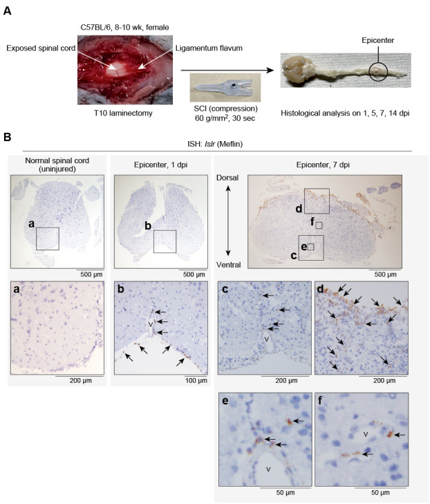
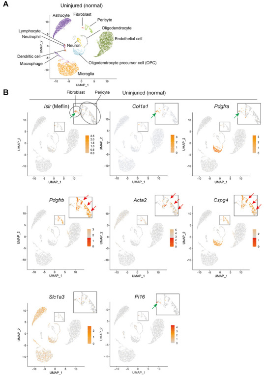
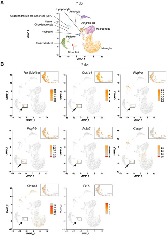
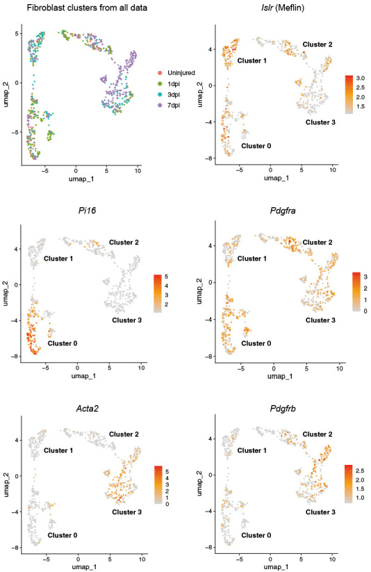
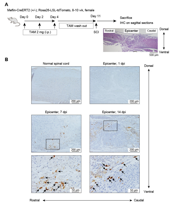
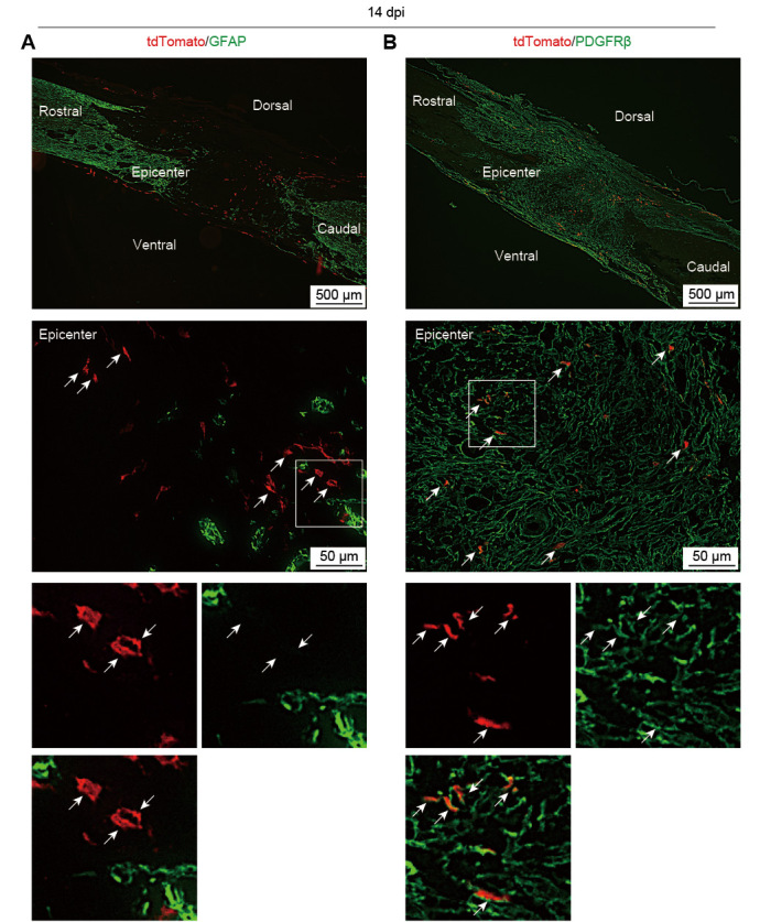
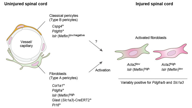
Scar formation after spinal cord injury (SCI) hampers axonal regeneration and functional recovery. Previous studies have shown that scar formation is attributable to both gliosis and fibrosis, the latter requiring fibroblast proliferation and extracellular matrix deposition. In this setting, there are essentially two cell types generating new fibroblasts: pericytes and tissue-resident fibroblasts. Here, we showed that Meflin, a glycosylphosphatidylinositol-anchored protein (a specific marker of fibroblasts across multiple organs) is expressed by fibroblasts in the normal mouse spinal cord. An in situ hybridization analysis showed that Meflin+ cells arose from the meninges and perivascular region of the spinal parenchyma after spinal cord compression injury. That finding was corroborated by single-cell transcriptomic data from normal and injured mouse spinal cords. Interestingly, Meflin+ cells are positive for the fibroblast markers collagen type I and platelet-derived growth factor receptor (PDGFR) α but not for pericyte markers such as PDGFRβ and chondroitin sulfate proteoglycan 4 in the normal spinal cord. Those findings are consistent with the recent view that tissue-resident fibroblasts play a central role in many other types of fibrotic disease. A lineage-tracing experiment using a knock-in mouse line that expressed inducible Cre recombinase under the control of the Meflin promoter showed that Meflin+ cells yield PDGFRβ+ myofibroblasts but not glial cells positive for glial fibrillary acidic protein. These findings suggest that the Meflin+ population contains the cells of origin of myofibroblasts that are involved in scar formation after SCI.

Keywords: Islr; Meflin; fibrosis; immunoglobulin superfamily containing leucine rich repeat; spinal cord injury.


Conflict of interest statement

The authors declare no conflicts of interest.

Figures

Fig. 1
Fig. 1
Meflin+ cells appear around the perivascular area and the meninges after induction of SCI Fig. 1A: A diagram showing the experimental process for the induction of SCI by the extradural clip compression method. See Materials and Methods for details of the protocol. Fig. 1B: Tissue sections prepared from uninjured spinal cord at the T10 level (left) and the epicenter of injured spinal cord at 1 (middle) and 7 (right) dpi were stained for Meflin (Islr) mRNA by ISH. The boxed areas (af) are magnified in lower panels. Arrows denote Meflin+ cells. V: vessel SCI: spinal cord injury ISH: in situ hybridization Islr: immunoglobulin superfamily containing leucine rich repeat
Fig. 2
Fig. 2
Meflin expression in fibroblasts in the normal mouse spinal cord Fig. 2A: A publicly available data set based upon a single-cell transcriptomic analysis of cells isolated from the normal mouse spinal cord (GSE162610) was analyzed, and the selected cell clusters were visualized by the UMAP algorithm. Fig. 2B: The data were examined for the expression of Meflin (Islr) and marker genes of fibroblasts (Col1a1, Pdgfra), conventional pericytes (Pdgfrb, Acta2, Cspg4), type A pericytes (Slc1a3) and universal fibroblasts (Pi16). The fibroblast clusters positive for Meflin (Islr), Col1a1, Pdgfra and Pi16 and the pericyte clusters positive for Pdgfrb, Acta2 and Cspg4 are indicated by green and red arrows, respectively. Islr: immunoglobulin superfamily containing leucine rich repeat Col1a1: collagen type I alpha 1 chain Pdgfra: platelet derived growth factor receptor alpha Pdgfrb: platelet derived growth factor receptor beta Acta2: actin alpha 2, smooth muscle Cspg4: chondroitin sulfate proteoglycan 4 Slc1a3: solute carrier family 1 member 3 Pi16: peptidase inhibitor 16 UMAP: Uniform Manifold Approximation and Projection
Fig. 3
Fig. 3
Meflin expression in fibroblasts in the injured mouse spinal cord Fig. 3A: The data set representing a single-cell transcriptomic analysis of cells isolated from the mouse spinal cord at 7 dpi (GSE162610). The selected cell clusters were visualized by the UMAP algorithm. Fig. 3B: The data were examined for the expression of Meflin (Islr) and marker genes of fibroblasts (Col1a1, Pdgfra), myofibroblasts (Pdgfrb, Acta2), conventional pericytes (Cspg4), type A pericytes (Slc1a3) and universal fibroblasts (Pi16). Arrows indicate the fibroblast clusters. Islr: immunoglobulin superfamily containing leucine rich repeat Col1a1: collagen type I alpha 1 chain Pdgfra: platelet derived growth factor receptor alpha Pdgfrb: platelet derived growth factor receptor beta Acta2: actin alpha 2, smooth muscle Cspg4: chondroitin sulfate proteoglycan 4 Slc1a3: solute carrier family 1 member 3 Pi16: peptidase inhibitor 16 UMAP: Uniform Manifold Approximation and Projection
Fig. 4
Fig. 4
Features of fibroblasts from both normal and injured spinal cords Col1a1+ fibroblasts were collected from a data set of a single-cell transcriptomic analysis performed on all cells collected from a normal spinal cord and injured spinal cords after the induction of SCI (1, 3, and 7 dpi). The identified fibroblast clusters (clusters 0, 1, 2, and 3) were visualized with the UMAP algorithm, followed by an expression analysis of the indicated genes. Islr: immunoglobulin superfamily containing leucine rich repeat Pi16: peptidase inhibitor 16 Pdgfra: platelet derived growth factor receptor alpha Acta2: actin alpha 2, smooth muscle Pdgfrb: platelet derived growth factor receptor beta UMAP: Uniform Manifold Approximation and Projection
Fig. 5
Fig. 5
Analysis of Meflin lineage cells after SCI induction in a Meflin reporter mouse line Fig. 5A: Female mice (8-10-week-old Meflin-CreERT2; LSL-tdTomato) were administered TAM (2 mg) three times every other day, followed by a one-week washout period and the induction of SCI. Fig. 5B: Sagittal sections prepared from the spinal cords at 0 (normal spinal cord), 1, 7, and 14 dpi were stained for tdTomato by IHC to visualize Meflin lineage cells. Boxed areas are magnified in lower panels. Arrows denote tdTomato+ cells. TAM: tamoxifen i.p.: intraperitoneal injection SCI: spinal cord injury IHC: immunohistochemistry dpi: days post-injury
Fig. 6
Fig. 6
Meflin lineage cells become myofibroblasts following spinal cord injury Sagittal sections prepared from spinal cords at 14 dpi were stained by IF for tdTomato (red, arrows) and either GFAP (A) or PDGFRβ (B) (green). Boxed areas are magnified in lower panels. Arrows denote tdTomato+ cells. dpi: days post-injury GFAP: glial fibrillary acidic protein PDGFRβ: platelet derived growth factor receptor beta IF: Immunofluorescence
Fig. 7
Fig. 7
Schematic illustration of heterogeneous features of stromal cells in normal and injured spinal cords The data presented here suggest that Meflin is preferentially expressed by Col1a1+Pdgfra+ fibroblasts, which substantially overlap with type A pericytes, but not Cspg4+Pdgfrb+ classic pericytes in the normal spinal cord. The single-cell transcriptomic analysis and clustering showed fibroblast activation retaining Meflin expression in the injured spinal cord, where an inverse correlation between Meflin and Acta2 was observed. The activation of fibroblasts, but not classic pericytes, by spinal cord injury was proven by the lineage tracing experiment performed in the present study. Cspg4: chondroitin sulfate proteoglycan 4 Pdgfrb: platelet derived growth factor receptor beta Islr: immunoglobulin superfamily containing leucine rich repeat Col1a1: collagen type I alpha 1 chain Pdgfra: platelet derived growth factor receptor alpha Glast: Glutamate/Aspartate Transporter 1 Slc1a3: solute carrier family 1 member 3 Pi16: peptidase inhibitor 16 Acta2: actin alpha 2, smooth muscle

Similar articles

References

    1. Quadri SA, Farooqui M, Ikram A, et al. Recent update on basic mechanisms of spinal cord injury. Neurosurg Rev. 2020;43(2):425–441. doi: 10.1007/s10143-018-1008-3 - DOI - PubMed
    1. Segi N, Nakashima H, Machino M, et al. Epidemiology of Cervical Fracture/Cervical Spinal Cord Injury and Changes in Surgical Treatment Modalities in Elderly Individuals During a 10-year Period: A Nationwide Multicenter Study in Japan. Global Spine J. 2024;14(5):1583–1594. doi: 10.1177/21925682231151643 - DOI - PMC - PubMed
    1. Burda JE, Sofroniew MV. Reactive gliosis and the multicellular response to CNS damage and disease. Neuron. 2014;81(2):229–248. doi: 10.1016/j.neuron.2013.12.034 - DOI - PMC - PubMed
    1. Gaudet AD, Fonken LK. Glial Cells Shape Pathology and Repair After Spinal Cord Injury. Neurotherapeutics. 2018;15(3):554–577. doi: 10.1007/s13311-018-0630-7 - DOI - PMC - PubMed
    1. Bradbury EJ, Burnside ER. Moving beyond the glial scar for spinal cord repair. Nat Commun. 2019;10(1):3879. doi: 10.1038/s41467-019-11707-7 - DOI - PMC - PubMed

LinkOut - more resources