. 2025 Aug 5:8:0815.
doi: 10.34133/research.0815. eCollection 2025.

Fibroblast Growth Factor 21 Promotes Vascular Smooth Muscle Cell Contractile Polarization via p38 Mitogen-Activated Protein Kinase-Promoted Serum Response Factor Phosphorylation

Affiliations

Fibroblast Growth Factor 21 Promotes Vascular Smooth Muscle Cell Contractile Polarization via p38 Mitogen-Activated Protein Kinase-Promoted Serum Response Factor Phosphorylation

Mengmeng Zhu et al. Research (Wash D C). .

Abstract

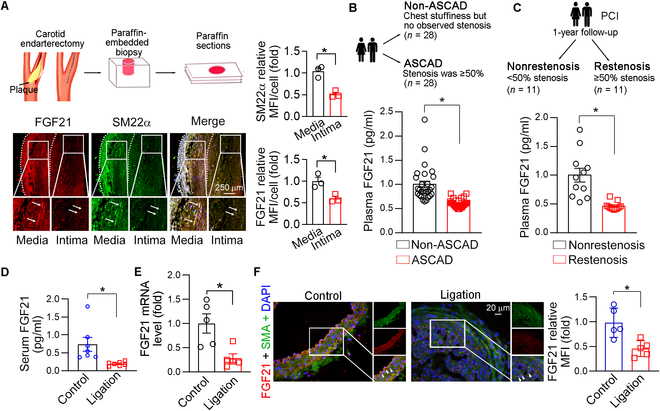
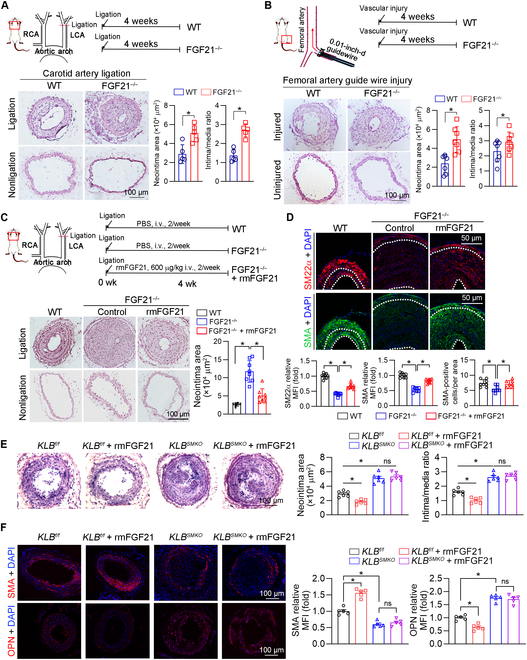
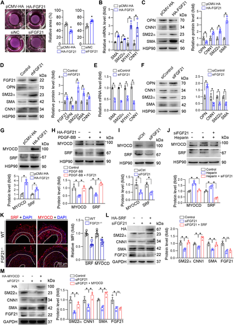
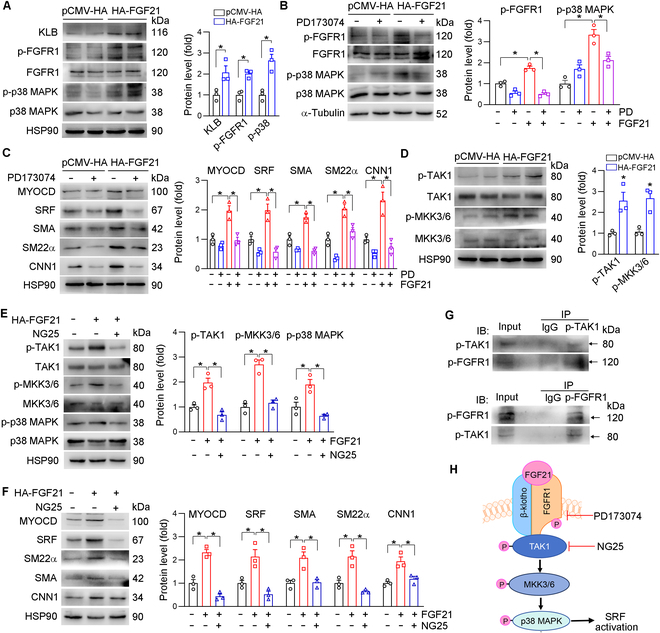
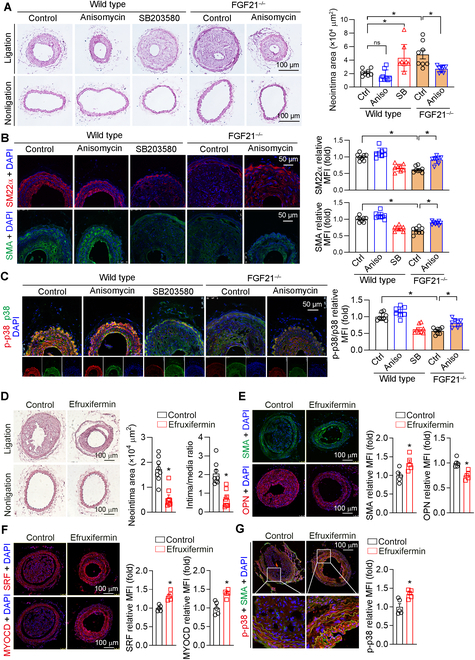
Phenotypic abnormalities in vascular smooth muscle cells (VSMCs) are believed to play essential roles in the progression of vascular diseases. Here, we explored the impact of fibroblast growth factor 21 (FGF21) on the phenotypic transition of VSMCs. Our findings revealed that FGF21 expression was substantially down-regulated in both human and mouse neointimal regions. Additionally, plasma FGF21 levels were lower in patients with atherosclerotic coronary artery disease (ASCAD) compared to those without ASCAD. Similarly, patients with restenosis exhibited reduced FGF21 levels compared to those without restenosis. In vivo, FGF21 deficiency accelerated intimal hyperplasia and decreased the number of contractile VSMCs in mouse neointima. However, hepatocyte-specific FGF21 knockout had no effect on ligation-induced intimal hyperplasia. Conversely, administration of recombinant FGF21 protein reduced neointima formation. This effect was abolished in mice with β-klotho VSMC-specific knockout, suggesting a direct effect of FGF21 on VSMCs. In vitro, FGF21 could promote the contractile phenotype transition of human aortic smooth muscle cells under basal or platelet-derived growth factor-BB incubation conditions. Furthermore, FGF21 activation led to the phosphorylation of p38 mitogen-activated protein kinase (p38 MAPK), which subsequently formed a complex with the serum response factor (SRF)-myocardin complex. This complex increased the phosphorylation of SRF at serine 224, thereby enhancing the transcription activation of the SRF-myocardin complex. Finally, we revealed that treatment with the FGF21 analog efruxifermin or activation of p38 MAPK using anisomycin effectively inhibited neointima formation. Taken together, these results indicate that modulating FGF21 or its subsequent signal pathways could serve as a therapeutic strategy for vascular diseases characterized by abnormal VSMC phenotypic transition.


Conflict of interest statement

Competing interests: The authors declare that they have no competing interests.

Figures

Fig. 1.
Fig. 1.
Fibroblast growth factor 21 (FGF21) expression was significantly down-regulated in neointimal hyperplasia. (A) Paraffin sections of the carotid artery were prepared and used to determine the expression of FGF21 and smooth muscle 22α (SM22α) by immunofluorescence staining (n = 3). Bars: 250 μm. (B) Plasma FGF21 levels were determined by enzyme-linked immunosorbent assay (ELISA) in atherosclerotic coronary artery disease (ASCAD) patients and non-ASCAD patients (n = 28). (C) FGF21 levels in the plasma of patients who underwent percutaneous coronary intervention (PCI) with and without restenosis were determined via ELISA (n = 11). (D to F) Carotid artery ligation was performed in wild-type (WT) mice. Serum FGF21 levels were determined by ELISA (n = 7) (D). FGF21 messenger RNA (mRNA) expression was determined by quantitative polymerase chain reaction (qPCR) (n = 5) (E). The control and ligated carotid artery cross-sections were subjected to immunofluorescence staining to determine FGF21 and smooth muscle α-actin (SMA) expression (n = 5) (F). Data information: Data are expressed as mean ± SD. Student t test, *P < 0.05. MFI, mean fluorescence intensity.
Fig. 2.
Fig. 2.
FGF21 inhibits neointimal formation by binding to its receptor in the aorta. (A and B) Carotid artery ligation (n = 5) and femoral artery guidewire injury (n = 7) were performed in WT and FGF21−/− mice. Both the left and right carotid arteries and the femoral artery were collected, and cross-sections were prepared for hematoxylin and eosin (HE) staining for morphological analysis with quantitative analysis of the neointima and media areas. Data information: Data are expressed as mean ± SD. Student t test, *P < 0.05. (C and D) Left carotid artery ligation was performed on female C57BL/6J and FGF21−/− mice (n = 8), and FGF21−/− mice were intravenously injected with recombinant mouse FGF21 protein (rmFGF21; 600 μg/kg) twice a week for 4 weeks. At the end of the experiment, carotid artery samples were individually collected and used for the following assay: (C) HE staining for morphological analysis with quantitative analysis of neointima and media areas. (D) The expression of SMA and SM22α in neointimal areas was determined by immunofluorescence staining. (E and F) Female KLBf/f and KLBSMKO mice (n = 6) were subjected to left carotid artery ligation, and rmFGF21 (600 μg/kg) was intravenously injected twice a week for 4 weeks. At the end of the experiment, carotid artery samples were individually collected and used for the following assay: (E) HE staining for morphological analysis with quantitative analysis of neointima and media areas. (F) The expression of SMA and osteopontin (OPN) in neointimal areas was determined by immunofluorescence staining (n = 5). Data information: The data are expressed as mean ± SD. Student t test (2 groups), 1-way analysis of variance (ANOVA) or 2-way ANOVA followed by Tukey’s test (more than 2 groups), *P < 0.05; ns, not significant. RCA, right carotid artery; LCA, left carotid artery; PBS, phosphate-buffered saline; i.v., intravenous.
Fig. 3.
Fig. 3.
FGF21 promotes vascular smooth muscle cell (VSMC) contractile phenotype switching through up-regulation of serum response factor (SRF) and myocardin (MYOCD). Human aortic smooth muscle cells (HASMCs) were transfected with pCMV-hemagglutinin (HA)/HA-FGF21 or siCtrl/siFGF21 for 24 h in serum-free medium and cultured with complete medium for 24 h. Then, the HASMCs were harvested and mixed with collagen at a ratio of 1:4 for another 48 h, after which the size of the collagen gel was evaluated (n = 3) (A). FGF21, SMA, SM22α, myosin heavy chain 11 (MYH11), and calponin 1 (CNN1) mRNA levels were determined by qPCR (n = 3) (B and E); FGF21, SMA, CNN1, SM22α, and OPN protein expression was determined by Western blotting (n = 3) (C and F). (D) HASMCs were treated with recombinant human FGF21 (rhFGF21) protein (0.5 mg/ml) in serum-free medium for 24 h. The protein expression of FGF21, SM22α, SMA, and CNN1 was determined by Western blotting (n = 3). (G to J) HASMCs in a 6-well plate were transfected with pCMV-HA/HA-FGF21 or siCtrl/siFGF21 for 24 h. Then, the cells were treated with or without platelet-derived growth factor-BB (PDGF-BB; 40 ng/ml) or heparin (100 μg/ml) for 24 h. The expression of SRF and MYOCD was determined by Western blotting (n = 3). (K) The expression of SRF and MYOCD in the left carotid artery was determined by immunofluorescence staining and quantification of the MFI (n = 5). (L and M) HASMCs in a 6-well plate were transfected with siCtrl/siFGF21 and HA-SRF or HA-MYOCD for 24 h. The expression of HA, SM22α, CNN1, and SMA was determined by Western blotting (n = 3). Data information: Data are expressed as mean ± SD. Student t test (2 groups) or one-way ANOVA followed by Tukey’s test (more than 2 groups), *P < 0.05.
Fig. 4.
Fig. 4.
FGF21 promotes phosphorylation of the SRF Ser224 site by p38 mitogen-activated protein kinase (p38 MAPK), which in turn promotes recruitment of downstream contractile genes. (A) HASMCs in a 6-well plate were transfected with pCMV-HA/HA-FGF21 for 24 h. The expression of p38 MAPK and p-p38 MAPK was determined by Western blotting (n = 3). (B) The expression of p38 MAPK and p-p38 MAPK in the aortas of the WT and FGF21−/− mice subjected to carotid artery ligation was determined by Western blotting (n = 3). (C and D) HASMCs were transfected with pCMV-HA or HA-FGF21 for 24 h in serum-free medium. Then, the cells were treated with SB203580 (10 μmol/l) for 12 h. The expression of SRF, MYOCD, p38 MAPK, p-p38 MAPK, SMA, CNN1, and SM22α was determined by Western blotting (n = 3). (E) HASMCs were transfected with pCMV-HA or HA-MYOCD for 24 h, after which the cells were transferred to complete medium for 24 h. The resulting cell lysates were subjected to immunoprecipitation (IP) with anti-p-p38 MAPK, anti-SRF, or anti-MYOCD antibodies. The pulled-down complexes and input cell lysates were analyzed by Western blotting with the anti-p-p38 MAPK, anti-SRF, or anti-MYOCD antibodies. (F) HASMCs were transfected with pCMV-HA, HA-SRF, or HA-SRF plus siFGF21 for 24 h in serum-free medium, after which the cells were switched to complete medium for 24 h. The IP experiment was performed with HA-tagged magnetic beads, followed by immunoblotting with anti-MYOCD, SRF, and p38 MAPK antibodies (n = 3). (G) Chromatin was isolated from HASMCs with or without rhFGF21 treatment (0.5 mg/ml). After determination of input, IP was conducted with normal immunoglobulin G (IgG) or SRF antibodies, followed by qPCR. *P < 0.05 versus the corresponding control (n = 3). (H) HASMCs were transfected with pCMV-HA or HA-SRF for 24 h in serum-free medium, after which the cells were switched to complete medium and cultured for 24 h. An IP experiment was performed with HA-tagged magnetic beads, followed by immunoblotting with anti-phospho-serine or anti-phospho-threonine antibody. (I) HASMCs were transfected with pCMV-HA or HA-FGF21 for 24 h in serum-free medium, after which the cells were switched to complete medium and cultured for 24 h. IP experiments were performed with protein A/G magnetic beads, IgG, and anti-SRF antibody, followed by immunoblotting with anti-phospho-serine and anti-SRF antibodies. (J) Molecular docking of SRF and p38 MAPK proteins. (K and L) HASMCs were transfected with HA-SRF for 24 h in serum-free medium, after which the cells were switched to complete medium supplemented with anisomycin for 24 h. The cell lysate was separated via sodium dodecyl sulfate–polyacrylamide gel electrophoresis (SDS-PAGE). The band corresponding to the molecular weight of SRF was cut and digested to carry out a liquid chromatography–tandem mass spectrometry (LC-MS/MS) assay. (M) HASMCs were transfected with HA-SRF, HA-SRF S224A, or HA-SRF S224D for 24 h in serum-free medium, after which the cells were switched to complete medium for 24 h. The protein expression of SRF, MYOCD, HA, SMA, SM22α, CNN1, and OPN was determined by Western blotting (n = 3). Data information: Data are expressed as mean ± SD. Student t test (2 groups) or one-way ANOVA followed by Tukey’s test (more than 2 groups), *P < 0.05; ns, not significant. IB, immunoblotting.
Fig. 5.
Fig. 5.
FGF21 activates p38 MAPK through the β-klotho (KLB)/FGFR1–transforming growth factor-β-activated kinase 1 (TAK1)–mitogen-activated protein kinase kinase 3/6 (MKK3/6) pathway. HASMCs were transfected with pCMV-HA or HA-FGF21 for 24 h, after which the cells were switched to complete medium for 24 h. The expression of p38 MAPK, p-p38 MAPK, FGFR1, p-FGFR1, and KLB in (A) or MKK3/6, p-MKK3/6, TAK1, and p-TAK1 in (D) was detected by Western blotting (n = 3). (B and C) HASMCs were transfected with pCMV-HA or HA-FGF21 for 24 h. Then, the cells were treated with PD173074 (10 nmol/l) for 24 h. The expression of p-FGFR1, FGFR1, p-p38 MAPK, and p38 MAPK (B) and MYOCD, SRF, SMA, SM22α, and CNN1 (C) was determined by Western blotting (n = 3). (E and F) After HA-FGF21 transfection for 24 h, HASMCs were treated with NG25 (10 μmol/l) for 24 h. The expression of p-TAK1, TAK1, p-MKK3/6, MKK3/6, p-p38 MAPK, and p38 MAPK (E) and SMA, SM22α, CNN1, MYOCD, and SRF (F) was determined by Western blotting (n = 3). (G) HASMCs were transfected with HA-FGF21 for 24 h, followed by culture in complete medium for 24 h. The cell lysates were subjected to immunoprecipitation with anti-p-FGFR1 or anti-p-TAK1 antibodies. The pulled-down complexes and input cell lysates were analyzed by Western blotting with the anti-p-FGFR1 or anti-p-TAK1 antibodies. (H) Diagram of FGF21-activated p38 MAPK through the KLB/FGFR1–TAK1–MKK3/6 pathway. Data information: Data are expressed as mean ± SD. Student t test (2 groups) or 1-way ANOVA or 2-way ANOVA followed by Tukey’s test (more than 2 groups), *P < 0.05; ns, not significant.
Fig. 6.
Fig. 6.
p38 MAPK activation and efruxifermin improves intimal hyperplasia. (A to C) Female C57BL/6J or FGF21−/− mice (n = 8) were subjected to left carotid artery ligation and then intraperitoneally injected with the vehicle (distilled water), SB203580 (10 mg/kg), or anisomycin (15 mg/kg) every day, respectively, for 4 weeks. At the end of the experiment, carotid artery samples were collected and used for the following assays. HE staining for morphological analysis with quantitative analysis of neointima and media areas (A). The expression of SMA, SM22α (B), p38 MAPK, and p-p38 MAPK (C) in neointima areas was determined by immunofluorescence staining. (D to G) Left carotid artery ligation was performed on female C57BL/6J mice (n = 8), which were then injected subcutaneously with physiological saline or efruxifermin (5 mg/kg) once a week for 4 weeks. At the end of the experiment, carotid artery samples were individually collected and used for the following assay: (D) HE staining for morphological analysis with quantitative analysis of neointima and media areas. (E to G) The expression of SMA and OPN (E), SRF and MYOCD (F), and p-p38 MAPK in SMA-positive cells (G) in neointimal areas was determined by immunofluorescence staining (n = 5). Data information: Data are expressed as mean ± SD. Student t test (2 groups) or 2-way ANOVA followed by Tukey’s test, *P < 0.05.

Similar articles

References

    1. Sahin B, Ilgun G. Risk factors of deaths related to cardiovascular diseases in World Health Organization (WHO) member countries. Health Soc Care Community. 2022;30(1):73–80. - PubMed
    1. Ke C, Lipscombe LL, Weisman A, Zhou L, Austin PC, Shah BR, Booth GL. Trends in the association between diabetes and cardiovascular events, 1994-2019. JAMA. 2022;328(18):1866–1869. - PMC - PubMed
    1. Wong M, Dai Y, Ge J. Pan-vascular disease: What we have done in the past and what we can do in the future? Cardiol Plus. 2024;9(1):1–5. - PMC - PubMed
    1. Chareonrungrueangchai K, Wongkawinwoot K, Anothaisintawee T, Reutrakul S. Dietary factors and risks of cardiovascular diseases: An umbrella review. Nutrients. 2020;12(4):1088. - PMC - PubMed
    1. Miano JM, Fisher EA, Majesky MW. Fate and state of vascular smooth muscle cells in atherosclerosis. Circulation. 2021;143(21):2110–2116. - PMC - PubMed

LinkOut - more resources