

Renal epidermoid cyst mimicking renal tuberculous abscess: a case report
- PMID: 40766064
- PMCID: PMC12321514
- DOI: 10.3389/fmed.2025.1632764
Renal epidermoid cyst mimicking renal tuberculous abscess: a case report
Abstract
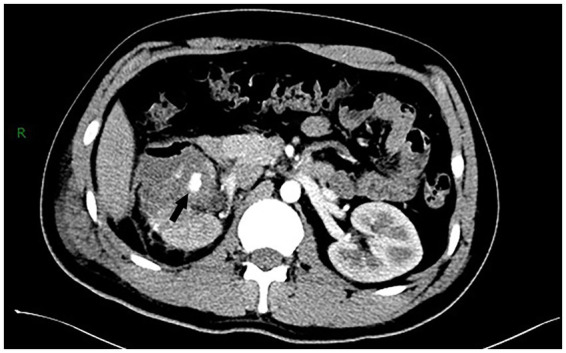
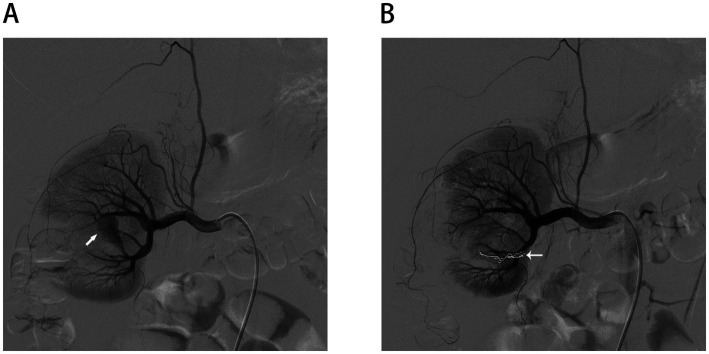
Renal epidermoid cysts (RECs) are exceedingly rare benign cystic lesions, with only 15 histologically confirmed cases reported worldwide to date. Due to their non-specific clinical and radiological features, they are often misdiagnosed preoperatively as infectious or neoplastic conditions. Here, we report a 25-year-old man in whom a complex renal cyst was incidentally identified during a routine health examination. Retrospectively, the patient reported mild urinary frequency and low-grade fever. Imaging suggested a non-enhancing heterogeneous cyst in the lower pole of the right kidney. Laparoscopic partial nephrectomy was performed, revealing abundant yellow-white caseating material intraoperatively, prompting empirical anti-tuberculosis therapy in the context of regional endemicity. However, histopathological analysis confirmed a diagnosis of RECs, and anti-tuberculous treatment was subsequently withdrawn. On postoperative day 5, the patient developed gross hematuria due to a renal artery pseudoaneurysm, which was successfully managed with selective arterial embolization. This case highlights the diagnostic challenges posed by atypical cystic renal lesions and underscores the importance of integrating imaging, intraoperative findings, and histopathology. Including RECs in the differential diagnosis may prevent unnecessary antituberculous therapy and overtreatment.
Keywords: case report; cystic renal lesion; empirical anti-tuberculosis therapy; partial nephrectomy; pseudoaneurysm; renal epidermoid cyst; renal tuberculous abscess.
Copyright © 2025 Gao, Luo, Zhu, Liu, Li, Chen, Wang, Zhou, Li, Liang and Chen.
Conflict of interest statement
The authors declare that the research was conducted in the absence of any commercial or financial relationships that could be construed as a potential conflict of interest.
Figures


References
-
- Moch H, Amin MB, Berney DM, Comperat EM, Gill AJ, Hartmann A, et al. The 2022 World Health Organization classification of tumours of the urinary system and male genital organs-part a: renal, penile, and testicular tumours. Eur Urol. (2022) 82:458–68. doi: 10.1016/j.eururo.2022.06.016, PMID: - DOI - PubMed
Publication types
LinkOut - more resources
Full Text Sources