

Noninvasive Ultrasound Imaging in Juvenile Idiopathic Arthritis: Diagnostic and Findings on the Temporomandibular Joint-A Prospective Study
- PMID: 40766257
- PMCID: PMC12324909
- DOI: 10.1155/ijod/9491663
Noninvasive Ultrasound Imaging in Juvenile Idiopathic Arthritis: Diagnostic and Findings on the Temporomandibular Joint-A Prospective Study
Abstract
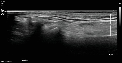
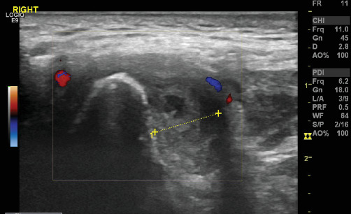
Introduction: Juvenile idiopathic arthritis (JIA) is a chronic autoimmune condition. The temporomandibular joint (TMJ) is one of the most affected joints in JIA. It can bring significant symptoms and impairments if not treated, and routinely instrumental exams are necessary to track its progress during the visits. The purpose of this study was to determine the efficiency in tracking the status of TMJ involvement with ultrasound (US) imaging in patients with a diagnosis of JIA and to assess its effectiveness in detecting different alterations. Materials and Methods: Inclusion criteria included patients previously diagnosed with JIA to be recruited in this prospective observational study. Each patient underwent detailed US evaluation of the TMJ to assess for various pathological changes, including condylar profile alterations, erosive phenomena, bone apposition, osteophyte formation, disc displacement, and soft tissue changes. The assessment was performed by two expert blinded operators. The US findings were compared with clinical manifestations and conventional imaging, for sensitivity, specificity, and predictive values. Results: A total of 46 patients divided into 39 female and 7 males, between 7 and 19 years were recruited. Of the recruited patients, 15% showed discordance and were asymptomatic, while 85% of the patients showed at least one joint manifestations. Sensitivity, specificity, and negative predictive value (NPV) of US for detecting TMJ pathology were calculated using conventional imaging as the reference standard. Conclusions: US showed a good concordance with traditional diagnosis, however it does not substitute traditional imaging for diagnosis. US demonstrated potential to be a reference noninvasive tool for monitoring TMJ secondary lesions in JIA and for monitoring during routine visits, offering advantages, such as noninvasiveness, cost-effectiveness, and real-time dynamic imaging capabilities.
Keywords: diagnosis; juvenile idiopathic arthritis; temporomandibular joint; ultrasound.
Copyright © 2025 Marco Farronato et al. International Journal of Dentistry published by John Wiley & Sons Ltd.
Conflict of interest statement
The authors declare no conflicts of interest.
Figures

Similar articles
-
123I-MIBG scintigraphy and 18F-FDG-PET imaging for diagnosing neuroblastoma.Cochrane Database Syst Rev. 2015 Sep 29;2015(9):CD009263. doi: 10.1002/14651858.CD009263.pub2. Cochrane Database Syst Rev. 2015. PMID: 26417712 Free PMC article.
-
Dorsal Subluxation of the First Metacarpal During Thumb Flexion is an Indicator of Carpometacarpal Osteoarthritis Progression.Clin Orthop Relat Res. 2023 Jun 1;481(6):1224-1237. doi: 10.1097/CORR.0000000000002575. Epub 2023 Mar 6. Clin Orthop Relat Res. 2023. PMID: 36877171 Free PMC article.
-
Comparison between ultrasound and magnetic resonance imaging of the temporomandibular joint in juvenile idiopathic arthritis: A systematic review.J Oral Rehabil. 2023 Oct;50(10):1082-1092. doi: 10.1111/joor.13529. Epub 2023 Jun 23. J Oral Rehabil. 2023. PMID: 37301975
-
The Black Book of Psychotropic Dosing and Monitoring.Psychopharmacol Bull. 2024 Jul 8;54(3):8-59. Psychopharmacol Bull. 2024. PMID: 38993656 Free PMC article. Review.
-
Variation within and between digital pathology and light microscopy for the diagnosis of histopathology slides: blinded crossover comparison study.Health Technol Assess. 2025 Jul;29(30):1-75. doi: 10.3310/SPLK4325. Health Technol Assess. 2025. PMID: 40654002 Free PMC article.
References
-
- Abramowicz S., Levy J. M., Prahalad S., Travers C. D., Angeles-Han S. T. Temporomandibular Joint Involvement in Children With Juvenile Idiopathic Arthritis: A Preliminary Report. Oral Surgery, Oral Medicine, Oral Pathology and Oral Radiology . 2019;127(1):19–23. doi: 10.1016/j.oooo.2018.07.008. - DOI - PubMed
-
- Farronato M., Cavagnetto D., Abate A., Cressoni P., Fama A., Maspero C. Assessment of Condylar Volume and Ramus Height in JIA Patients With Unilateral and Bilateral TMJ Involvement: Retrospective Case-Control Study. Clinical Oral Investigations . 2020;24(8):2635–2643. doi: 10.1007/s00784-019-03122-5. - DOI - PubMed
LinkOut - more resources
Full Text Sources
