. 2025 Jul 22:15:1582996.
doi: 10.3389/fonc.2025.1582996. eCollection 2025.

The protease-activated receptors are expressed in glioblastoma and differentially modulate adherent versus stem-like growth of LN-18 GBM cells

Affiliations

The protease-activated receptors are expressed in glioblastoma and differentially modulate adherent versus stem-like growth of LN-18 GBM cells

Sandra Bien-Möller et al. Front Oncol. .

Abstract

Background: Glioblastoma (GBM) remains the most aggressive and common malignant brain tumor in adults, often accompanied by venous thromboembolism due to hypercoagulability. Protease-activated receptors (PAR1-4) are thought to influence GBM progression, which in this study led to examine their expression in both tissue from GBM patients and in a GBM cell model.

Methods: Using quantitative PCR and immunoblot analyses, we investigated the expression of PAR1-4 in human GBM samples compared to non-malignant brain and evaluated its role in patient survival. In addition, the expression of PAR1-4 in adherent LN-18 GBM cells in comparison to their stem cell-like neurosphere counterparts was analyzed. Finally, the influence of PAR1-4 modulation by specific agonists and antagonists on cell viability was investigated using this GBM cell model.

Results: PAR1-4 mRNA levels were significantly higher in GBM than in non-tumoral brain tissue, though this did not affect patient survival. Notably, PAR4 protein levels were lower in GBM, while PAR1, 2, and 3 were unchanged. However, high PAR1 protein levels were linked to poorer patient survival, with a similar trend observed for PAR4, though not statistically significant. Patients with high levels of both PAR1 and PAR3 or PAR4 faced an even greater risk of poor outcomes, but the most severe prognosis was seen in those patients with high PAR3 and PAR4 protein level. In stem-like LN-18 GBM neurospheres, PAR1-4 mRNA was significantly increased, with PAR3 protein elevated and PAR4 reduced. Inhibition of PAR1, PAR2, or PAR4 reduced the viability of adherent GBM cells but not stem-like neurospheres.

Conclusion: These findings suggest that PARs impact GBM patient survival and that tumor stem cells may respond differently to PAR inhibition compared to conventional tumor cells.

Keywords: PARs; glioblastoma; neurospheres; protease activated receptors; stem-like cells.


Conflict of interest statement

The authors declare that the research was conducted in the absence of any commercial or financial relationships that could be construed as a potential conflict of interest.

Figures
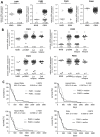
Figure 1
Figure 1
PAR1–4 mRNA expression in GBM specimen and its association with patients´ overall survival time. (A) Comparison of PAR1, PAR2, PAR3 and PAR4 mRNA expression in non-malignant brain (NMB, n=7) and all analyzed GBM samples (both primary and relapsed GBM, n=118). (B) Subdivision of GBM specimen in primary GBM (prGBM, n=78), first (1st, n=33) and second (2nd, n=7) relapses and comparison with NMB. (A+B) Gene expression was measured by qPCR. Each mRNA level of the target genes (PAR1-4) was normalized to the mean of GAPDH and β-actin using the 2-ΔΔct method. Data are shown as scatter plots representing the median as horizontal bars. Mann Whitney U test, *p < 0.05 and ***p < 0.001 for (A) and OneWay ANOVA/Kruskal Wallis test with Dunn’s Multiple Comparison Test, *p < 0.05, **p < 0.01 and *** p < 0.001 for (B). (C) Kaplan Meier survival analyses of PAR1–4 mRNA expression in GBM patients. Association of the relative mRNA expression of each single PAR receptor with the survival time of patients with primary GBM. The patients were divided into two subgroups depending on the median gene expression. No significant association was found.
Figure 2
Figure 2
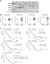
Protein expression of PAR1–4 in GBM patients´ samples. (A) Representative immunoblots of PAR1–4 in GBM tissue in comparison to non-malignant brain samples (NMB). Detection of GAPDH was used as loading control for normalization of the respective PAR protein level to a housekeeping protein. (B) Relative protein expression of PAR1–4 in GBM and NMB after densitometric evaluation and normalization to GAPDH. Data are shown as scatter plots representing the median as horizontal bars. Mann Whitney U test, ***p < 0.001. (C) Kaplan Meier survival analyses of PAR1–4 protein expression in GBM patients. Association of the relative protein expression of each single PAR receptor with the survival time of patients with primary GBM. The patients were divided into two subgroups depending on the median protein expression. The protein content of the respective PARs was determined by western blotting and normalized to the housekeeping protein GAPDH. Log-rank (Mantel-Cox) Test, *p < 0.05.
Figure 3
Figure 3
PAR1–4 mRNA and protein content in adherent and neurospheric LN-18 GBM cells. (A) Comparative PAR1–4 mRNA expression of both adherent (LN-18-adh, grey bars) and neurospheric (LN-18-NS, black bars) LN-18 cells of the passages one to four (Pass. 1-4) as well as of parental LN-18 cells (LN-18-par, white bars). Gene expression was measured by qPCR. Each mRNA level of the target genes (PAR1-4) was normalized to the mean of 18S rRNA, GAPDH and β-actin using the 2-ΔΔct method, n=4. (B) Comparative PAR1–4 protein content of both adherent (LN-18-adh, grey bars) and neurospheric (LN-18-NS, black bars) LN-18 cells of passage three. The protein content of the respective PARs was determined by western blotting and normalized to the housekeeping protein GAPDH. Representative immunoblots are shown above the diagram, n=3-4. OneWay ANOVA with Bonferroni’s Multiple Comparison Test, *p < 0.05, **p < 0.01 and ***p < 0.001 LN-18-adh vs. LN-18-NS; #p < 0.05, ##p < 0.01 and ###p < 0.001 LN-18-adh vs. LN-18-NS.
Figure 4
Figure 4
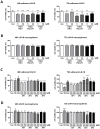
Impact of PAR subtype specific inhibitors and agonists on viability of LN-18 GBM cells. Adherent and neurospheric LN-18 cells of passage 1 or 3 were treated for 48 or 72h with the respective compounds followed by measurement of cell viability using the Resazurin assay. (A+B) Treatment of adherent (A) and neurospheric (B) LN-18 cells with inhibitors of PAR1 (RWJ), PAR2 (FSLLRY) and PAR4 (tcY) (each 5 and 20 µM), n=3-4. (C+D) Incubation of adherent (C) and neurospheric (D) LN-18 cells with Thrombin (TB, 30 U/ml), Factor Xa (FXa, 30 nM) or PAR subtype specific agonists (AP1 to AP4, each 100 and 200 µM), n=3-4. OneWay ANOVA Dunnett’s Multiple Comparison Test, *p < 0.05, **p < 0.01 and ***p < 0.001 vs. Con.

References

    1. Stupp R, Mason WP, van den Bent MJ, Weller M, Fisher B, Taphoorn MJB, et al. Radiotherapy plus concomitant and adjuvant temozolomide for glioblastoma. N Engl J Med. (2005) 352:987–96. doi: 10.1056/NEJMoa043330, PMID: - DOI - PubMed
    1. Ostrom QT, Price M, Neff C, Cioffi G, Waite KA, Kruchko C, et al. CBTRUS statistical report: primary brain and other central nervous system tumors diagnosed in the United States in 2015-2019. Neuro-oncology. (2022) 24:v1–v95. doi: 10.1093/neuonc/noac202, PMID: - DOI - PMC - PubMed
    1. Pan E, Tsai JS, Mitchell SB. Retrospective study of venous thromboembolic and intracerebral hemorrhagic events in glioblastoma patients. Anticancer Res. (2009) 29:4309–13., PMID: - PubMed
    1. Krenzlin H, Lorenz V, Danckwardt S, Kempski O, Alessandri B. The importance of thrombin in cerebral injury and disease. Int J Mol Sci. (2016) 17. doi: 10.3390/ijms17010084, PMID: - DOI - PMC - PubMed
    1. Nierodzik ML, Karpatkin S. Thrombin induces tumor growth, metastasis, and angiogenesis: Evidence for a thrombin-regulated dormant tumor phenotype. Cancer Cell. (2006) 10:355–62. doi: 10.1016/j.ccr.2006.10.002, PMID: - DOI - PubMed

LinkOut - more resources