[Preprint]. 2025 Jul 30:2025.07.30.667713.
doi: 10.1101/2025.07.30.667713.

The atypical adhesion GPCR ADGRA1 controls hippocampal inhibitory circuit function

Affiliations

The atypical adhesion GPCR ADGRA1 controls hippocampal inhibitory circuit function

Baris Tosun et al. bioRxiv. .

Abstract

Neural circuits contain a diverse array of inhibitory interneurons that control information processing. The cell surface receptors and signaling pathways that modulate cell type specific inhibitory synaptic function are unclear. Here, we identify the atypical adhesion GPCR ADGRA1 as essential for hippocampal PV and SST inhibitory synaptic function. ADGRA1 is selectively enriched in hippocampal PV and SST interneurons and localizes to a subset of synapses. ADGRA1 deletion in PV and SST interneurons impairs inhibitory synaptic inputs onto Dentate Gyrus granule cells and generates deficits in learning and memory. ADGRA1 engages several downstream G proteins, notably Gα13, a pathway important for the establishment of hippocampal PV interneuron synaptic networks. These results identify an orphan receptor pathway selective for specific inhibitory synapse subtypes and expand our understanding of the signaling mechanisms that establish hippocampal inhibitory circuits.


Conflict of interest statement

Conflict of Interest The authors declare no conflict of interest.

Figures

Figure 1:
Figure 1:. The atypical orphan aGPCR Adgra1 is enriched in hippocampal interneurons
A, representative sections from mice harboring the Cre-inducible RiboTag (HA-Rpl22) allele, vGAT-Cre, and the Ai14 reporter immunolabeled for HA together with DAPI. B, RiboTag experiments from ribosomal bound mRNA from postnatal day (P30) hippocampus for indicated aGPCRs. The relative enrichment of each target in the HA RiboTag immunoprecipitation was compared to the input sample from the same tissue. C, qPCR controls verifying enrichment of interneuronal transcripts from vGAT-Cre RiboTag experiments. Probes for interneurons (Pvalb, SST, Slc32a1/vGAT) were compared to probes for glial (Gfap) or excitatory neuron (Slc17a7/vGlut1) transcripts. D & E, Adgra1 is expressed in sparse subpopulations of cells in the postnatal hippocampus. D, representative RNA in situ of the P30 hippocampus labeled for Adgra1 together with DAPI. E, representative high-magnification images of RNA in situs for Adgra1 in the dentate gyrus (DG), CA3, and CA1. F & G, Adgra1 is enriched in hippocampal PV and SST-positive interneurons. F, RNA in situ hybridizations for Adgra1 together with PV. G, similar to F, except for in situ hybridizations for Adgra1 and SST. Numerical data are means ± SEM from 6 independent biological replicates (mice). See Figure S1 and S2 for additional analysis of Adgra1 expression in the brain.
Figure 2:
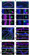
Figure 2:. Adgra1 is selectively expressed in PV- and SST-positive hippocampal interneurons and is a synaptic GPCR
A & B, Adgra1 is absent from hippocampal CCK- and Calb2-positive interneurons. A, representative RNA in situ for Adgra1 together with CCK in the P30 hippocampus. B, similar to A, except for RNA in situs probing for Adgra1 and Calb2. C-F, immunocytochemistry for HA-tagged ADGRA1 in primary hippocampal neurons. C, representative primary hippocampal neurons transduced with lentivirus expressing HA-ADGRA1 and immunolabeled for surface HA followed by presynaptic Bassoon and the somatodendritic marker MAP2. D, example dendrite immunolabeled for surface HA-ADGRA1 followed by Bassoon and MAP2. E, immunocytochemistry for surface HA-ADGRA1 and intracellular SHANK2 and MAP2. F, immunocytochemistry for surface HA-ADGRA1 and intracellular Gephyrin and MAP2. See Figure S1 and S2 for additional analysis of Adgra1 expression in the brain and Figure S3 for quantification of ADGRA1/synaptic marker colocalization.
Figure 3:
Figure 3:. Neuronal morphology, synapse density, and synaptic transmission are preserved in dentate gyrus (DG) granule cells (GCs) lacking ADGRA1
A, experimental diagram focused on the hippocampal DG circuit. Adgra1 cKO mice were crossed to the EMX1-Cre driver line to delete ADGRA1 from excitatory neurons. B-D, morphological analyses of DG GCs. GCs were filled with biocytin during electrophysiological recordings and dendritic spine density and dendritic arborization subsequently analyzed. B, representative spine images from Ctl or EMX-cKO GCs. C, quantification of dendritic spine densities in GCs. D, quantification of average total dendrite length from DG GCs. E-G, analysis of mEPSCs from Ctl or EMX-cKO GCs. E, representative mEPSC traces. F, cumulative probability plot of inter-event intervals and summary graph [inset] of the mean mEPSC frequency. G, cumulative probability plot and summary graph [inset] of mEPSC amplitude measurements. H-J, analysis of mIPSCs from Ctl or EMX-cKO GCs. H, representative mIPSC traces. I, cumulative probability plot of inter-event intervals and summary graph [inset] of the mean mIPSC frequency. J, cumulative probability plot and summary graph [inset] of mIPSC amplitude measurements. K-M, immunohistochemical analysis of synapses in the DG. K, immunolabeling for presynaptic Bassoon together with postsynaptic excitatory Homer1 in the hippocampal DG molecular layer (ML), granule cell layer (GCL), or CA3 stratum lucidum. L, similar to K, except for immunolabeling for presynaptic Syn1/2 and postsynaptic excitatory SHANK2. M, similar to K, except for presynaptic inhibitory vGAT or synaptoporin (SPO), a marker of DG large mossy fiber terminals (LMTs). Numerical data are means ± SEM or cumulative histograms. See Figure S4 for characterization of the Adgra1 cKO mouse allele, Figure S5 for additional electrophysiological and morphological parameters, and Figure S6 for quantification of immunohistochemistry. Control (Ctl) mice were Adgra1 cKO and EMX-cKO were EMX1-Cre/Adgra1 cKO littermates. Statistical significance was determined via two-tailed t-test or Kolmogorov-Smirnov test (cumulative histograms).
Figure 4:
Figure 4:. ADGRA1 is essential for inhibitory synaptic function in hippocampal PV interneurons
A, diagram of experimental approach to determine ADGRA1 function in PV inhibitory DG circuits. B-D, analysis of spontaneous mIPSCs from Ctl or PV-cKO GCs. B, representative mIPSC traces from GCs. C, cumulative probability plot of inter-event intervals and summary graph [inset] of the mean mIPSC frequency. D, cumulative probability plot and summary graph [inset] of mIPSC amplitude measurements. E-H, evaluation of eIPSCs in Ctl or PV-cKO GCs. E, representative eIPSC traces. F, eIPSC amplitude in response to increasing extracellular stimulation. G, representative eIPSC paired-pulse ratio (PPR) traces. H, quantification of PPR from GCs in Ctl or PV-cKO mice. Numerical data are means ± SEM or cumulative histograms. See Figure S7 for characterization of PV-Cre and SST-Cre lines and Figure S8 for additional electrophysiological parameters. Control (Ctl) mice were Adgra1 cKO and PV-cKO were PV-Cre/Adgra1 cKO littermates. Statistical significance was determined via two-tailed t-test or Kolmogorov-Smirnov test (*, p<0.05; ***, p<0.001).
Figure 5:
Figure 5:. ADGRA1 deletion in PV interneurons impairs learning and memory
A, representative images of Syt2-labeled presynaptic PV terminals in the indicated hippocampal sub-regions. B, quantification of Syt2 puncta density in the indicated hippocampal sub-regions. C, analysis of Syt2-positive puncta density on PV-Ai14 labeled soma. D & E, assessment of open field behavior in Ctl or PV-cKO mice. D, average distance travelled over a 60-minute open field trial. E, distance travelled over time during the 60-minute open field trial. F & G, cued learning from Pavlovian fear conditioning studies. F, quantification of percent time spent freezing before or after presentation of the cue stimulus following fear conditioning. G, percent time spent freezing during the habituation period or presentation of the cue. Numerical data are means ± SEM. See Figure S9 for additional behavioral characterization of PV-cKO mice. Control (Ctl) mice were Adgra1 cKO and PV-cKO were PV-Cre/Adgra1 cKO littermates. Statistical significance was determined via two-tailed t-test, one-way ANOVA with post hoc Tukey test or two-way ANOVA (**, p<0.01).
Figure 6:
Figure 6:. Adgra1 deletion in SST interneurons reduces inhibitory input onto DG GCs
A, diagram of experimental approach to determine ADGRA1 function in SST inhibitory DG circuits. B-D, analysis of spontaneous mIPSCs from Ctl or SST-cKO GCs. B, representative mIPSC traces from GCs. C, cumulative probability plot of inter-event intervals and summary graph [inset] of the mean mIPSC frequency. D, cumulative probability plot and summary graph [inset] of mIPSC amplitude measurements. E-H, eIPSC measurements in Ctl or SST-cKO GCs. E, representative eIPSC traces. F, eIPSC amplitude in response to increasing extracellular stimulation. G, representative eIPSC paired-pulse ratio (PPR) traces. H, quantification of PPR from GCs in Ctl or SST-cKO mice. I & J, assessment of open field behavior in Ctl or SST-cKO mice. I, average distance travelled over a 60-minute open field trial. J, distance travelled over time during the 60-minute open field trial. K & L, cued learning from Pavlovian fear conditioning. K, quantification of percent time spent freezing before or after presentation of the cue stimulus following fear conditioning. L, percent time spent freezing during the habituation period or presentation of the cue. Numerical data are means ± SEM or cumulative histograms. See Figure S10 for additional electrophysiological parameters and Figure S11 for additional behavior studies. Control (Ctl) mice were Adgra1 cKO and SST-cKO were SST-Cre/Adgra1 cKO littermates. Statistical significance was determined via two-tailed t-test or Kolmogorov-Smirnov test (*, p<0.05; ***, p<0.001).
Figure 7:
Figure 7:. ADGRA1 engages several G proteins including Gα13
A, diagram of ADGRA1 protein domains compared to standard aGPCRs. B, TRUPATH BRET2 analysis of full-length ADGRA1. A LPHN3 construct with either the Gαi1 or Gα13 TRUPATH sensors were used as negative or positive controls, respectively. C, similar to B, except using an ADGRA1 mutant lacking the short N-terminal extracellular sequence prior to the 7-TM GPCR. D-G, ADGRA1 plasmid dose-response curves using indicated TRUPATH BRET2 biosensors. D, ADGRA1 plasmid dose-response experiments for GαoB. E, similar to D, except for Gα11. F, similar to D, except for Gα15. G, similar to D, except for Gα13. H, representative immunocytochemistry for surface HA-ADGRA1 and intracellular Gα13 and MAP2 in indicated conditions. I, representative MAP2-labeled dendrite co-labeled for surface HA and intracellular Gα13. Numerical data are means ± SEM. See Figure S12 for additional characterization of ADGRA1 constructs and quantification of ADGRA1/Gα13 co-localization. Statistical significance was determined via one-way ANOVA with post hoc Tukey test (**, p<0.01; ***, p<0.001).

References

    1. Fishell G. & Kepecs A. Interneuron Types as Attractors and Controllers. Annu Rev Neurosci 43, 1–30 (2020). - PMC - PubMed
    1. Pelkey K. A. et al. Hippocampal GABAergic Inhibitory Interneurons. Physiol Rev 97, 1619–1747 (2017). - PMC - PubMed
    1. Pieraut S. et al. Experience-dependent remodeling of basket cell networks in the dentate gyrus. Neuron 84, 107–122 (2014). - PMC - PubMed
    1. Tzilivaki A. et al. Hippocampal GABAergic interneurons and memory. Neuron 111, 3154–3175 (2023). - PMC - PubMed
    1. Anderson G. R. et al. Postsynaptic adhesion GPCR latrophilin-2 mediates target recognition in entorhinal-hippocampal synapse assembly. J Cell Biol 216, 3831–3846 (2017). - PMC - PubMed

Publication types

LinkOut - more resources