[Preprint]. 2025 Aug 3:2025.08.03.668300.
doi: 10.1101/2025.08.03.668300.

Covalent Degraders of Immune Regulatory Transcription Factors IRF8 and IRF5

Affiliations

Covalent Degraders of Immune Regulatory Transcription Factors IRF8 and IRF5

Thang Cong Do et al. bioRxiv. .

Abstract

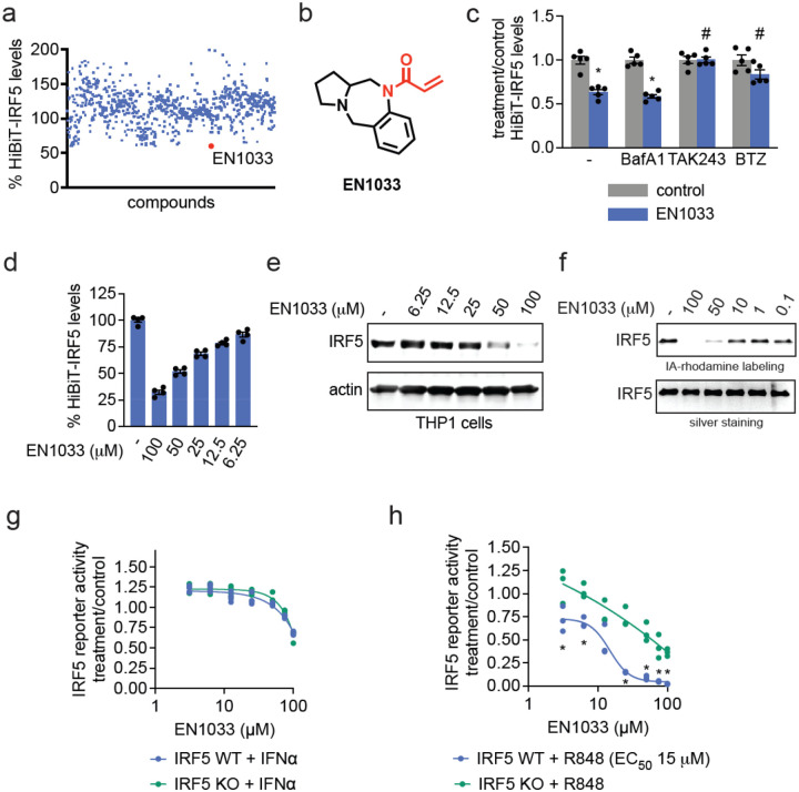
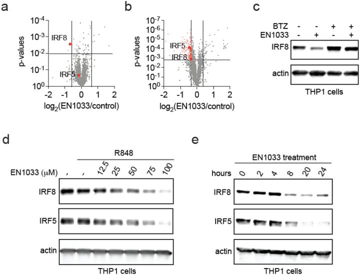
Transcription factors are among the most challenging targets for drug discovery due to their lack of classical binding pockets and high degree of intrinsic disorder, despite the therapeutic importance of many of these proteins. IRF5 and IRF8 are key transcriptional regulators of innate immune signaling that orchestrate pro-inflammatory gene expression programs in response to stimuli such as toll-like receptor activation, making them central players in autoimmune and inflammatory diseases. Despite their therapeutic interest, direct targeting of IRF5 and IRF8 has remained challenging. Here, we screened a library of cysteine-reactive covalent ligands to identify hits that could degrade IRF5. We identified acrylamide EN1033 as the top hit, which not only led to IRF5 loss in a proteasome-dependent manner but also bound directly and covalently to the IRF5 protein, inhibiting IRF5-specific transcriptional activity in a macrophage cell line. Upon further analysis, however, we found that EN1033 not only engaged and degraded IRF5 but also more robustly and rapidly engaged and degraded a related inflammatory transcription factor, IRF8. We further demonstrated that EN1033 destabilized and degraded IRF5 and IRF8 by covalently targeting C28 and C223, respectively, as evidenced by the attenuation of their degradation through mutagenesis of these cysteines. We also found that IRF8 loss led to the downregulation and inhibition of IRF5 activity, suggesting a crosstalk between these two transcription factors, which are both targeted by EN1033. Overall, we identify an early-stage pathfinder molecule that covalently targets IRF8 and IRF5, thereby degrading these transcription factors and inhibiting their pro-inflammatory transcriptional activity.

Keywords: IRF5; IRF8; activity-based protein profiling; chemoproteomics; covalent; cysteine; monovalent degraders; targeted protein degradation.


Conflict of interest statement

Competing Financial Interests Statement HC, LYC, CL, MLA, JG, SS, NSA, SK are Octant Bio employees. DKN is a co-founder, shareholder, and member of the scientific advisory board for Frontier Medicines and Zenith. DKN is a member of the board of directors for Vicinitas Therapeutics. DKN is also a member of the scientific advisory boards of The Mark Foundation for Cancer Research, American Association for Cancer Research, Photys Therapeutics, Oerth Bio, Apertor Pharmaceuticals, Axiom Therapeutics, and Ten30 Biosciences. DKN is also an Investment Advisory Partner for a16z Bio, an Advisory Board member for Droia Ventures, and an iPartner for The Column Group.

Figures

Figure 1.
Figure 1.. Screening for a Covalent IRF5 Degrader.
(a) Covalent ligand screen in THP1 cells overexpressing a HiBiT-tagged IRF5. HiBiT-IRF5-expressing THP1 cells were treated with DMSO vehicle or covalent ligand (50 μM) for 24 h, after which HiBiT-IRF5 was detected by luminescence through detection with LgBiT. Individual compounds, structures, and data points are shown in Table S1. (b) Structure of top hit EN1033. Cysteine-reactive acrylamide warhead highlighted in red. (c) Ubiquitin and proteasome-dependence of HiBiT-IRF5 loss. HiBiT-IRF5-expressing THP1 cells were pre-treated with DMSO vehicle or lysosomal v-ATPase inhibitor Bafilomycin A (BafA), E1 ubiquitin conjugating enzyme inhibitor TAK243, and proteasome inhibitor BTZ (100 nM) for 1 h prior to treatment with DMSO vehicle or EN1033 (50 μM) for 24 h, after which HiBiT-IRF5 was detected by luminescence through detection with LgBiT. (d) Dose-response of HiBiT-IRF5 loss. HiBiT-IRF5-expressing THP1 cells were treated with DMSO vehicle or EN1033 for 24 h, after which HiBiT-IRF5 was detected by luminescence through detection with LgBiT. (e) Dose-response of IRF5 loss. THP1 cells were treated with DMSO vehicle or EN1033 for 24 h after which IRF5 and loading control actin levels were assessed by SDS/PAGE and Western blotting. (f) Gel-based ABPP of EN1033 against pure IRF5 protein. IRF5 protein was pre-incubated with DMSO vehicle or EN1033 for 1 h prior to treatment with IA-rhodamine (100 nM) for 30 min. IA-rhodamine labeling was assessed by SDS/PAGE and in-gel fluorescence and loading was assessed by silver staining. (g, h) IRF5 luciferase reporter activity. IRF5 wildtype (WT) or knockout (KO) THP1 cells expressing an IRF5 luciferase reporter were stimulated with IFNα (1000 U/mL) (g) or R848 (10 μM) (h) and treated with DMSO or vehicle EN1033 for 24 h, after which luciferase activity was assessed. Data in (c,d,g,h) are from n=3–4 biologically independent replicates per group. Graphs in (c,d) show individual replicate values and average ± sem. Blots and gels in (e,f) are representative of n=3 biologically independent replicates per group. Significance expressed as *p<0.05 compared to vehicle-treated controls in (c) and compared to EN1033 treatment in THP1 KO cells in (h) and #p<0.05 compared to cells treated with EN1441 alone.
Figure 2.
Figure 2.. Validation of EN1033 as an IRF5 Degrader and Discovery of Effects on IRF8.
(a, b) Quantitative tandem mass tagging (TMT)-based proteomic profiling of EN1033 treatment. THP1 cells were treated with DMSO vehicle or EN1033 (100 μM) for 15 h (a) or 24 h (b). IRF5 and IRF8 are highlighted. (c) Proteasome-dependence of IRF8 degradation. THP1 cells were pre-treated with DMSO vehicle or BTZ (500 nM) 1 h prior to treatment with DMSO vehicle or EN1033 (100 μM) for 18 h, after which IRF8 and loading control actin levels were assessed by SDS/PAGE and Western blotting. (d) Dose-response of IRF5 and IRF8 degradation. THP1 cells were stimulated with DMSO or R848 (10 μM) for 1 h prior to treatment of cells with DMSO vehicle or EN1033 for 24 h, after which IRF8, IRF5, and loading control actin levels were assessed by SDS/PAGE and Western blotting. (e) Time-course of IRF5 and IRF8 degradation. THP1 cells were treated with EN1033 (100 μM) and IRF8, IRF5, and loading control actin levels were assessed by SDS/PAGE and Western blotting. Data in (a,b) are from n=3 biologically independent replicates per group and proteomic data can be found in Table S2. Blots in (c,d,e) are representative of n=3 biologically independent replicates per group.
Figure 3.
Figure 3.. Further Characterization of EN1033 with an Alkyne Functionalized Probe and Through Chemoproteomic Profiling.
(a) Structure of alkyne functionalized probe TH3–189 with the acrylamide warhead highlighted in red. (b) Dose-response of IRF5 and IRF8 loss. THP1 cells were treated with DMSO vehicle or EN1033 for 24 h after which IRF8, IRF5, and loading control actin levels were assessed by SDS/PAGE and Western blotting. (c,d) IRF8 and IRF5 pure protein labeling by TH3–189. Pure IRF8 (c) or IRF5 (d) protein was labeled with DMSO vehicle or TH3–189 (100 μM) for 1 h, after which an azide-functionalized rhodamine handle was appended onto probe-labeled proteins by CuAAC. Proteins were resolved by SDS/PAGE, and TH3–189 labeling was assessed by in-gel fluorescence, and loading was assessed by silver staining. (e,f) IRF8 and IRF5 pulldown with TH3–189 probe in cells. THP1 cells were treated with DMSO vehicle or TH3–189 (100 μM) for 4 h, after which probe-modified proteins from resulting lysates were appended to an azide-functionalized biotin enrichment handle by CuAAC, avidin-enriched, eluted, and IRF8, IRF5, and loading control actin levels from input and pulldown eluate were assessed by SDS/PAGE and Western blotting. (g) Chemoproteomic profiling of TH3–189 targets. THP1 cells were treated with DMSO vehicle or TH3–189 (100 μM) for 4 h, after which probe-modified proteins from resulting lysates were appended to an azide-functionalized biotin enrichment handle by CuAAC, avidin-enriched, eluted, tryptically digested, and analyzed by LC-MS/MS. Highlighted in red are proteins that were significantly enriched compared to the DMSO vehicle, and IRF8 is highlighted. (h) isoDTB-ABPP analysis of EN1033. THP1 cells were treated with DMSO vehicle or EN1033 (100 μM) for 6 h, after which the resulting lysates were labeled with an alkyne-functionalized iodoacetamide probe (IA-alkyne) (200 μM) for 1 h. Probe-labeled proteins were then appended to an isotopically light or heavy azide-functionalized desthiobiotin handle, enriched by streptavidin beads, eluted, tryptically digested, and analyzed by LC-MS/MS. Shown in red are cysteines in proteins that were significantly engaged with C223 or IRF8, highlighted in red. Blots and gels shown in (b-f) are representative of n=3 biologically independent replicates per group. Data in (g,h) are from n=3 biologically independent replicates per group and chemoproteomic data can be found in Table S3 and Table S4, respectively.
Figure 4.
Figure 4.. Assessing Mechanism of IRF5 and IRF8 Degradation by EN1033.
(a) CETSA analysis of EN1033. THP1 cells were treated with DMSO vehicle or EN1033 (100 μM) for 4 h, after which cell lysates were heated to the designated temperatures, insoluble proteins were precipitated, and IRF8, IRF5, and control protein actin levels were assessed by Western blotting and quantified by densitometry. (b,c,d,e) IRF5 or IRF8 degradation by EN1033 in FLAG-IRF5 or FLAG-IRF8 WT or mutant expressing cells. HEK293T cells expressing FLAG-IRF5 WT, C28S, C121S, or C28S/C121S mutant were treated with DMSO vehicle or EN1033 (50 μM) for 24 h (b,c), or FLAG-IRF8 WT, C223S, C385S, or C223S/C385S mutant (d,e) were treated with DMSO vehicle or EN1033 (50 μM) for 18 h, after which FLAG-IRF5, FLAG-IRF8, or loading control actin levels were assessed by SDS/PAGE and Western blotting and quantified in (c,e). Data in (a-e) are from n=3 biologically independent replicates per group. Graphs in (a) show average ± sem. Bar graphs in (c,e) show individual replicate values and average ± sem. Significance in (a,c,e) is shown as *p<0.05 compared to vehicle-treated controls for each group and #p<0.05 compared to EN1033-treated FLAG-IRF5 WT or FLAG-IRF8 WT-expressing cells.
Figure 5.
Figure 5.. Transcriptomic profiling of EN1033.
THP1 cells were stimulated with R848 (10 μM) and treated with DMSO vehicle or EN1033 (50 μM) for 24 h, after which RNA from cells was subjected to RNA sequencing and quantification. Shown in (a) are significantly altered transcripts with EN1033 treatment, with representative IRF5 and IRF8 target genes designated with gene names. Pathway analysis from significantly downregulated genes is shown in (b). Full data are presented in Table S5. Data are from n=3 biologically independent replicates per group. P-values less than 1e-500 were capped at 1e-500.

References

    1. Bushweller J. H. Targeting transcription factors in cancer - from undruggable to reality. Nat. Rev. Cancer 19, 611–624 (2019). - PMC - PubMed
    1. Henley M. J. & Koehler A. N. Advances in targeting ‘undruggable’ transcription factors with small molecules. Nat. Rev. Drug Discov. 20, 669–688 (2021). - PubMed
    1. Negishi H., Taniguchi T. & Yanai H. The Interferon (IFN) Class of Cytokines and the IFN Regulatory Factor (IRF) Transcription Factor Family. Cold Spring Harb. Perspect. Biol. 10, a028423 (2018). - PMC - PubMed
    1. Taniguchi T., Ogasawara K., Takaoka A. & Tanaka N. IRF family of transcription factors as regulators of host defense. Annu. Rev. Immunol. 19, 623–655 (2001). - PubMed
    1. Yu X. et al. Interferon regulatory factor 5: a potential target for therapeutic intervention in inflammatory diseases. Front. Immunol. 16, 1535823 (2025). - PMC - PubMed

Publication types

LinkOut - more resources