[Preprint]. 2025 Sep 24:2025.08.01.668160.
doi: 10.1101/2025.08.01.668160.

Hemoglobin alpha regulates T-lymphocyte activation and mitochondrial function

Affiliations

Hemoglobin alpha regulates T-lymphocyte activation and mitochondrial function

Emily C Reed et al. bioRxiv. .

Abstract

We have recently discovered hemoglobin alpha a1 (Hbα-a1 mRNA and Hbα protein) in T-lymphocytes and previously reported that its expression was sensitive to mitochondrial redox perturbations. However, outside of its occurrence and basic characterization, the functional role of Hbα in T-lymphocytes remained unknown. Herein, we identify Hbα in both CD4+ and CD8+ T-lymphocyte subsets, and found its expression is highly dynamic, differs between the two subtypes, and is dependent upon activation stage. Further, the loss of Hbα by use of a novel T-lymphocyte-specific Hbα knock-out mouse impairs mitochondrial function, dysregulates cytokine production, and lowers the activation threshold primarily in CD4+ T-lymphocytes, indicating a critical role for Hbα within this subset. While these data suggested the loss of Hbα in T-lymphocytes may promote aberrant activation of autoreactive T-lymphocytes, surprisingly, we discovered that mice lacking Hbα in T-lymphocytes exhibited reduced severity of experimental autoimmune encephalomyelitis (EAE) compared to wild-type control animals. Interestingly, T-lymphocytes lacking Hbα in vivo appeared to function identically to wild-type controls, which did not explain the protection against EAE. In contrast, T-lymphocyte Hbα knock-out mice displayed significantly reduced levels of circulating immunoglobulins and CD40L expression compared to their wild-type counterparts during EAE, suggesting possible impaired intercellular communication. These data elucidate a previously unrecognized role for Hbα in T-lymphocyte function, which may have implications for hemoglobin-related diseases (i.e., hemoglobinopathies).

Keywords: EAE; Hemoglobinopathy; Immune; Inflammation; redox.


Conflict of interest statement

Conflict of Interest Statement: The authors have declared that no conflict of interest exists.

Figures
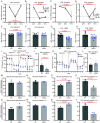
Figure 1:
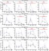
Figure 1:. Activation state of T-lymphocytes determines intracellular Hbα levels and impact on mitochondria.
A-D: Hbα-a1 mRNA (N=7) and Hbα protein expression (N=7) over 24, 48, 72-hour time points after activation of CD4+ (A-B) and CD8+ (C-D) T-lymphocytes compared to naive T-lymphocytes (N=6). E-H: Splenic CD4+ and CD8+ T-lymphocytes were isolated and activated for 72 hours, then assessed by flow cytometric analysis of MitoSOX (E-F) and TMRE MFI (G-H). I-L: Splenic CD4+ and CD8+ T-lymphocytes were isolated and activated for 72 hours, then assessed using Seahorse mitochondrial stress test (N=5). R+A = rotenone + antimycin A. M-T: Splenic T-lymphocytes were activated for 72 hours, then puromycin MFI was measured via SCENITH protocol in CD4+ and CD8+ T-lymphocytes to obtain calculations graphed (reported in percentage). Statistics measured by a one-way ANOVA with Dunnett’s multiple comparisons test, unpaired Student’s t-test, or two-way ANOVA with Šídák’s multiple comparisons test where appropriate.
Figure 2:
Figure 2:. Activated HbKO T-lymphocytes exhibit an accelerated and enhanced proinflammatory phenotype.
A-H: Extracellular cytokine protein measured from CD4+ (A-D) and CD8+ (E-H) T-lymphocytes at 24, 48, 72 hours post-activation (pg/mL per 106 cells) (N=5). I-P: Extracellular cytokine protein measured from CD4+ (I-L) and CD8+ (M-P) T-lymphocytes treated for 24 hours with anti-CD28 and varying concentrations of anti-CD3 (N=3). Statistics measured using unpaired Student’s t-test, mixed-effects analysis, or two-way ANOVA with Šídák’s multiple comparisons test where appropriate.
Figure 3:
Figure 3:. HbKO EAE animals exhibit better disease phenotypes but similar inflammatory profiles as WT EAE animals.
A: Schematic of 28-day EAE experimental design. B-C: EAE severity scores (0–5) and weights (g) over 28-day incubation period (WT N=14, HbKO N=10). D-G: Protein concentration (pg/mL) of cytokines in plasma. H-K: Percentage of CD4+ and CD8+ present in the spleen and inguinal lymph nodes at day 28. L-O: Percentage of splenic CD4+ polarized T-lymphocytes. P-S: Splenocytes restimulated with 10 μg/mL MOG35–55 for 72 hours, then assessed for extracellular cytokine protein concentration (pg/mL per 106 cells). Statistics measured by two-way ANOVA with Šídák’s multiple comparisons test or Student’s t-test where appropriate.
Figure 4:
Figure 4:. Loss of Hbα impairs T-lymphocyte intercellular communication.
A-H: Splenic T-lymphocytes were isolated and activated with a 1:1 ratio of Dynabeads for 72 hours, replated into transwell inserts over control, RANTES, or CXCL12 media for 4 hours, then total cell counts (A-D) and ratio of cells migrated over control well migration (E-H) were assessed by flow cytometry. I-J: Relative IgM and lambda immunoglobulin concentrations in plasma from 14-day EAE animals (AU: arbitrary units). K-L: Splenic T-lymphocytes were isolated and assessed at 4-hour activated (L) and 24-hour activated (K) for receptor expression by flow cytometry. Statistics measured using Student’s t-test. Statistics (only significant shown) were measured by two-way ANOVA with Šídák’s multiple comparisons test or student’s t-test where appropriate.

References

    1. Ohyagi Y., Yamada T., Goto I., Hemoglobin as a novel protein developmentally regulated in neurons. Brain Res 635, 323–327 (1994). - PubMed
    1. Richter F., Meurers B. H., Zhu C., Medvedeva V. P., Chesselet M. F., Neurons express hemoglobin alpha- and beta-chains in rat and human brains. J Comp Neurol 515, 538–547 (2009). - PMC - PubMed
    1. Schelshorn D. W. et al. , Expression of hemoglobin in rodent neurons. J Cereb Blood Flow Metab 29, 585–595 (2009). - PubMed
    1. Liu L., Zeng M., Stamler J. S., Hemoglobin induction in mouse macrophages. Proc Natl Acad Sci U S A 96, 6643–6647 (1999). - PMC - PubMed
    1. Nishi H. et al. , Hemoglobin is expressed by mesangial cells and reduces oxidant stress. J Am Soc Nephrol 19, 1500–1508 (2008). - PMC - PubMed

Publication types