[Preprint]. 2025 Aug 1:2025.07.31.666823.
doi: 10.1101/2025.07.31.666823.

A microsporidial deubiquitinase blocks ubiquitin transfer from adenylated E1 to human UBE2K ubiquitin conjugating enzyme

Affiliations

A microsporidial deubiquitinase blocks ubiquitin transfer from adenylated E1 to human UBE2K ubiquitin conjugating enzyme

Tomiwa Lawal et al. bioRxiv. .

Abstract

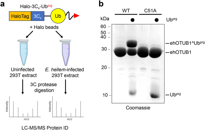
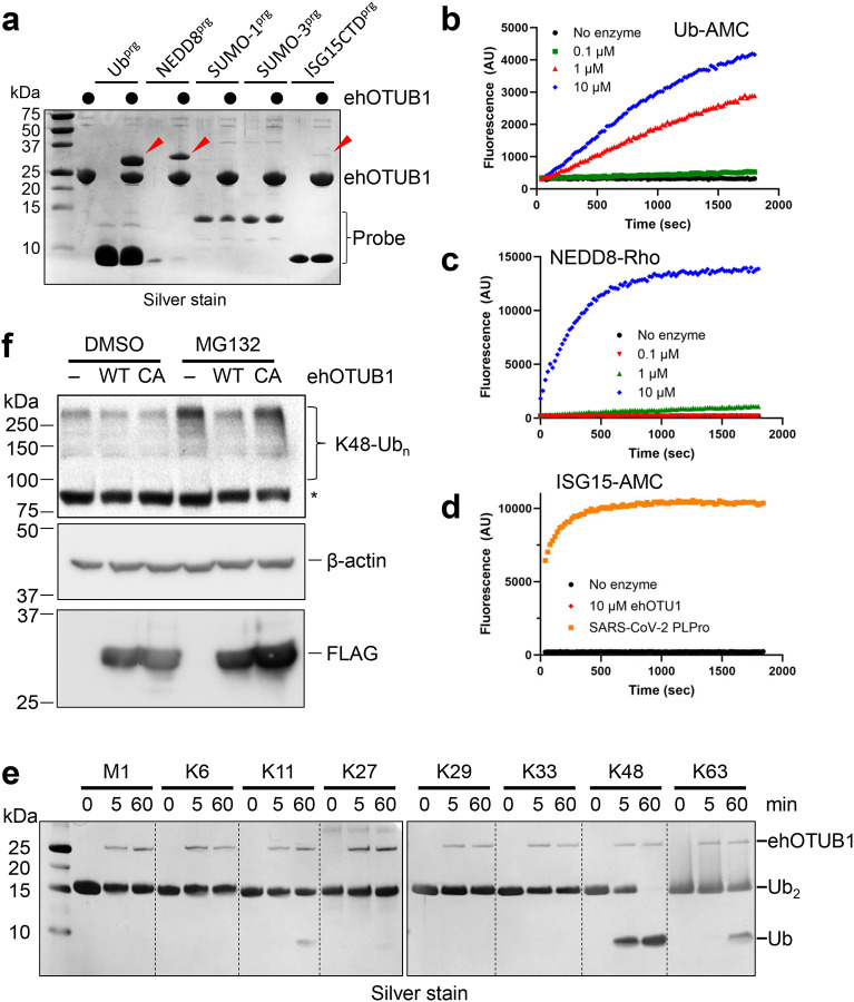
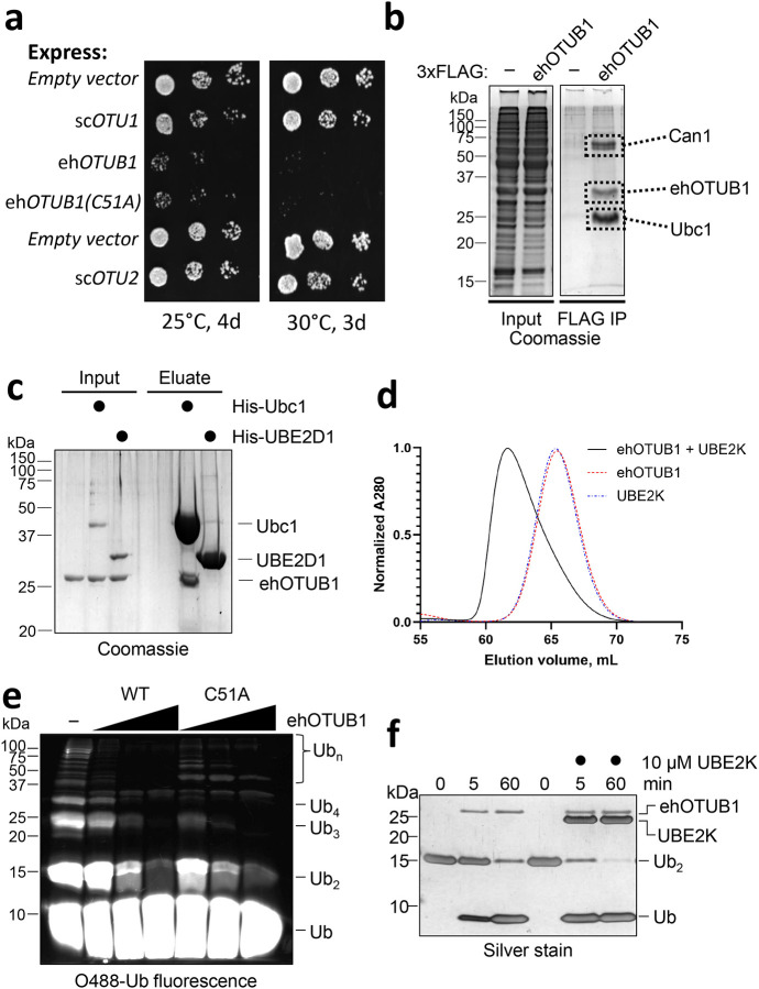
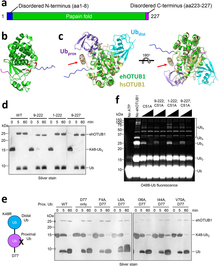
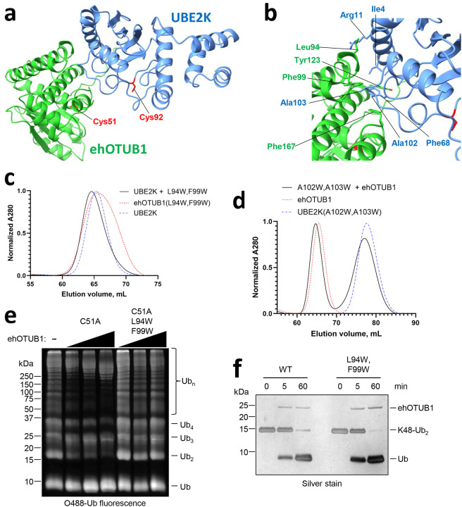
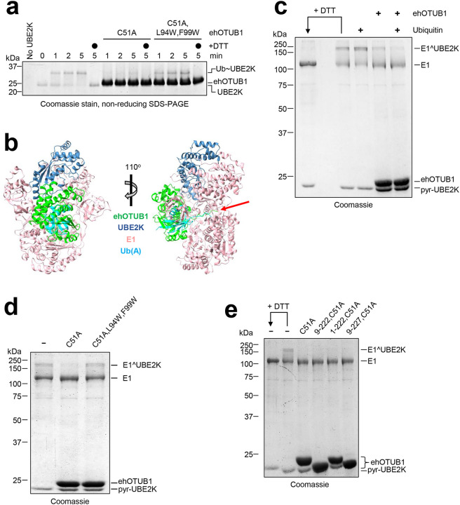
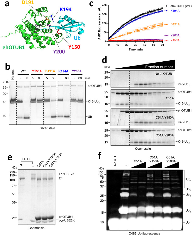
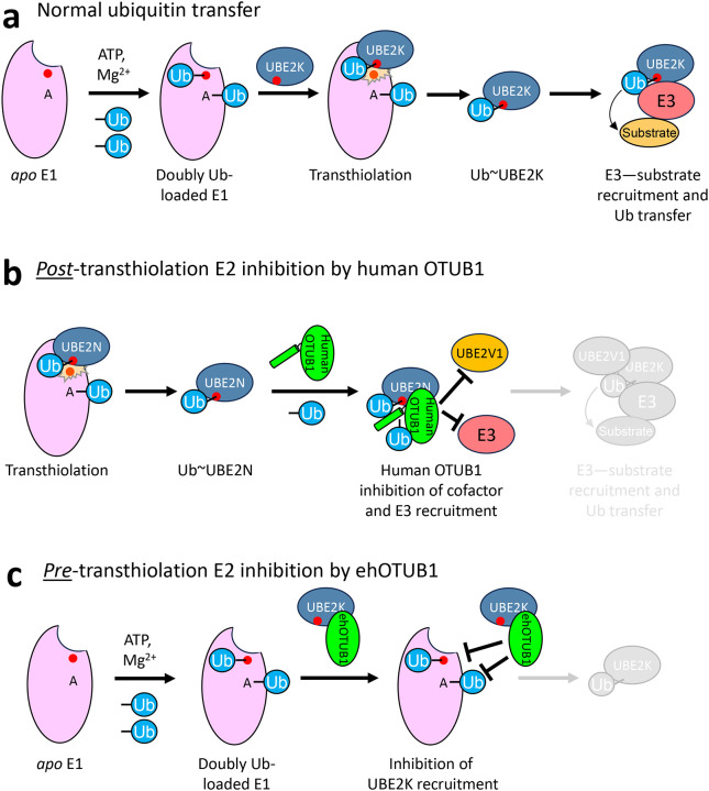
Intracellular pathogens frequently subjugate the ubiquitin system to evade host immune defenses and establish intracellular replication niches. Microsporidia are obligate intracellular animal parasites that typically cause self-limiting infections in humans, but can sometimes cause fatal disseminated disease. At present, the ubiquitin system of microsporidia is virtually unexplored. Here, we discover a likely effector deubiquitinase (DUB) of the otubain subgroup from the human pathogenic microsporidian Encephalitozoon hellem, which we designate ehOTUB1. We find that ehOTUB1 selectively binds the human ubiquitin conjugating (E2) enzyme UBE2K and inhibits its ubiquitin conjugation activity independent of ehOTUB1 DUB activity. We show that ehOTUB1 obstructs docking of UBE2K onto ubiquitin E1 enzyme via steric conflict with ubiquitin in the E1 adenylation site to prevent ubiquitin transfer to UBE2K. This unconventional mechanism of E2 inhibition expands the known repertoire by which pathogens manipulate ubiquitin signaling, and suggests that direct inhibition of E2 enzymes may be a broader function of otubain subfamily DUBs than originally appreciated.


Conflict of interest statement

Competing interests The authors have no competing interests with the content of this work or with the journal.

Figures

Figure 1:
Figure 1:. Identification of EHEL_050640 as a putative microsporidial deubiquitinating enzyme.
A, Scheme for covalent trapping and mass spectrometric identification of microsporidial deubiquitinating enzymes. A ubiquitin propargylamide suicide probe immobilized via an N-terminal Halo tag was incubated with extracts of uninfected or E. hellem infected 293T cells, and after extensive washing, ubiquitin and any covalently bound proteins were eluted using human rhinovirus 3C protease for identification via LC-MS/MS. B, Purified recombinant WT ehOTUB1, but not ehOTUB1(C51A), reacted with ubiquitin propargylamide (Ubprg).
Figure 2:
Figure 2:. Characterization of EHEL_050640/ehOTUB1.
A, ehOTUB1 was reacted with propargylamides of the indicated ubiquitin-like proteins to identify possible substrates. Red arrowheads indicate ehOTUB1 covalently modified with the ubiquitin-like protein probes. B-D, The indicated concentrations of ehOTUB1 were incubated with fluorogenic substrates ubiquitin-AMC (Ub-AMC) (B), NEDD8-rhodamine 110 (C), or ISG15-AMC (D) and fluorescence liberated was measured over time. SARS-CoV-2 papain-like protease (PLPro; 100 nM) was included as a positive control for ISG15-AMC hydrolysis. E, 500 nM ehOTUB1 was incubated with 20 μM Ub2 harboring the indicated linkages for 0, 5, or 60 minutes before quenching of the reactions and analysis by SDS-PAGE. F, Flp-In T-REx 293 cells were transfected with plasmids encoding the indicated forms of ehOTUB1, and after 24 hours, were treated with 20 μM MG132 or DMSO control for 4 hours prior to analysis. An asterisk indicates an apparent nonspecific band.
Figure 3:
Figure 3:. Identification of Ubc1/UBE2K as a target of ehOTUB1-mediated inhibition.
A, Cells expressing the indicated proteins as N-terminal 3xFLAG fusions from high-copy vector p424GPD were spotted in six-fold serial dilutions before incubation as shown. B, ehOTUB1 copurifies from yeast with Ubc1. 3xFLAG-ehOTUB1 and interacting proteins were immunopurified from wild type yeast by 3xFLAG affinity, and the boxed areas were sent for protein identification by liquid chromatography-mass spectrometry. The most abundant protein in each band is shown. The input and FLAG IP are non-adjacent lanes from the same gel. C, Ubc1 interacts with ehOTUB1 in vitro. Untagged ehOTUB1 was incubated with buffer, His-tagged Ubc1, or His-tagged human UBE2D1 before Ni-NTA affinity purification and SDS-PAGE. D, Human UBE2K binds ehOTUB1. Equal amounts of ehOTUB1, UBE2K, or ehOTUB1-UBE2K mixture were separated by gel filtration. The leftward shift in the elution volume for the mixture indicates direct interaction. E, ehOTUB1 inhibits formation of K48-linked polyubiquitin chains by UBE2K in a manner independent of ehOTUB1 catalytic activity. Ubiquitination reactions containing 1 μM E1, 5 μM UBE2K, and fluorescent ubiquitin were incubated for two hours in the presence or absence of 1 to 4-fold molar excess of ehOTUB1 or ehOTUB1(C51A) to UBE2K, followed by analysis by SDS-PAGE and fluorescence imaging. F, UBE2K does not interfere with ehOTUB1 deubiquitination activity. 500 nM ehOTUB1 was preincubated with 10 μM human UBE2K prior to addition of 20 μM K48-Ub2 for the indicated times.
Figure 4:
Figure 4:. The N- and C-termini of ehOTUB1 are dispensable for ehOTUB1 functions.
A, Domain architecture of ehOTUB1. B, An AlphaFold prediction of the ehOTUB1 structure indicates the extreme N- and C-termini (blue and magenta, respectively) are likely to be disordered. The residues comprising the catalytic triad are shown in red stick mode. C, Superimposition of ehOTUB1 colored as in (A) with human OTUB1 (tan) bound to two ubiquitin molecules (cyan and purple). The human OTUB1 N-terminal helix that binds the proximal ubiquitin is indicated with a red arrow. D, N- and C-terminal truncation mutants of ehOTUB1 cleave K48-Ub2 as well as WT ehOTUB1. K48-Ub2 (20 μM) was incubated with 1 μM of the indicated ehOTUB1 proteins at 37°C for the times shown. E, ehOTUB1 DUB activity is unaffected by mutation of common contact patches on the proximal Ub. As in (D), but 500 nM ehOTUB1 was used. F, N- and C-terminal truncation mutants of ehOTUB1 inhibit UBE2K as well as WT. Reactions were performed as in Fig. 3E.
Figure 5:
Figure 5:. A mutagenesis-validated AlphaFold model of the ehOTUB1–UBE2K complex.
A, AlphaFold model of ehOTUB1 (green) in complex with UBE2K (cornflower blue) indicates that the catalytic cysteines (red sticks) of both enzymes remain exposed. B, Detail of the predicted hydrophobic interface between ehOTUB1 and UBE2K. Residues at the interface between the two proteins are indicated. Catalytic cysteines are shown as red sticks. C and D, Mutation of residues at the predicted ehOTUB1-UBE2K interface disrupt their interaction. Equal amounts of the indicated WT and mutant proteins or mixtures thereof were separated by gel filtration. Note that introduction of tryptophan residues into UBE2K caused enhanced hydrophobic interactions with the gel filtration resin that slowed its migration relative to the WT protein. E, The ehOTUB1(L94W,F99W) mutant retains WT DUB activity. F, UBE2K ubiquitin conjugation activity is unaffected by the ehOTUB1(C51A,L94W,F99W) mutant.
Figure 6:
Figure 6:. ehOTUB1 interferes with recruitment of UBE2K to ubiquitin-loaded E1 enzyme.
A, UBE2K ubiquitin thioester (Ub~UBE2K) formation is blocked by ehOTUB1(C51A) but not by ehOTUB1(C51A,L94W,F99W). B, The ehOTUB1–UBE2K AlphaFold model was superimposed onto Ubc4 (omitted for clarity) from PDB 4II2. E1 and the ubiquitin in the adenylation site are shown in pink and cyan, respectively). The potential accessing of the E1 adenylation site by the ehOTUB1 N-terminal region is indicated with a red arrow. C, Inhibition of E1-UBE2K crosslinking by ehOTUB1 is enhanced in the presence of Ub. E1 and disulfide-activated UBE2K were pre-incubated with ubiquitin and ehOTUB1, respectively, where indicated, before mixing and separation by non-reducing SDS-PAGE. The position of the E1-UBE2K disulfide is indicated as E1ÛBE2K. D, The ehOTUB1(C51A,L94W,F99W) mutant fails to suppress the E1-UBE2K crosslink. E, The ehOTUB1 N- and C-termini are dispensable for inhibition of E1-UBE2K crosslinking.
Figure 7:
Figure 7:. Docking of ubiquitin into the ehOTUB1 active site is dispensable for UBE2K inhibition.
A, AlphaFold model of ehOTUB1 (green) in complex with distal ubiquitin (cyan), with residues predicted to contact ubiquitin highlighted. B, The indicated ehOTUB1 proteins (500 nM) were incubated with 20 μM K48-Ub2 for the times shown before analysis by SDS-PAGE. C, Ub-AMC hydrolysis assay of WT and mutant ehOTUB1. The indicated ehOTUB1 proteins (500 nM) were incubated with 500 nM Ub-AMC, and fluorescence was monitored over time. D, The Y150A and Y200A ehOTUB1 mutations abrogate interaction with K48-Ub2. K48-Ub2 (80 μM) was incubated with the indicated ehOTUB1 proteins (90 μM) for one hour at room temperature before separation by gel filtration. E, Y150A and Y200A ehOTUB1 mutants retain the ability to block E1-UBE2K interaction as visualized by crosslinking. F, Y150A and Y200A ehOTUB1 mutants retain the ability to block ubiquitin conjugation by UBE2K.
Figure 8:
Figure 8:. Comparison of pre- and post-transthiolation E2 inhibition mechanisms.
A, Cartoon of E2 transthiolation and subsequent E3-mediated ubiquitin transfer to substrates. B, Human OTUB1 mediates E2 inhibition at post-transthiolation steps due to its preference for thioesterified E2, conferred by its N-terminal ubiquitin-binding helix. This configuration blocks recruitment of cofactor UBE2V1 and E3 ligases. C, Pre-transthiolation inhibition of UBE2K by ehOTUB1. Steric conflict mediated in part between ehOTUB1 and ubiquitin bound in the E1 adenylation site prevents transthiolation of UBE2K.

Similar articles

References

    1. Han B., Pan G. & Weiss L. M. Microsporidiosis in Humans. Clin Microbiol Rev 34, e0001020 (2021). 10.1128/CMR.00010-20 - DOI - PMC - PubMed
    1. Bojko J. et al. Microsporidia: a new taxonomic, evolutionary, and ecological synthesis. Trends Parasitol 38, 642–659 (2022). 10.1016/j.pt.2022.05.007 - DOI - PubMed
    1. Stentiford G. D. et al. Microsporidia - Emergent Pathogens in the Global Food Chain. Trends Parasitol 32, 336–348 (2016). 10.1016/j.pt.2015.12.004 - DOI - PMC - PubMed
    1. Ruan Y. et al. The largest meta-analysis on the global prevalence of microsporidia in mammals, avian and water provides insights into the epidemic features of these ubiquitous pathogens. Parasit Vectors 14, 186 (2021). 10.1186/s13071-021-04700-x - DOI - PMC - PubMed
    1. Orenstein J. M. et al. Fatal pulmonary microsporidiosis due to encephalitozoon cuniculi following allogeneic bone marrow transplantation for acute myelogenous leukemia. Ultrastruct Pathol 29, 269–276 (2005). 10.1080/01913120590951257 - DOI - PubMed

Publication types

LinkOut - more resources