[Preprint]. 2025 Jul 31:2025.07.30.667769.
doi: 10.1101/2025.07.30.667769.

Modulation of the 14-3-3σ/C-RAF "auto"inhibited complex by molecular glues

Affiliations

Modulation of the 14-3-3σ/C-RAF "auto"inhibited complex by molecular glues

Markella Konstantinidou et al. bioRxiv. .

Abstract

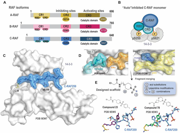
Molecular glues, compounds that bind cooperatively at protein-protein interfaces are revolutionizing chemical biology and drug discovery, allowing the modulation of traditional "undruggable" targets. Here, we focus on the native protein-protein interaction (PPI) of C-RAF, a key component of the MAPK signaling pathway, with the scaffolding protein 14-3-3. Although extensive drug discovery efforts have focused on the MAPK pathway, its central role in oncology and developmental disorders (RASopathies), still requires alternative approaches, moving beyond direct kinase inhibition. Indeed, stabilization of native PPIs is a relatively unexplored territory in this pathway. The function of C-RAF is regulated on multiple levels including dimerization, phosphorylation and complex formation with the hub protein 14-3-3. 14-3-3 prevents C-RAF activation by molecular recognition and binding at the phospho-serine 259. We used a fragment-merging approach to design a molecular glue scaffold that would bind to the composite surface of the 14-3-3/C-RAF "auto"inhibited complex. The synthesized molecular glues stabilized the 14-3-3/C-RAF complex up to 300-fold in biophysical assays; their glue-based mechanism of action was confirmed with several crystal structures of ternary complexes. Selectivity among the other RAF isoforms and other RAF phosphorylation sites was evaluated with biophysical assays. The best compounds showed excellent selectivity among a broad panel of 80 14-3-3 clients. Validation in cell assays showed on-target engagement, enhanced phosphorylation levels of the C-RAF pS259 site, reduced RAF dimerization and reduced ERK phosphorylation. Overall, this approach enables chemical biology studies on a C-RAF site that is intrinsically disordered prior to 14-3-3 binding and has not been targeted previously. These molecular glues will be useful as chemical probes and starting points for further drug discovery efforts to elucidate the effect of native PPI stabilization in the MAPK pathway with applications in oncology and RASopathies.

Keywords: 14-3-3; C-RAF; PPI; covalent; fragment merging; induced proximity; molecular glue; stabilizer.


Conflict of interest statement

The authors declare the following competing interest: M.R.A., C.O., and L.B. are founders of Ambagon Therapeutics.

Figures

Figure 1.
Figure 1.. RAF isoforms, 14-3-3/C-RAF pS259 complex and fragment merging approach.
A) Inhibiting and activating sites on A-, B- and C-RAF. B) Proposed model for the “auto”-inhibited C-RAF monomer complex bound to a 14-3-3 dimer. C) Crystal structure of 14-3-3σ (gray surface) bound to a phospho-C-RAF259 peptide (cyan surface). C38 (14-3-3) is shown as yellow sticks (PDB: 9EW7. D) Left: crystal structure of 1074202/14-3-3σ/ERα (PDB 8AWG). Right: crystal structure of TC521/14-3-3/p65 (PDB 6YOW). E) Top: Fragment merging approach towards a designed scaffold for the 14-3-3σ/C-RAF pS259 complex. Bottom: Representative examples and their binding mode (14-3-3 is omitted for clarity, C38 is shown as yellow sticks, and C-RAF as cyan sticks).
Figure 2.
Figure 2.. SAR and crystal structures of selected phenyl analogs.
A) MS bar graphs at 1 μM. For each compound, time course experiments were performed with measurements at 1h, 8h, 16h and 24h. C-RAF259 data are shown with different colors for each compound, and apo data in black. B) Bar graphs of FA compound titration pEC50 values after overnight incubation. C-RAF259 data are shown with different colors for each compound. C) Top: Crystal structures of 12 (pink sticks) and 21 (light purple sticks) with 14-3-3σ (white surface) and C-RAF pS259 10-mer peptide (blue sticks). Bottom: overlays of 12 with 21 (left), and 12 with a binary complex (PDB 3IQU, right). D) Left: crystal structures of 12 (pink sticks)/22 (turquoise sticks)/23 (purple sticks) with 14-3-3σ and C-RAF259 (overlay). Middle: detailed interactions of 23/14-3-3σ/C-RAF259 10-mer (left) and 12-mer (right). Interacting water molecules are shown as red spheres. Left: overlay of the structures.
Figure 3.
Figure 3.. SAR and crystal structures of selected phenyl and spiro analogs.
A) MS bar graphs at 1 μM. For each compound, time course experiments were performed with measurements at 1h, 8h, 16h and 24h. C-RAF259 data are shown with different colors for each compound, and apo data in black. B) Bar graphs of FA compound titration pEC50 values after overnight incubation. C-RAF259 data are shown with different colors for each compound. C) Top: Crystal structures of 29 (yellow sticks) and 37 (green sticks) with 14-3-3σ (white surface) and C-RAF pS259 10-mer peptide (blue sticks). Bottom: crystal structure of 70 (salmon sticks) with 14-3-3σ/C-RAF259 10-mer and overlay with 37. Interacting water molecules are shown as red spheres.
Figure 4.
Figure 4.. SAR and crystal structures of aryl and spiro/fused rings combinations.
A) MS bar graphs at 1 μM. For each compound, time course experiments were performed with measurements at 1h, 8h, 16h and 24h. C-RAF259 data are shown with different colors for each compound, and apo data in black. B) Bar graphs of FA compound titration pEC50 values after overnight incubation. C-RAF259 data are shown with different colors for each compound. C) Top: Crystal structure of 78 (light green sticks) with 14-3-3σ/C-RAF259 10-mer and overlay with the 83 and 86 crystal structures. Bottom: crystal structure of 78 (light green sticks) with 14-3-3σ/C-RAF259 12-mer (left) and overlay of the two ternary complexes (right).
Figure 5.
Figure 5.. Crystallography and SPR with RAF isoforms.
A) Crystal structures of 23 (top) and 78 (bottom) with 14-3-3σ/C-RAF-259 (left), 14-3-3σ/A-RAF214 (middle) and 14-3-3σ/B-RAF365 (right) 12-mer peptides. Interacting water molecules are shown as red spheres. B) SPR data of C-RAF (blue), A-RAF (yellow), and B-RAF (pink), 12-mer peptides binding to 14-3-3σ (top panel), and 14-3-3σ covalently bound to molecular glue 23 (parameters shown mean ± SD, n=2).
Figure 6.
Figure 6.. Evaluation of MGs in cell assays.
A) 14-3-3σ/C-RAF NanoBRET schematic (left) and dose-response curves in HEK293T cells. B) Co-IP western blots and quantification. C) Protection of phosphorylation of the C-RAF pS259 site in HEK293T cells. D) N-RAS/C-RAF NanoBRET; 100% was considered the DMSO mBU and 0% the mBU value of NRAS/C-RAF R89L, which does not bind to C-RAF. E) C-RAF/C-RAF homodimers NanoBRET dose-response curves. F) Lumit immunoassay for pERK levels in dose-responses with 23. G) 14-3-3σ/A-RAF NanoBRET. H) 14-3-3σ/B-RAF NanoBRET. I) NanoBRET for heterodimer formation for A-RAF/C-RAF and B-RAF/C-RAF, compared to C-RAF homodimers NanoBRET for 23.

Similar articles

References

    1. Dhillon A. S.; Hagan S.; Rath O.; Kolch W. MAP Kinase Signalling Pathways in Cancer. Oncogene 2007, 26 (22), 3279–3290. 10.1038/sj.onc.1210421. - DOI - PubMed
    1. Fernandez-Medarde A.; Santos E. Ras in Cancer and Developmental Diseases. Genes & Cancer 2011, 2 (3), 344–358. 10.1177/1947601911411084. - DOI - PMC - PubMed
    1. Davies H.; Bignell G. R.; Cox C.; Stephens P.; Edkins S.; Clegg S.; Teague J.; Woffendin H.; Garnett M. J.; Bottomley W.; Davis N.; Dicks E.; Ewing R.; Floyd Y.; Gray K.; Hall S.; Hawes R.; Hughes J.; Kosmidou V.; Menzies A.; Mould C.; Parker A.; Stevens C.; Watt S.; Hooper S.; Wilson R.; Jayatilake H.; Gusterson B. A.; Cooper C.; Shipley J.; Hargrave D.; Pritchard-Jones K.; Maitland N.; Chenevix-Trench G.; Riggins G. J.; Bigner D. D.; Palmieri G.; Cossu A.; Flanagan A.; Nicholson A.; Ho J. W. C.; Leung S. Y.; Yuen S. T.; Weber B. L.; Seigler H. F.; Darrow T. L.; Paterson H.; Marais R.; Marshall C. J.; Wooster R.; Stratton M. R.; Futreal P. A. Mutations of the BRAF Gene in Human Cancer. Nature 2002, 417 (6892), 949–954. 10.1038/nature00766. - DOI - PubMed
    1. Zhang W. BRAF Inhibitors: The Current and the Future. Current Opinion in Pharmacology 2015, 23, 68–73. 10.1016/j.coph.2015.05.015. - DOI - PubMed
    1. Drosten M.; Barbacid M. Targeting the MAPK Pathway in KRAS-Driven Tumors. Cancer Cell 2020, 37 (4), 543–550. 10.1016/j.ccell.2020.03.013. - DOI - PubMed

Publication types