[Preprint]. 2025 Aug 1:2025.07.29.664810.
doi: 10.1101/2025.07.29.664810.

Discovery of chromatin-based determinants of azacytidine and decitabine anti-cancer activity

Affiliations

Discovery of chromatin-based determinants of azacytidine and decitabine anti-cancer activity

Rishi V Puram et al. bioRxiv. .

Abstract

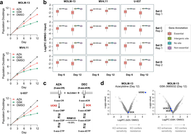
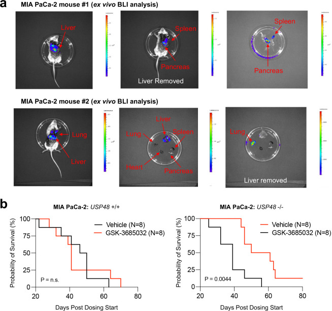
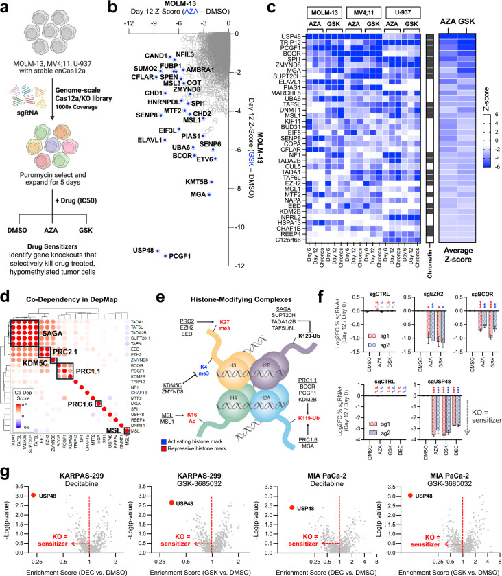
The DNA-incorporating nucleoside analogs azacytidine (AZA) and decitabine (DEC) have clinical efficacy in blood cancers, yet the precise mechanism by which these agents kill cancer cells has remained unresolved - specifically, whether their anti-tumor activity arises from conventional DNA damage or DNA hypomethylation via DNA methyltransferase 1 (DNMT1) inhibition. This incomplete mechanistic understanding has limited their broader therapeutic application, particularly in solid tumors, where early clinical trials showed limited efficacy. Here, through the assessment of drug sensitivity in over 600 human cancer models and comparison to a non-DNA-damaging DNMT1 inhibitor (GSK-3685032), we establish DNA hypomethylation, rather than DNA damage, as the primary killing mechanism of AZA and DEC across diverse cancer types. In further support of an epigenetic killing mechanism, CRISPR drug modifier screens identified a core set of chromatin regulators, most notably the histone deubiquitinase USP48, as AZA and DEC protective factors. We show that USP48 is recruited to newly hypomethylated CpG islands and deubiquitinates non-canonical histones, establishing USP48 as a key molecular link between the two components of epigenetic gene regulation: DNA methylation and chromatin modification. Furthermore, loss of USP48, which occurs naturally through biallelic deletions in human cancers, sensitized both hematologic and solid tumors to DNMT1 inhibition in vitro and in vivo. Our findings elucidate the epigenetic mechanism of action of AZA and DEC and identify a homeostatic link between DNA methylation and chromatin state, revealing new therapeutic opportunities for DNMT1 inhibitors in solid tumors.


Conflict of interest statement

COMPETING INTERESTS STATEMENT R.V.P., M.S., and T.R.G. receive research funding unrelated to this work from Calico Life Sciences LLC. T.R.G. is a paid advisor and/or equity holder in Dewpoint Therapeutics, Sherlock Biosciences, Amplifyer Bio, and Braidwell, Inc.. S.A.C. is a member of the scientific advisory boards of Kymera, PTM BioLabs, MobilION, and PrognomIQ.

Figures

Extended Data Figure 1:
Extended Data Figure 1:. PRISM screens with AZA, DEC, and GSK-3685032.
a. PRISM screen: >600 barcoded human cancer cell lines were pooled and treated with AZA (n=3), DEC (n=3), or GSK-3685032 (n=3) in a 7-point dose response (0.25–20 μM for AZA, 0.05–5 μM for DEC, and 0.25–20 μM for GSK-3685032). Viability for each cell line was determined based on relative barcode abundance with drug treatment compared to a DMSO control. b. Analysis of protein expression features that predict sensitivity to AZA. c. Analysis of RNA expression features that predict sensitivity to DEC or AZA. d-f. Viability of KARPAS-299 (lymphoid), HCT116 (colorectal), and OC314 (ovarian) cell lines following 6-day treatment with AZA (n=3) or DEC (n=3), compared to DMSO (n=3). g. Histograms of genome-wide cytosine methylation values in MOLM-13 cells after 3-day treatment with 1 μM GSK-3685032 (n=1) or DMSO (n=1). h. Lineage analysis of GSK-3685032 killing activity in the PRISM screen. Heatmap indicates the percentage of sensitive cell lines (Log2FC<−2) for each lineage and drug dose. i. Viability of KARPAS-299, HCT116, and OC314 cell lines following 6-day treatment with GSK-3685032 (n=3) compared to DMSO (n=3). j. AUC correlation analysis of AZA and GSK-3685032 across the cancer cell lines in PRISM. k. Viability histograms for >400 cell lines treated with 220 nM DEC (n=3) (upper panel) or 830 nM GSK-3685032 (n=3) (lower panel) for 6 or 10 days in an extended timecourse PRISM screen. l. Relative viability of MOLM-13 cells treated with ARA-C (n=3) or DEC (n=3) across a dose range for 3 or 6 days. *p < 0.05, **p < 0.01, ***p < 0.001, determined by a two-sided Student’s t test (compared to the DMSO control). Mean values are shown unless otherwise specified, and error bars represent ± SEM.
Extended Data Figure 2:
Extended Data Figure 2:. CRISPR drug modifier screens with AZA and GSK-3685032.
a. Intra-screen growth curves of MOLM-13, MV4;11, or U-937 AML cell lines treated with AZA, GSK-3685032, or DMSO. b. Activity of sgRNAs predicted to target essential genes, intergenic sites (control), no genomic sites (control), or non-essential genes for each cell line (MOLM-13, MV4;11, or U-937), timepoint (day 6 or 12), sgRNA library set (set C or D), and replicate (replicate 1 or 2). Across all screens, sgRNAs targeting essential genes were depleted more than the control or non-essential sgRNA classes. c. Biochemical pathway for AZA and DEC cellular uptake and bioactivation. AZA and DEC enter the cell via the equilibrative nucleoside transporter SLC29A1 and are phosphorylated by UCK2 or DCK, respectively. 5-aza-CDP is converted to a deoxyribose form (5-aza-dCDP) by ribonucleotide reductase (RRM1/2), thereby enabling DNA incorporation. d. Volcano plots of MOLM-13 CRISPR drug modifier screens. Genetic deletion of UCK2 (but not DCK) rescues killing by AZA. Neither UCK2 knockout nor DCK knockout rescues killing by GSK-3685032.
Extended Data Figure 3:
Extended Data Figure 3:. USP48 loss sensitizes cancer cells to DNMT1 inhibitor-induced cell death.
a. Competition assays with MOLM-13 cells expressing sgRNAs targeting USP48 (or control sgRNAs) after treatment with 500 nM AZA (n=3), 50 nM DEC (n=3), 350 nM GSK-3685032 (n=3), or DMSO (n=3). b. Annexin-V flow cytometry assays in USP48-intact (sgCTRL_35) or USP48-deleted (sgUSP48_06 or sgUSP48_80) MOLM-13 cells after 48-hr treatment with AZA (n=3), DEC (n=3), or DMSO (n=3). c. Dose-response curves of USP48-intact (sgCTRL_35) or USP48-deleted (sgUSP48_06 or sgUSP48_80) MOLM-13 cells after 72-hr treatment with chemotherapeutic agents (cytarabine, paclitaxel, or vinblastine) (n=3). d. Dose-response curves with USP48-intact (sgCTRL_35) or USP48-deleted (sgUSP48_06 or sgUSP48_80) AML cell lines treated with DEC (n=3) or GSK-3685032 (n=3) for 72 hrs. *p < 0.05, **p < 0.01, ***p < 0.001, determined by an unpaired, two-sided Student’s t-test. Mean values are shown unless otherwise specified, and error bars represent ± SEM.
Extended Data Figure 4:
Extended Data Figure 4:. USP48 is a catalytically-active DUB that targets variant histones.
a. ConSurf amino acid residue conservation score projected onto the AF2 structural model of USP48. The peptidase domain and catalytic cysteine (Cys-98) of USP48 are highly conserved from yeast to humans. b. CpG island enrichment analysis of USP48 genomic binding sites in MOLM-13 cells. c. Methyl-seq analysis of novel USP48 genomic binding sites gained in GSK-3685032-treated MOLM-13 cells. d. CTCF motif analysis of the MOLM-13 ATAC-seq profiles. e. Western blot analysis of DNMT1 protein levels in isogenic USP48-intact (sgCTRL_35) and USP48-deleted (sgUSP48_80) MOLM-13 cells after 48-hr treatment with GSK-3685032. f. Whole proteome profiling of USP48-deleted (sgUSP48_06) cells compared to USP48-intact (sgCTRL_35) cells for MOLM-13 (n=2), MV4;11 (n=3), or MIA PaCa-2 (n=3), highlighting preserved DNMT1 protein stability in the absence of USP48. g. Ubiquitylome (normalized to proteome) profiling in MOLM-13 (n=2) or MV4;11 (n=3) following USP48 deletion (sgUSP48_06) compared to intact USP48 (sgCTRL_35). h. IP-MS strategy to identify chromatin-based interacting partners of USP48 using distinct USP48 isoforms. The long isoform (IsoT7) of USP48 is a nuclear protein, while the short isoform (IsoT2) is a cytoplasmic protein and can be used as a control. i. IP-MS volcano plots of the interacting partners of the short isoform (n=3) or long isoform (n=3) of USP48. MACROH2A1 selectively binds the USP48 long isoform.
Extended Data Figure 5:
Extended Data Figure 5:. In vivo metastatic pancreatic cancer model.
a. Ex vivo BLI reveals extensive metastatic tumor formation in NSG mice following intracardiac injection of MIA PaCa-2 pancreatic cancer cells. b. Kaplan-Meier survival analysis of USP48 +/+ or USP48 −/− MIA PaCa-2 metastatic tumor cohorts treated with vehicle or GSK-3685032 (30 mg/kg BID).
Figure 1:
Figure 1:. DNA hypomethylation via DNMT1 degradation is the principal killing mechanism of AZA and DEC in diverse cancers.
a. Upper panel: Chemical structures of AZA and DEC, highlighting the shared nitrogen substitution in the fifth position of their pyrimidine rings and distinct sugar backbones. Lower panel: AUC correlation analysis of AZA and DEC across all cell lines in the PRISM screen. b. Left panel: The de novo methyltransferases DNMT3A and DNMT3B act on unmethylated DNA, whereas the maintenance methyltransferase DNMT1 and its binding partner UHRF1 act on hemimethylated DNA during DNA replication. Right panel: Analysis of protein expression features across the PRISM cell lines predictive of DEC sensitivity. c. Lineage analysis of AZA and DEC killing activity in the PRISM screen. Heatmap indicates the percentage of sensitive cell lines (Log2FC<−2) within each lineage for the lowest four doses of each drug. d. Comprehensive profiling of the proteome (left) and ubiquitylome (normalized to proteome) (right) in MV4;11 leukemia cells following treatment with 250 nM GSK-3685032 (n=3) for 48 hrs, as compared to DMSO (n=3). Each datapoint represents a protein group (see Methods). e. Western blot analysis of DNMT1 protein and γ-H2A.X levels in MOLM-13 cells following 48-hr treatment with AZA, DEC, or GSK-3685032. f. 5-methylcytosine DNA dot blot of MOLM-13 cells after 72-hr treatment with AZA or GSK-3685032. g. AUC correlation analysis of DEC and GSK-3685032 across the cancer cell lines in PRISM. h. Viability histograms for >500 cell lines treated with DEC (n=3) (upper panel) or GSK-3685032 (n=3) (lower panel) for 6 or 10 days in an extended timecourse PRISM screen.
Figure 2:
Figure 2:. Chromatin-based modifiers of DNMT1 inhibitor anti-cancer activity.
a. Genome-scale CRISPR-Cas12a screening strategy to identify genetic modifiers of DNMT1 inhibition. b. Z-score analysis of gene knockouts that sensitize MOLM-13 cells to AZA (n=2) compared to GSK-3685032 (n=2) at day 12. c. Heatmap of top drug sensitizers across different cell lines (MOLM-13, MV4;11, or U-937), timepoints (days 6 or 12), and anchor drugs (AZA or GSK-3685032). Chronos Z-scores represent a combined metric of gene knockout activity over time. Genes are ranked by average Z-score, and factors with known chromatin-based functions are annotated. d. Co-dependency analysis of top-scoring epigenetic hits from the CRISPR screens reveals distinct histone modifying complexes. A positive co-dependency score for a gene pair indicates that the individual gene knockouts produce a similar killing pattern across the >1,000 cell lines profiled in DepMap. e. Nucleosome schematic depicts histone modifications targeted by the chromatin complexes identified in the co-dependency analysis. f. MOLM-13 cell competition assays for validation of individual sgRNAs targeting components of PRC1.1 (BCOR) and PRC2.1 (EZH2) or a novel histone deubiquitinase (USP48). EZH2 (n=2), BCOR (n=3), and USP48 (n=3) knockout cells, but not cells expressing a control sgRNA (n=3), have a competitive disadvantage following treatment with a DNMT1 inhibitor compared to DMSO. g. CRISPR-Cas9 drug modifier screens with a ubiquitin-proteasome system focused sgRNA library in KARPAS-299 (lymphoid) or MIA PaCa-2 (pancreatic) cell lines treated with either DEC (n=3) or GSK-3685032 (n=3). *p < 0.05, **p < 0.01, ***p < 0.001, determined by an unpaired, two-sided Student’s t-test. Mean values are shown unless otherwise specified, and error bars represent ± SEM.
Figure 3:
Figure 3:. USP48 deletion synergizes with DNMT1 inhibitors to induce tumor cell death.
a. USP48-intact (sgCTRL_35) or USP48-deleted (sgUSP48_06 or sgRNA_80) MOLM-13 cells were validated for knockout by western blot and treated with compounds (n=3) in the GSK-3685032 chemical series for 72 hrs in a dose response. Compounds vary in their biochemical potency for DNMT1 inhibition. Doxorubicin (n=3) was used as a control for conventional DNA damage. b. Western blot validation of USP48 degradation in MOLM-13 cells with USP48-dTAG add-back. c. USP48-dTAG add-back cells were pre-treated with DMSO (n=3) or 1 μM GSK-3685032 (n=3) for 3 days to induce DNA hypomethylation, and dTAGV-1 (0, 0.5, 1, or 2 μM) was then added to induce USP48 degradation. Viable cells were counted following the addition of dTAGV-1. d. Competition assays with three unique pairs of control sgRNA (co-expression of BFP) and USP48 sgRNA (co-expression of RFP657) in OCI-AML2 (TP53-intact), MV4;11 (TP53-intact), and U-937 (TP53-null) cells treated with DMSO (n=3) or 500 nM GSK-3685032 (n=3) over 10 days. A reduction in RFP657+ cells indicates a competitive disadvantage for USP48 sgRNA-expressing cells compared to control sgRNA-expressing cells. e. Western blot analysis of P53 and P21 expression in isogenic TP53 +/+ and TP53 −/− MOLM-13 cells treated with 5 μM Nutlin-3 for 24 hrs. f. Competition assays with control sgRNA (co-expression of BFP) and USP48 sgRNA (co-expression of RFP657) in isogenic TP53 +/+ and TP53 −/− MOLM-13 cells treated with DMSO (n=3) or GSK-3685032 (n=3). g. Experimental approach to test the combination effect of USP48 deletion and GSK-3685032 (n=3) across 219 solid tumor cell lines in PRISM. h. Identification of a subset of solid tumor cell lines with greater sensitivity to the USP48 knockout and GSK-3685032 combination compared to the single perturbations. i. Dose-response curves for isogenic USP48-intact (sgCTRL_35) or USP48-deleted (sgUSP48_06) MIA PaCa-2, NCIH-1299, or KNS60 cells after 6-day treatment with GSK-3685032 (n=3). *p < 0.05, **p < 0.01, ***p < 0.001, determined by an unpaired, two-sided Student’s t-test. Mean values are shown unless otherwise specified, and error bars represent ± SEM.
Figure 4:
Figure 4:. USP48 is a molecular sensor linking DNA hypomethylation and histone deubiquitination.
a. AF2 model of full-length human USP48 protein. The N-terminal peptidase domain, three DUSP domains, and a C-terminal ubiquitin-like (UBL) domain are highlighted in cyan, green, and pink, respectively. b. Superposition of the catalytic domains of USP48 (rainbow) and USP7 (cyan) in complex with ubiquitin (magenta surface) (PDB: 5KYE). The well-conserved catalytic triad (Cys-98, His-353, and Asn-370) is illustrated. c. In vitro K63-linked tetra-ubiquitin kinetic DUB assay with His6-USP48. Addition of DUB inhibitors (PR-619 or 17e) blocks tetra-ubiquitin hydrolysis. d. Immunofluorescence images depicting cytoplasmic localization of the short USP48 isoform (WT_IsoT2) and nuclear localization of long USP48 isoform (WT_IsoT7) in MIA PaCa-2 cells. e. Rescue of USP48 deletion with WT_IsoT7 in MIA PaCa-2 cells after treatment with GSK-3685032 (n=3). WT_IsoT2 and a catalytic dead version of the long USP48 isoform (C98S_IsoT7) fail to rescue. f. Heatmaps of USP48 ChIP-seq peaks in USP48-intact (sgCTRL_35) (n=2) and USP48-deleted (sgUSP48_80) (n=2) MOLM-13 cells. Additional heatmaps show CpG density and mCG content across 4-kb windows centered on the identified USP48 ChIP-seq peaks. USP48 selectively binds hypomethylated CpG islands. g. Whole proteome profiling of MOLM-13 cells treated with 100 nM GSK-3685032 (n=2) compared to DMSO (n=2). h. Genome browser tracks for USP48 ChIP-seq peaks (blue) and mCG values (yellow) in MOLM-13 cells treated with DMSO or 1 μM GSK-3685032. USP48 localizes to newly hypomethylated CpG islands in GSK-3685032-treated cells. i. ATAC-seq profiles in MOLM-13 cells (n=2) showing predicted nucleosome enrichment across 4-kb windows for the identified USP48 binding sites. j. Ubiquitylome (normalized to proteome) profiling in MIA PaCa-2 cells reveals increased ubiquitination of the variant histone MACROH2A1 in USP48-deleted (sgUSP48_06) (n=3) cells compared to USP48-intact (sgCTRL_35) (n=3) cells. Venn diagram illustrates ubiquitinated proteins (and target lysine sites) that are upregulated (Log2FC>5) in the ubiquitylome following USP48 deletion (sgUSP48_06), and the intersection across three cell lines (MIA PaCa-2, MV4;11, and MOLM-13). MACROH2A1 Lys-134 is the single shared target across the three cell models. k. Mechanistic model: USP48 functions as a molecular sensor of DNA hypomethylation. USP48 is specifically recruited to hypomethylated CpG islands where it deubiquitinates non-canonical histones, most notably MACROH2A1, thereby providing a molecular link between DNA hypomethylation and histone modification.
Figure 5:
Figure 5:. USP48 copy number loss is frequent in human tumors and sensitizes to DNMT1 inhibition in vivo.
a. Frequency of shallow or deep deletions of USP48 across non-redundant human tumor genomic datasets in cBioPortal. b. MOLM-13 systemic AML model of USP48 +/+ or USP48 −/− leukemias treated with vehicle or GSK-3685032 (30 mg/kg BID). c. Kaplan-Meier survival analysis of USP48 +/+ or USP48 −/− MOLM-13 leukemias treated with vehicle or GSK-3685032 (30 mg/kg BID). d. Left panel: BLI measurements of all mice in the USP48 +/+ or USP48 −/− MOLM-13 leukemia cohorts while on treatment. Right panel: BLI images for 5 representative mice in the USP48 −/− cohort at day 10 after treatment initiation. e. MIA PaCa-2 pancreatic cancer metastasis model. f. Left panel: BLI measurements for all mice in the USP48 +/+ or USP48 −/− MIA PaCa-2 tumor cohorts while on treatment with vehicle or GSK-3685032. Right panel: BLI images for 5 representative mice in the USP48 −/− cohort at day 21 after treatment initiation.

References

    1. Fenaux P. et al. Azacitidine prolongs overall survival compared with conventional care regimens in elderly patients with low bone marrow blast count acute myeloid leukemia. J Clin Oncol 28, 562–569 (2010). 10.1200/JCO.2009.23.8329 - DOI - PubMed
    1. DiNardo C. D. et al. Azacitidine and Venetoclax in Previously Untreated Acute Myeloid Leukemia. N Engl J Med 383, 617–629 (2020). 10.1056/NEJMoa2012971 - DOI - PubMed
    1. Montesinos P. et al. Ivosidenib and Azacitidine in IDH1-Mutated Acute Myeloid Leukemia. N Engl J Med 386, 1519–1531 (2022). 10.1056/NEJMoa2117344 - DOI - PubMed
    1. Sorm F., Piskala A., Cihak A. & Vesely J. 5-Azacytidine, a new, highly effective cancerostatic. Experientia 20, 202–203 (1964). 10.1007/BF02135399 - DOI - PubMed
    1. Sorm F. & Vesely J. The Activity of a New Antimetabolite, 5-Azacytidine, against Lymphoid Leukaemia in Ak Mice. Neoplasma 11, 123–130 (1964). - PubMed

Publication types