

doi: 10.1016/j.bvth.2024.100034.
eCollection 2025 Feb.
Podoplanin and microthrombi in lung injury
Affiliations
- PMID: 40766876
- PMCID: PMC12320407
- DOI: 10.1016/j.bvth.2024.100034
Item in Clipboard
Podoplanin and microthrombi in lung injury
Blood Vessel Thromb Hemost.
.
No abstract available
Conflict of interest statement
Conflict-of-interest disclosure: The authors declare no competing financial interests.
Figures
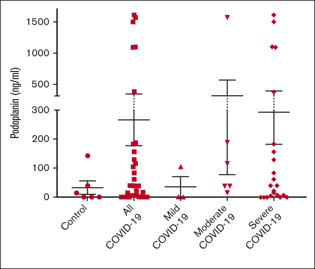
PDPN plasma level in patients with COVID-19. PDPN plasma levels and the severity of COVID-19 as defined by the National Institutes of Health criteria.
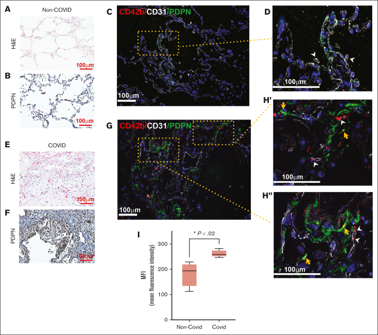
Expression of PDPN in the lung in COVID-19 pneumonia. Tissue specimens from the lungs of patients with COVID-19 pneumonia and patients without COVID-19 obtained during autopsy were examined. (A) Hematoxylin and eosin (H&E) and (B) immunohistochemistry stain for PDPN of the non–COVID-19 lung. Immunofluorescence staining of non–COVID-19 lungs with (C) lower original magnification (×100) and (D) higher original magnification (×200) showing few platelets in the lung parenchyma inside blood vessels (white arrowheads). (E) H&E and (F) immunohistochemistry stain for PDPN of COVID-19 lungs. Immunofluorescence staining of COVID-19 lungs at (G) lower original magnification (×100) and (H′-H′′) higher original magnification (×200). Platelets inside blood vessels are indicated with white arrowheads, and those colocalized with PDPN with yellow arrowheads. (I) Expressions of PDPN in COVID-19 and non–COVID-19 lungs are compared as mean fluorescence intensity (MFI) of immunofluorescence-stained slides. The box and whisker graph shows minimum to maximum values in each group (non–COVID-19: 182.5 ± 24.8, COVID-19: 261.7 ± 7.42; n = 4 images per group; P = .02, unpaired t test).
Similar articles
-
Podoplanin-positive cancer-associated fibroblast recruitment within cancer stroma is associated with a higher number of single nucleotide variants in cancer cells in lung adenocarcinoma.J Cancer Res Clin Oncol. 2018 May;144(5):893-900. doi: 10.1007/s00432-018-2619-3. Epub 2018 Mar 6. J Cancer Res Clin Oncol. 2018. PMID: 29511884 Free PMC article.
-
12/15-Lipooxygenase Inhibition Reduces Microvessel Constriction and Microthrombi After Subarachnoid Hemorrhage in Mice.Transl Stroke Res. 2025 Aug;16(4):1156-1172. doi: 10.1007/s12975-024-01295-0. Epub 2024 Sep 19. Transl Stroke Res. 2025. PMID: 39294532 Free PMC article.
-
12/15-Lipooxygenase Inhibition Reduces Microvessel Constriction and Microthrombi after Subarachnoid Hemorrhage in Mice.Res Sq [Preprint]. 2024 Jun 12:rs.3.rs-4468292. doi: 10.21203/rs.3.rs-4468292/v1. Res Sq. 2024. Update in: Transl Stroke Res. 2025 Aug;16(4):1156-1172. doi: 10.1007/s12975-024-01295-0. PMID: 38947083 Free PMC article. Updated. Preprint.
-
A role of platelet C-type lectin-like receptor-2 and its ligand podoplanin in vascular biology.Curr Opin Hematol. 2024 May 1;31(3):130-139. doi: 10.1097/MOH.0000000000000805. Epub 2024 Feb 6. Curr Opin Hematol. 2024. PMID: 38359177 Review.
-
Systematic Review of Microthrombi in COVID-19 Autopsies.Acta Haematol. 2021;144(5):476-483. doi: 10.1159/000515104. Epub 2021 Apr 19. Acta Haematol. 2021. PMID: 33873184 Free PMC article.
References
LinkOut - more resources
Full Text Sources