Non-contrast enhanced functional lung MRI in children: systematic review
- PMID: 40766916
- PMCID: PMC12322896
- DOI: 10.3389/fped.2025.1568172
Non-contrast enhanced functional lung MRI in children: systematic review
Abstract
Objectives: Magnetic resonance imaging (MRI) of the lung is well suited for repeated measurements especially in children due to the absence of ionizing radiation. Furthermore, non-contrast-enhanced (NCE) functional MRI techniques provide localized functional information on ventilation and perfusion without specialized set-ups (e.g., hyperpolarized gases) using standard clinical MRI systems. Current NCE-MRI techniques in the pediatric setting are matrix-pencil decomposition (MP)-MRI, phase-resolved functional lung (PREFUL)-MRI, self-gated non-contrast-enhanced functional lung (SENCEFUL)-MRI and Fourier decomposition (FD)-MRI. In this article, we comprehensively discuss these innovative techniques.
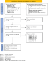
Study design: We review relevant functional NCE-MRI techniques based on a systematic literature research in MEDLINE, Embase, Cochrane Library, ClinicalTrials.gov and ICTRP. Core concepts were: 1. Aspects regarding lungs 2. MP-, PREFUL-, SENCEFUL and FD-MRI, and 3. children. Consecutively, we included 30 reports.
Results: Functional NCE-MRI in the pediatric setting has been successfully validated and used in observational studies covering a great variety of lung diseases. In contrast to initial implementation studies additionally reporting on clinical findings, later studies focus primarily on clinical topics. Heterogeneous study designs and examination protocols hamper the direct comparability between the different NCE-MRI techniques in terms of their performance against current functional imaging standards or specific objectives.
Conclusion: Their easy applicability makes NCE-MRI techniques highly attractive for widespread clinical use. Following successful implementation studies, still varying test protocols and approaches for calculating outcome values must next be compared and standardized.
Keywords: MP-MRI; MRI; children; functional lung MRI; lung function; lung imaging; pulmonology.
© 2025 Streibel, Bauman, Bieri, Pusterla, Stranzinger, Curdy, Latzin and Kieninger.
Conflict of interest statement
OB has received money from the Swiss National Science Foundation (SNF 320030_149576). PL received within the past 36 months payment by: Grants or contracts (Vertex and OM Pharma – paid to his institution), Payment or honoraria for lectures, presentations, speakers bureaus, manuscript writing or educational events (Vertex, Vifor, OM Pharma – paid to his institution and to him), Participation on a Data Safety Monitoring Board or Advisory Board (Polyphor, Vertex, OM Pharma, Vifor – paid to his institution and to him, Santhera (DMC), Allecra, Sanofi Aventis – paid to him). EK has within the past 36 months received Speaker Honorar by Sanofi Aventis and Vertex. The remaining authors declare that the research was conducted in the absence of any commercial or financial relationships that could be construed as a potential conflict of interest.
Figures


Similar articles
-
[Volume and health outcomes: evidence from systematic reviews and from evaluation of Italian hospital data].Epidemiol Prev. 2013 Mar-Jun;37(2-3 Suppl 2):1-100. Epidemiol Prev. 2013. PMID: 23851286 Italian.
-
Patient navigator programmes for children and adolescents with chronic diseases.Cochrane Database Syst Rev. 2024 Oct 9;10(10):CD014688. doi: 10.1002/14651858.CD014688.pub2. Cochrane Database Syst Rev. 2024. PMID: 39382077
-
Imaging modalities for the detection of posterior pelvic floor disorders in women with obstructed defaecation syndrome.Cochrane Database Syst Rev. 2021 Sep 23;9(9):CD011482. doi: 10.1002/14651858.CD011482.pub2. Cochrane Database Syst Rev. 2021. PMID: 34553773 Free PMC article.
-
Carbon dioxide detection for diagnosis of inadvertent respiratory tract placement of enterogastric tubes in children.Cochrane Database Syst Rev. 2025 Feb 19;2(2):CD011196. doi: 10.1002/14651858.CD011196.pub2. Cochrane Database Syst Rev. 2025. PMID: 39968844
-
Management of urinary stones by experts in stone disease (ESD 2025).Arch Ital Urol Androl. 2025 Jun 30;97(2):14085. doi: 10.4081/aiua.2025.14085. Epub 2025 Jun 30. Arch Ital Urol Androl. 2025. PMID: 40583613 Review.
References
Publication types
LinkOut - more resources
Full Text Sources

