. 2025 Aug 20;73(33):20792-20809.
doi: 10.1021/acs.jafc.5c04312. Epub 2025 Aug 6.

Multimodal Inhibition of Pectobacterium brasiliense Virulence by the Citrus Flavanone Naringenin

Affiliations

Multimodal Inhibition of Pectobacterium brasiliense Virulence by the Citrus Flavanone Naringenin

Manoj Pun et al. J Agric Food Chem. .

Abstract

Naringenin, a flavanone from citrus, was studied for its ability to reduce virulence in Pectobacterium, a phytopathogen causing soft rot disease in crop plants. Naringenin downregulated quorum sensing (QS) and suppressed critical virulence determinants in Pectobacterium brasiliense Pb1692, including plant cell wall-degrading enzymes, bacterial motility, and biofilm formation, consequently reducing disease symptoms in two host plants. Molecular docking simulations revealed a plausible binding mode for naringenin within the QS protein ExpI, which were maintained during microsecond-long Molecular Dynamics simulations. These simulations provided atomic-scale insight into specific interactions and estimated binding free energies, supporting naringenin's QS inhibition mode of action. In contrast, S-adenosyl methionine, the natural ligand of ExpI, was unable to maintain a stable binding mode in the ExpI site during simulations. Beyond QS disruption, naringenin induced reactive oxygen species accumulation and compromised DNA repair, indicating a multimodal mechanism of action. Despite these promising findings, naringenin's limited aqueous solubility challenges practical applications.

Keywords: Pectobacterium brasiliense; ROS; molecular docking; molecular dynamics; naringenin; quorum sensing inhibitor.


Figures

1
1
Pectobacterium brasiliense Pb1692 growth curves following exposure to naringenin over 24 h. Pb1692 cultures were treated with naringenin (0.5–2.0 mM), water (dH2O) or DMSO (0.8%) as controls (A). Bacterial growth occurred at 24 h following exposure to Naringenin. The data points and standard error mean (SEM) represent 4 replicates per treatment from three independent experiments (B).
2
2
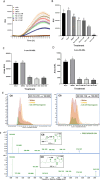
Luminescence intensity (RLU = LU/OD600) of Escherichia coli pSB401 induced by eAHL (positive control) and supernatants from Pectobacterium carotovorum Pb1692 treated with naringenin (0.5–2 mM), water, or 0.8% DMSO (controls) was measured every 0.5 h over 18 h (A). Bar graph shows RLU at 18 h for increasing naringenin concentrations (B). Quantitative levels of 3-oxo-C6-HSL (C) and 3-oxo-C8-HSL (D) in Pb1692 suspensions with naringenin are shown. Extracted ion chromatograms (EICs) for 3-oxo-C6-HSL and 3-oxo-C8-HSL ([MH+]: 214.019 and 242.138 to 102.055 fragment ion) display retention time (x-axis) and signal intensity (y-axis) (E). High-resolution fragmentation spectra by collision-induced dissociation, with precursor ions marked by blue diamonds, are shown for both AHLs and metabolites (F). Data represent the SEM of 8 replicates per treatment from two independent experiments. Statistical differences were analyzed by one-way ANOVA with Tukey–Kramer HSD. Bars that are labeled with a different letter are considered significantly different (p < 0.05).
3
3
Visualization of violacein (purple pigment) production by CV026 in response to AHLs from DH5α/pGEM expI (top panel, inner circle), Pb1692 (WT) (middle panel, inner circle), and DH5α/pGEM (negative control, bottom panel). Central paper discs were treated with 30 μL of dH2O, 0.8% DMSO, or naringenin (0.5–2.0 mM). For rescue, 30 μL of 500 nM 3-oxo-C6-HSL (eAHL) was added after 2 mM naringenin. The outer circle (3 cm diameter) contained CV026; the inner circle (1.5 cm diameter) contained test strains.
4
4
Swarming motility (A) and swimming motility (B) of Pb1692 after treatment with naringenin (0.5, 1, and 2 mM), dH2O, or 0.8% DMSO (controls). Representative images of swarming and swimming plates after 24 h at 28 °C (C). Biofilm formation in liquid yeast extract medium after 72 h at 28 °C, quantified by absorbance of crystal violet at 550 nm (D). Each bar represents mean ± SEM; motility assays: 3 independent experiments, 8 replicates per treatment (n = 24); biofilm assays: 2 independent experiments, 8 replicates each (n = 16). Statistical differences were analyzed by one-way ANOVA with Tukey–Kramer HSD; bars with different letters indicating significant differences (p < 0.05).
5
5
Effect of naringenin concentrations (0.5–2 mM) on pectate lyase (Pel) (A), polygalacturonase (Peh) (B), and protease (Prt) (C) activities in Pb1692. Bars show mean ± SEM (% of dH2O control, n = 16). One-way ANOVA and Tukey–Kramer HSD were used to analyze differences. Different letters indicate significant differences (p < 0.05).
6
6
Effect of naringenin (0.5, 1, and 2 mM) on expression of quorum sensing (expI, and expR), PCWDE (Pel, Peh, and Prt) and motility (motA, flhC, and fliA) genes in Pectobacterium brasiliense Pb1692 measured by qPCR after 8 h growth in LB at 28 °C. dH2O or 0.8% DMSO were used as controls. Bars show means + SE of 6 replicates per treatment with 4 biological repeats. Different letters indicate significant differences (P < 0.05; n = 24).
7
7
Effect of naringenin (0.5–2 mM) on decay of potato tubers (A) and calla lily leaf discs (B) infected by Pb1692. dH2O in minimal media (M9) and 0.8% DMSO served as the controls. Bars show mean ± SEM of percent tissue decay or infected area from two independent experiments (8 replicates/treatment). One-way ANOVA with Tukey–Kramer HSD; different letters indicate significant differences (p < 0.05).
8
8
Expression level of RecA (A) ROS formation (B), and PPO activity (D) in Pectobacterium brasiliense Pb1692 after exposure to naringenin (0.5–2 mM). dH2O or 0.8% DMSO served as controls. Bars show mean + SE from three independent experiments (8 replicates/treatment). IVIS fluorescence image of ROS in 96-well plates (C). One-way ANOVA with Tukey–Kramer HSD was used for (A,B). Student’s t test was used for (D). Different letters indicate significant differences (p < 0.05).
9
9
Interactions between SAM and ExpI during the two MD simulations, initiated from two distinct initial poses in the presence of the acyl chain (AC). During the simulations the position of the AC was restrained via two hydrogen bonds to the backbone of Phe102 and Arg101. (A) Interaction map for the trajectory initiated from Pose 1. (B) Interaction map for the trajectory initiated form Pose 2. Note: it is possible to have interactions with >100% as some residues may have multiple interactions of a single type with the same ligand atom. For example, the Arg side chain has four H-bond donors that can all hydrogen-bond to a single H-bond acceptor. For a hydrogen bond to be considered valid, the distance between the hydrogen atom and the acceptor atom (H···A) must be less than 2.8 Å, the angle between the donor atom, the hydrogen atom, and the acceptor (D–H···A) must exceed 120.0°, and the angle formed by H···A–B must be greater than 90.0°. In face-to-face pi–pi interactions, the maximum distance between the centroids of the rings is 4.4 Å with a maximum angle of 30° between the ring planes. For edge-to-face pi–pi interactions, the maximum distance between the centroids of the ring is 5.5 Å, again, with a maximum angle of 30° between the ring planes. In pi–cation interactions, the maximum distance between the center of the cation and the centroid of the ring is 6.6 Å, and the maximum angle between the normal to the plane of the ring and the line between the cation center and the ring center is 30°.
10
10
Interactions between naringenin and ExpI during the two MD simulations, initiated from two distinct initial poses in the absence (A,B) and presence (C,D) of an acyl chain (AC). During the simulations the position of the AC was restrained via two hydrogen bonds to the backbone of Phe102 and Arg101. (A) Note: it is possible to have interactions with >100% as some residues may have multiple interactions of a single type with the same ligand atom. For example, the Arg side chain has four H-bond donors that can all hydrogen-bond to a single H-bond acceptor.

Similar articles

References

    1. Gustafsson, J. ; Cederberg, C. ; Sonesson, U. ; Emanuelsson, A. . The Methodology of the FAO Study: Global Food Losses and Food Waste–Extent, Causes and Prevention–FAO, 2011; SIK Institutet för livsmedel och bioteknik, 2013.
    1. Charkowski A. O.. The Changing Face of Bacterial Soft-Rot Diseases. Annu. Rev. Phytopathol. 2018;56:269–288. doi: 10.1146/annurev-phyto-080417-045906. - DOI - PubMed
    1. Ma B., Hibbing M. E., Kim H. S., Reedy R. M., Yedidia I., Breuer J., Breuer J., Glasner J. D., Perna N. T., Kelman A., Charkowski A. O.. Host range and molecular phylogenies of the soft rot enterobacterial genera pectobacterium and dickeya. Phytopathology. 2007;97(9):1150–63. doi: 10.1094/PHYTO-97-9-1150. - DOI - PubMed
    1. Yin H., Deng Y., Wang H., Liu W., Zhuang X., Chu W.. Tea polyphenols as an antivirulence compound Disrupt Quorum-Sensing Regulated Pathogenicity of Pseudomonas aeruginosa. Sci. Rep. 2015;5:16158. doi: 10.1038/srep16158. - DOI - PMC - PubMed
    1. Tapia-Rodriguez M. R., Bernal-Mercado A. T., Gutierrez-Pacheco M. M., Vazquez-Armenta F. J., Hernandez-Mendoza A., Gonzalez-Aguilar G. A., Martinez-Tellez M. A., Nazzaro F., Ayala-Zavala J. F.. Virulence of Pseudomonas aeruginosa exposed to carvacrol: alterations of the Quorum sensing at enzymatic and gene levels. Journal of cell communication and signaling. 2019;13(4):531–537. doi: 10.1007/s12079-019-00516-8. - DOI - PMC - PubMed

MeSH terms