. 2025 Aug 19;97(32):17490-17500.
doi: 10.1021/acs.analchem.5c02338. Epub 2025 Aug 6.

Quantitative HILIC-Q-TOF-MS Analysis of Glycosaminoglycans and Non-reducing End Carbohydrate Biomarkers via Glycan Reductive Isotopic Labeling

Affiliations

Quantitative HILIC-Q-TOF-MS Analysis of Glycosaminoglycans and Non-reducing End Carbohydrate Biomarkers via Glycan Reductive Isotopic Labeling

Amrita Basu et al. Anal Chem. .

Abstract

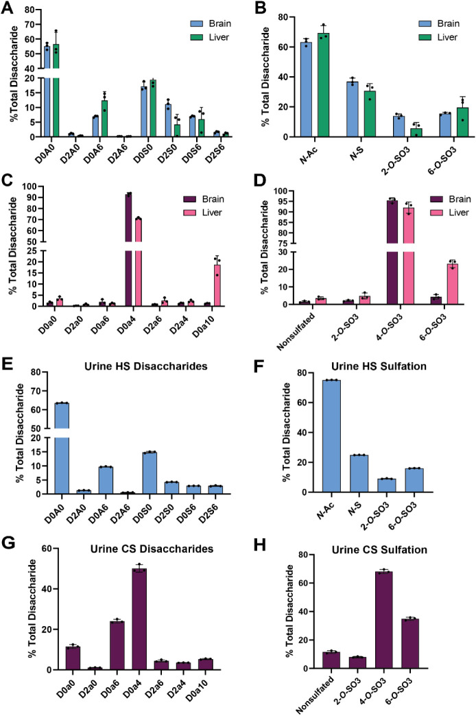
Glycosaminoglycans (GAGs) are linear, heterogeneous polysaccharides expressed on all animal cells. Sulfated GAGs, including heparan sulfate (HS) and chondroitin/dermatan sulfate (CS/DS), are involved in numerous physiological and pathological processes; therefore, precise and robust analytical methods for their characterization are essential to correlate structure with function. In this study, we developed a method utilizing hydrophilic interaction liquid chromatography coupled with time-of-flight mass spectrometry (HILIC-Q-TOF-MS) and glycan reductive isotopic reducing end labeling (GRIL) for the quantitative compositional analysis of HS and CS/DS polysaccharides. Lyase-generated disaccharides and commercial standards were chemically tagged on the reducing end with aniline stable isotopes, thus enabling the absolute quantification of HS and CS/DS disaccharides in complex biological samples. In addition, we adapted this workflow, in conjunction with new synthetic carbohydrate standards, for the quantification of disease-specific non-reducing end (NRE) carbohydrate biomarkers that accumulate in patients with mucopolysaccharidoses (MPS), a subclass of lysosomal storage disorders. As a proof of concept, we applied this method to measure NRE biomarkers in patient-derived MPS IIIA and MPS IIID fibroblasts, as well as in cortex tissue from a murine model of MPS VII. Overall, this method demonstrates improved sensitivity compared to previous GRIL-LC/MS techniques and, importantly, avoids the use of ion-pairing reagents, which are undesirable in certain mass spectrometry instrumentation and contexts. By combining the benefits of HILIC separation with isotopic labeling, our approach offers a robust and accessible tool for the analysis of GAGs, paving the way for advancements in understanding GAG structure and function.


Figures

1
1
HILIC-Q-TOF-MS method development for disaccharide analysis of glycosaminoglycans. (A) Overview of the workflow for isolation, purification, and reductive isotopic labeling for HILIC-Q-TOF-MS analysis. (B) The extracted ion chromatogram (XIC) for each of a mixture of eight HS disaccharide standards. The disaccharide structure code is described in Table S1 and ref . (C) XIC for the free molecular ion for [13C6] aniline-labeled HS disaccharide, D0A0, [M-H]−1 (m/z = 461). (D) Sensitivity and linear range for HS species (D0A0) shown as a correlation between picomole (pmol) amount and relative mass abundance. (E) The extracted ion chromatogram (XIC) for each of a mixture of seven CS/DS disaccharide standards. (F) XIC for the free molecular ion for [13C6] aniline-labeled CS/DS disaccharide, D0a0, [M-H]−1 (m/z = 461). (G) Sensitivity and linear range for CS/DS species (D0a0) are shown as a correlation between the picomole amount and relative mass abundance.
2
2
HILIC-Q-TOF-MS analysis of commercial GAG standards. (A) XIC chromatogram for [12C6] aniline-tagged heparin disaccharides resolved by HILIC-Q-TOF-MS. (B) Corresponding mass spectra for D2S6 from the standard and heparin sample tagged with [13C6] or [12C6] aniline, respectively. (C) LC-MS quantification of disaccharides from heparin and CHO-S HS (n = 3 biological replicates). (D) XIC chromatogram for [12C6] aniline-tagged CHO-S HS disaccharides resolved by HILIC-Q-TOF-MS. (E) Corresponding mass spectra for D0A0 from the standard and CHO-S HS sample tagged with [13C6] or [12C6] aniline, respectively. (F) Sulfation per disaccharide for pharmaceutical heparin and CHO-S HS (n = 3 biological replicates). (G) XIC chromatogram for [12C6] aniline-tagged CS-A disaccharides resolved by HILIC-Q-TOF-MS. (H) Corresponding mass spectra for D0a0 from the standard and CS-A tagged with [13C6] or [12C6] aniline, respectively. (I) LC-MS quantification of disaccharides from CS-A (n = 3 biological replicates).
3
3
HILIC-Q-TOF-MS analysis of GAGs isolated from human cells. (A) LC-MS quantification of HS disaccharides from human malignant melanoma cells (A375) and human chondrocytes (TC28a2) (n = 3 biological replicates). (B) HS sulfation per disaccharide for A375 and TC28a2 cells (n = 3). (C) LC-MS quantification of total HS in A375 and TC28a2 cells (n = 3). (D) LC-MS quantification of CS/DS disaccharides from A375 and TC28a2 cells (n = 3). (E) CS/DS sulfation per disaccharide for A375 and TC28a2 cells (n = 3). (F) LC-MS quantification of total CS/DS in A375 and TC28a2 cells (n = 3).
4
4
HILIC-Q-TOF-MS analysis of GAGs isolated from murine tissue and human urine. (A) LC-MS quantification of HS disaccharides from NOD-SCID mouse whole brain and liver (n = 3 biological replicates). (B) HS sulfation per disaccharide for mouse whole brain and liver (n = 3). (C) LC-MS quantification of CS/DS disaccharides from mouse whole brain and liver (n = 3). (D) CS/DS sulfation per disaccharide for mouse whole brain and liver (n = 3). (E) LC-MS quantification of HS disaccharides from human urine (n = 3). (F) HS sulfation per disaccharide for human urine (n = 3). (G) LC-MS quantification of CS/DS disaccharides from human urine (n = 3). (H) CS/DS sulfation per disaccharide for human urine (n = 3).
5
5
HILIC-Q-TOF-MS analysis of non-reducing end (NRE) species from MPS IIIA/IIID fibroblasts and a mouse model of MPS VII. (A) XIC chromatogram for aniline-tagged N-sulfo-glucosamine (S0) monosaccharide resolved by HILIC-Q-TOF-MS. Inset: mass spectra for [12C6] (335 m/z) and [13C6] (341 m/z) aniline-tagged S0 shown in panel A. (B) LC-MS quantification of total HS levels from aged MPS IIIA patient-derived fibroblasts (GM06110) versus healthy control fibroblasts. (C) Analysis of N-sulfo-glucosamine (S0) NRE species in MPS IIIA and healthy fibroblasts. (D) XIC chromatogram for aniline-tagged glucosamine-6-sulfate (H6) monosaccharide resolved by HILIC-Q-TOF-MS. Inset: mass spectrum for [12C6] (335 m/z) and [13C6] (341 m/z) aniline-tagged H6 shown in panel D. (E) LC-MS quantification of total HS levels from aged MPS IIID patient-derived fibroblasts (GM05093) versus healthy control fibroblasts. (F) Quantification of glucosamine-6-sulfate (H6) NRE species in MPS IIID and healthy fibroblasts. (G) XIC chromatograph for aniline-tagged G0S0 NRE disaccharide resolved by HILIC-Q-TOF-MS. Inset: mass spectra for [12C6] (511 m/z) and [13C6] (517 m/z) aniline-tagged G0S0 shown in panel G. Respective sodium adducts (533, 539 m/z) are also included. (H) LC-MS quantification of total HS levels from wild-type and MPS VII mice (Gusb –/– ). (I) Quantification of G0S0 NRE species in wild-type and MPS VII (Gusb –/–) mice.

Similar articles

Cited by

  • Glycosaminoglycan-driven lipoprotein uptake protects tumours from ferroptosis.
    Calhoon D, Sang L, Ji F, Bezwada D, Hsu SC, Cai F, Kim N, Basu A, Wu R, Pimentel A, Brooks B, La K, Paulina Serrano A, Cassidy DL, Cai L, Toffessi-Tcheuyap V, Moussa ME, Uritboonthai W, Hoang LT, Kolli M, Jackson B, Margulis V, Siuzdak G, Brugarolas J, Corbin I, Pratt DA, Weiss RJ, DeBerardinis RJ, Birsoy K, Garcia-Bermudez J. Calhoon D, et al. Nature. 2025 Aug;644(8077):799-808. doi: 10.1038/s41586-025-09162-0. Epub 2025 Jun 11. Nature. 2025. PMID: 40500442

References

    1. Basu A., Patel N. G., Nicholson E. D., Weiss R. J.. Spatiotemporal diversity and regulation of glycosaminoglycans in cell homeostasis and human disease. Am. J. Physiol. Cell Physiol. 2022;322(5):C849–c864. doi: 10.1152/ajpcell.00085.2022. - DOI - PMC - PubMed
    1. Trowbridge J. M., Gallo R. L.. Dermatan sulfate: new functions from an old glycosaminoglycan. Glycobiology. 2002;12(9):117R–125R. doi: 10.1093/glycob/cwf066. - DOI - PubMed
    1. Mizumoto S., Yamada S.. Congenital Disorders of Deficiency in Glycosaminoglycan Biosynthesis. Front. Genet. 2021;12:12. doi: 10.3389/fgene.2021.717535. - DOI - PMC - PubMed
    1. Mizumoto S., Yamada S., Sugahara K.. Mutations in Biosynthetic Enzymes for the Protein Linker Region of Chondroitin/Dermatan/Heparan Sulfate Cause Skeletal and Skin Dysplasias. BioMed. Res. Int. 2015;2015:861752. doi: 10.1155/2015/861752. - DOI - PMC - PubMed
    1. Khan S. A., Nidhi F., Leal A. F., Celik B., Herreño-Pachón A. M., Saikia S., Benincore-Flórez E., Ago Y., Tomatsu S.. Glycosaminoglycans in mucopolysaccharidoses and other disorders. Adv. Clin Chem. 2024;122:1–52. doi: 10.1016/bs.acc.2024.06.011. - DOI - PubMed

LinkOut - more resources