. 2025 Sep 10;16(9):e0162825.
doi: 10.1128/mbio.01628-25. Epub 2025 Aug 6.

Genome-wide CRISPR screen reveals key role of sialic acids in PEDV and porcine coronavirus infections

Affiliations

Genome-wide CRISPR screen reveals key role of sialic acids in PEDV and porcine coronavirus infections

Guanghao Guo et al. mBio. .

Abstract

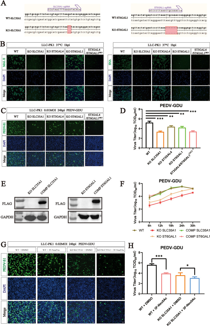
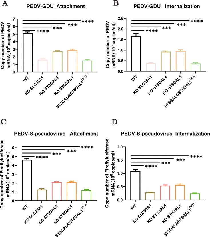
Porcine epidemic diarrhea virus (PEDV) is a globally distributed alphacoronavirus with economic importance that can cause severe watery diarrhea and even death in piglets. To identify host factors essential for PEDV infection, we performed a genome-wide CRISPR/Cas9 screen in human hepatocellular carcinoma cells (Huh7) using the highly virulent PEDV GIIb strain GDU. Several genes involved in the sialic acid and heparan sulfate biosynthesis pathway and cholesterol metabolism were highly enriched following PEDV selection. We validated that the host factor ST3 beta-galactoside alpha-2,3-sialyltransferase 4 (ST3GAL4), which catalyzes the transfer of sialic acid to sugar chains via α2,3-linked linkages, is important for PEDV infection. To systematically investigate the role of sialic acid in PEDV infection, we knocked out genes related to sialic acid synthesis. This led to a reduced abundance of sialic acid on the cell surface, which in turn inhibited PEDV adsorption and internalization. Furthermore, we found that both α2,3-linked and α2,6-linked sialic acids can serve as cellular attachment factors for PEDV. We conducted a glycan microarray screen to determine which sialoglycans are preferred by the PEDV spike protein. The results revealed that PEDV favors binding to α2,3-sialoglycans. Additionally, we found that not only current circulating PEDV strains but also other porcine coronaviruses rely on sialic acid for efficient infection. Collectively, our findings provide insights into critical host factors involved in PEDV infection and demonstrate that disrupting genes involved in sialic acid biosynthesis negatively affects the infectivity of multiple porcine enteric coronaviruses.IMPORTANCEA wide range of viruses utilize sialic acid as receptors. Sialic acid binding may serve as a key determinant of viral host range. Different viruses exhibit distinct preferences for specific types of sialic acid linkages. However, it remains unclear which specific subtypes of sialic acid are utilized during PEDV infection. In this study, we performed CRISPR-based genome-wide knockout screening and identified ST3GAL4 as a key host factor for PEDV infection. Furthermore, we found that both α2,3-linked and α2,6-linked sialic acids can function as attachment factors for PEDV infection. A glycan microarray screen revealed that PEDV S1 shows the strongest binding preference for α2,3-linked and α2,8-linked sialosides. Sialic acids were also implicated in infections by other porcine enteric coronaviruses. Overall, our findings advance our understanding of viral entry mechanisms of PEDV and other swine coronaviruses and may provide avenues for designing antiviral strategies.

Keywords: PEDV; SLC35A1; ST3GAL4; ST6GAL1; coronaviruses; sialic acid.


Conflict of interest statement

The authors declare no conflict of interest.

Figures

Fig 1
Fig 1
Genome-scale CRISPR screen identified host dependency factors for PEDV infection. (A) Overview of CRISPR screen in Huh7 cells. (B) Scatter plots reveal sgRNA-targeted sequence frequencies and the enrichment in control cells versus cells that survived PEDV infection. (C) KEGG pathway enrichment analysis for the top 50 sgRNA targets from the third PEDV challenge rounds. (D) PEDV infection of control and gene (DIPK2A, STGAL4, FOXA2, and PATZ1) KO LLC-PK1 cells. Cells were infected with PEDV (MOI = 0.1), and PEDV-infected cells were visualized by immunofluorescence using an S1-specific antibody. Scale bar = 100 µm. (E) TCID50 assay measuring PEDV titers in WT and KO LLC-PK1 cells. (F) qPCR quantification of PEDV N RNA in WT and KO LLC-PK1 cells. P values were determined by two-tailed unpaired t-tests. Error bars represent standard deviations from three independent experimental replicates. ns, no significant; *P < 0.05; **P < 0.01; ***P < 0.001; and ****P < 0.0001. Data are representative of at least three independent experiments.
Fig 2
Fig 2
ST3GAL4 is an important host factor in PEDV infection. (A) Alignment of the nucleic acid sequences of WT and ST3GAL4 KO LLC-PK1 cells. (B) Western blot analysis to detect the expression of ST3GAL4-flag in ST3GAL4 KO LLC-PK1 and KO-ST3GAL4 complement LLC-PK1 cells. (C) Cell viability detection in WT, ST3GAL4 KO, and KO-ST3GAL4 complement LLC-PK1 cells by a CCK-8 kit. (D) Immunofluorescence assays for detection of the PEDV in WT, ST3GAL4 KO, and KO-ST3GAL4 complement LLC-PK1 cells following infection with PEDV (MOI = 0.1) at 24 hpi. Scale bar, 100 µm. (E) The viral titers of PEDV in WT, ST3GAL4 KO, and KO-ST3GAL4 complement LLC-PK1 cells infected with PEDV (MOI = 0.1) at 24 hpi were evaluated by TCID50 assay. (F) One-step growth curves of WT, ST3GAL4 KO, and KO-ST3GAL4 complement LLC-PK1 cells infected with PEDV (MOI = 0.1) measured by TCID50 assay. P values were determined by two-tailed unpaired t-tests. Error bars represent standard deviations from three independent experimental replicates. ns, no significant; *P < 0.05; **P < 0.01; ***P < 0.001; and ****P < 0.0001. Data are representative of at least three independent experiments.
Fig 3
Fig 3
PEDV can use both α2,6-linked and α2,3-linked sialic acid to infect cells. (A) Alignment of the nucleic acid sequences of WT, SLC35A1, and ST6GAL1 knockout (KO) cells. (B) MAL II (binds to Sia-α2,3-galactose-GlcNAc) staining of WT and KO cells. SNA (binds to Sia-α2,6-galactose-GlcNAc) staining of WT and KO cells. Scale bar, 100 µm. (C) Immunofluorescence assays for detection of the PEDV in WT and KO cells following infection with PEDV (MOI = 0.1) at 24 hpi. Scale bar, 100 µm. (D) PEDV titers in WT and KO cells infected with PEDV (MOI = 0.1) at 24 hpi were evaluated by TCID50 assay. (E) Western blot analysis to detect the expression of SLC35A1-Flag in SLC35A1-KO and KO-SLC35A1 complement LLC-PK1 cells. Western blot analysis to detect the expression of ST6GAL1-Flag in ST6GAL1-KO and KO-ST6GAL1 complement LLC-PK1 cells. (F) One-step growth curves of WT, SLC35A1 KO, KO-SLC35A1 complement, ST6GAL1 KO, and KO-ST6GAL1 complement LLC-PK1 cells infected with PEDV (MOI = 0.1) measured by TCID50 assay. (G) Immunofluorescence assays for detection of the PEDV-S1 protein in cells following infection with PEDV (MOI = 0.2) at 24 hpi. Scale bar, 100 µm. (H) PEDV titers in cells infected with PEDV (MOI = 0.2) at 24 hpi were evaluated by TCID50 assay. P values were determined by two-tailed unpaired t-tests. Error bars represent standard deviations from three independent experimental replicates. ns, no significant; *P < 0.05; **P < 0.01; ***P < 0.001; and ****P < 0.0001. Data are representative of at least three independent experiments.
Fig 4
Fig 4
Sialic acid facilitates PEDV attachment and internalization. (A) The adsorption of PEDV on the WT, ST3GAL4 KO, and KO-ST3GAL4 complement LLC-PK1 cells, and the ST6GAL1 KO and KO-ST6GAL1 complement cells were evaluated by qRT‒PCR. The cells were infected with PEDV-GDU (MOI = 10) at 4°C for 1 h to evaluate the adsorption of PEDV. (B) The internalization of PEDV on the WT, ST3GAL4 KO, and KO-ST3GAL4 complement, and ST6GAL1 KO and KO-ST6GAL1 complement LLC-PK1 cells were evaluated by qRT‒PCR. The cells were incubated with PEDV (MOI = 10) at 4°C for 1 h, transferred to 37°C for 30 min and washed two times with acidic PBS (pH = 1.3) at 4°C to remove noninternalized virus particles. (C) The adsorption of PEDV on the WT, ST3GAL4 KO, and KO-ST3GAL4 complement LLC-PK1 cells, and the ST6GAL1 KO and KO-ST6GAL1 complement cells were evaluated by qRT‒PCR. The cells were infected with PEDV-S-pseudovirus (MOI = 10) at 4°C for 1 h to evaluate the adsorption of PEDV. (D) The internalization of PEDV on the WT, ST3GAL4 KO, KO-ST3GAL4 complement, and ST6GAL1 KO and KO-ST6GAL1 complement LLC-PK1 cells were evaluated by qRT‒PCR. The cells were incubated with PEDV-S-pseudovirus (MOI = 10) at 4°C for 1 h, transferred to 37°C for 30 min and washed two times with acidic PBS (pH = 1.3) at 4°C to remove noninternalized virus particles. P values were determined by two-tailed unpaired t-tests. Error bars represent standard deviations from three independent experimental replicates. ns, no significant; *P < 0.05; **P < 0.01; ***P < 0.001; and ****P < 0.0001. Data are representative of at least three independent experiments.
Fig 5
Fig 5
PEDV spike protein preferably binds α2,3-linked and α2,8-linked sialic acid. (A) The synthetic glycans 1–24 were used for microarray screening of the binding with PEDV S1. (B) Glycan microarray screen to identify which type of sialic acid for PEDV S1 binds. The average relative fluorescence value (RFU) was calculated by four independent replicates on the glycan microarray after the removal of the highest and lowest signals. The error bars indicate the standard deviation (SD) of RFU.
Fig 6
Fig 6
α2,3-linked and α2,6-linked sialic acids are essential for multiple coronaviruses infection. (A) Immunofluorescence assays (IFAs) for detection of the PEDV-S1 protein in WT and KO LLC-PK1 cells infected with PEDV(GII-b) (MOI = 0.1 at 24 hpi). Scale bar, 100 µm. Viral titers were measured by TCID50 assay. (B) IFAs for detection of the PEDV-S1 protein in WT and KO LLC-PK1 cells infected with PEDV(GII-c) (MOI = 0.1 at 24 hpi). Scale bar, 100 µm. Viral titers were measured by TCID50 assay. (C) IFAs for detection of the PDCoV-S1 protein in WT and KO LLC-PK1 cells infected with PDCoV (MOI = 0.1 at 24 hpi). Scale bar, 100 µm. Viral titers were measured by TCID50 assay. (D) IFAs for detection of the TGEV-S1 protein in WT and KO LLC-PK1 cells infected with TGEV (MOI = 0.1 at 24 hpi). Scale bar, 100 µm. Viral titers were measured by TCID50 assay. (E) IFAs for detection of the GFP protein in WT and KO LLC-PK1 cells infected with SADS-GFP (MOI = 0.1 at 24 hpi). Scale bar, 100 µm. Viral titers were measured by TCID50 assay. (F) Proposed model illustrating the different steps in synthesis of Sia and sialylated glycans. PEDV can use both α2,6-linked and α2,3-linked sialic acid to infect cells. P values were determined by two-tailed unpaired t-tests. Error bars represent standard deviations from three independent experimental replicates. ns, no significant; *P < 0.05; **P < 0.01; ***P < 0.001; and ****P < 0.0001. Data are representative of at least three independent experiments.

References

    1. Song D, Park B. 2012. Porcine epidemic diarrhoea virus: a comprehensive review of molecular epidemiology, diagnosis, and vaccines. Virus Genes 44:167–175. doi: 10.1007/s11262-012-0713-1 - DOI - PMC - PubMed
    1. Wang D, Fang L, Xiao S. 2016. Porcine epidemic diarrhea in China. Virus Res 226:7–13. doi: 10.1016/j.virusres.2016.05.026 - DOI - PMC - PubMed
    1. Pensaert MB, de Bouck P. 1978. A new coronavirus-like particle associated with diarrhea in swine. Arch Virol 58:243–247. doi: 10.1007/BF01317606 - DOI - PMC - PubMed
    1. Chen Q, Li G, Stasko J, Thomas JT, Stensland WR, Pillatzki AE, Gauger PC, Schwartz KJ, Madson D, Yoon K-J, Stevenson GW, Burrough ER, Harmon KM, Main RG, Zhang J. 2014. Isolation and characterization of porcine epidemic diarrhea viruses associated with the 2013 disease outbreak among swine in the United States. J Clin Microbiol 52:234–243. doi: 10.1128/JCM.02820-13 - DOI - PMC - PubMed
    1. Mole B. 2013. Deadly pig virus slips through US borders. Nature 499:388–388. doi: 10.1038/499388a - DOI - PubMed

MeSH terms

LinkOut - more resources