. 2025 Aug 6;20(8):e0329911.
doi: 10.1371/journal.pone.0329911. eCollection 2025.

Progressive chronic tissue loss disease in Siderastrea siderea on Florida's coral reef

Affiliations

Progressive chronic tissue loss disease in Siderastrea siderea on Florida's coral reef

Greta Smith Aeby et al. PLoS One. .

Abstract

Stony coral tissue loss disease (SCTLD) has devastated numerous species of corals across the Western Atlantic but one reef coral, Siderastrea siderea, displays unusual tissue loss lesions. We examined the dynamics of lesions in S. siderea from the cellular to the ecological level and compared the disease with SCTLD in other coral species. We tagged and monitored six S. siderea colonies with bleached lesions in Fort Lauderdale and 17 S. siderea colonies with purple lesions in the Florida Keys for 18 months. Lesions on most colonies showed progressive tissue loss with an average change in healthy tissue of +5.5% in Fort Lauderdale (some bleached lesions resolved) and -51.1% in the Florida Keys. Case fatality rate was zero for colonies within Fort Lauderdale and 5.9% for colonies in the Florida Keys. The disease remained on S. siderea throughout the study in the Florida Keys but fluctuated through time in Fort Lauderdale. Lesion morphologies and disease pathogenesis differed between regions which could be due to different disease agents, environmental co-factors, intrinsic differences among colonies or different stages of the same disease. S. siderea is known to be a species complex which might also explain differences in lesion morphologies and disease pathogenesis. Aquaria studies found S. siderea with lesions transmitted disease to S. siderea and Orbicella faveolata and that S. siderea was also susceptible to SCTLD. Unlike SCTLD in other species, treatment with antibiotics did not stop lesion progression in S. siderea. Histology on lesions indicated a disease process regardless of lesion morphology and was consistent with SCTLD. We cannot completely rule out SCTLD but based on the other components of disease pathogenesis (rate of tissue loss, lesion morphology, colony mortality, response to antibiotics) we conclude this could be a different disease, which we term Siderastrea sidera chronic tissue loss disease, consistent with accepted disease nomenclature.


Conflict of interest statement

The authors have declared that no competing interests exist.

Figures

Fig 1
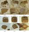
Fig 1. Gross lesions on S. siderea colonies Examples of S. siderea lesions on colonies tagged in Fort Lauderdale (A and B) and Florida Keys (C and D).
White areas on colonies in Fort Lauderdale indicates bleaching not tissue loss.
Fig 2
Fig 2. Schematic of the experimental design for transmission studies in aquaria.
Symbols with the same color indicate coral fragments were collected from the same colony. ‘H’ refers to an apparent healthy fragment and ‘D’ refers to a disease fragment displaying lesions.
Fig 3
Fig 3. Regional differences in disease prevalence through time for tagged colonies of diseased S. siderea.
(A) Prevalence of six colonies in Fort Lauderdale. (B) Prevalence of 17 colonies in the Florida Keys.
Fig 4
Fig 4. Representative photos of tagged S. siderea with lesions at Fort Lauderdale.
C54 (left) shows a bleached lesion that progressed to partial tissue loss. C56 (right) shows a bleached lesion that resolved through time.
Fig 5
Fig 5. Representative photos of tagged S. siderea with lesions in the Florida Keys.
C68 (far left panel) shows a colony with initial multi-focal bleached patches with some tissue loss (Nov 2019) that shows lesion resolution (no bleached spots) but with partial colony mortality (Dec 2020). C80 (middle panel) shows two colonies with initial tissue loss lesions and purple discoloration (Nov 2019) with both colonies progressing to near complete colony mortality (May 2021). C57 (far right panel) shows colony with initial multi-focal purple discoloration with some tissue loss (Nov 2019) that progresses to near complete mortality (May 2020).
Fig 6
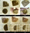
Fig 6. Gross pathological changes of post-fixed S. siderea collected from Florida’s Coral Reef.
(A) tissue loss lesions with no pigmentation. (B) tissue loss with bleached lesions. (C) tissue loss with purple discoloration. (D) purple discoloration. All scale bars = 2 mm.
Fig 7
Fig 7. Histopathology of S. siderea collected from Florida’s Coral Reef.
(A) Radial section across the oral cavity showing lytic necrosis lesions (black arrows) and sloughing of necrotic tissue into the oral cavity (red arrows) (H&E; scale bar = 500 µm). (B) Representative sagittal section along the septa showing border (white line) between surface bleaching (blue arrows) with reduced density of, loss of, or in situ necrosis of endosymbiotic Symbiodiniaceae with abnormal appearance (white arrows), and apparently healthy tissue (red arrows) with endosymbionts (black arrows). Note abnormal appearance of remnant endosymbionts (white hollow arrows) and general loss of cytoplasm or increased mucus (white space) in lesioned area (H&E; scale bar = 50 µm). Inset shown with enlarged abnormal endosymbiont (scale bar = 20 µm). (C) Periodic acid-Schiff stain (PAS) Sagittal section at the gastrodermal layer close to the surface area showing endosymbionts filled with PAS-positive (pink, red) starch granules (black arrows) (PAS-MY: scale bar = 5 µm). (D) Sagittal section of the mesenterial filament close to the surface area showing prominent coral-acid rich protein (CARP) granules (black arrow) in the calicodermal layer, possibly reacting to adjacent endoliths (red arrow) in the skeleton. Compare with apparent (subjective) thinner layer or presumptive lower density of CARPs (blue arrow) on the other side of the mesentery with less endoliths (H&E; scale bar = 500 µm).
Fig 8
Fig 8. Representative photos of disease progression in coral fragments used in transmission experiments using S. siderea.
A-C and G-I are diseased S. siderea and D-F and J-L are test fragments that developed lesions. Left panels shows the fragments at the start of the experiment. Middle panels show fragments on the date that contact was removed between healthy test fragments and the diseased S. siderea. Right panels show fragments on termination of the experiment. A-F shows transmission that resulted in bleaching. G-L shows transmission that resulted in progressive tissue loss.
Fig 9
Fig 9. Representative photos of disease progression in coral fragments used in transmission experiments using S. siderea and O. faveolata.
A-C and G-I are diseased S. siderea and D-F and J-L are test O. faveolata fragments that developed lesions. Left panels shows the fragments at the start of the experiment. Middle panels show fragments on the date that contact was removed between healthy test fragments and the diseased S. siderea. Right panels show fragments on termination of the experiment. A-F shows transmission from diseased S. siderea with a bleached lesion. G-L shows transmission from S. siderea with a lesion with purple pigmentation.
Fig 10
Fig 10. Response of S. siderea with lesions to treatment with antibiotics.
(n = 18 paired fragments).
Fig 11
Fig 11. Representative photos of the response of diseased S. siderea fragments to antibiotic treatment.
Coral fragments were split in half with one half treated with antibiotics and the other half left untreated as a control. Top row is of fragments at the start of the experiment and bottom row of fragments at the end of the experiment.

References

    1. Eakin CM, Sweatman HPA, Brainard RE. The 2014–2017 global-scale coral bleaching event: insights and impacts. Coral Reefs. 2019;38:539–45.
    1. Ward JR, Lafferty KD. The elusive baseline of marine disease: are diseases in ocean ecosystems increasing?. PLoS Biol. 2004;2(4):E120. doi: 10.1371/journal.pbio.0020120 - DOI - PMC - PubMed
    1. Vega Thurber RL, Silva D, Speare L, Croquer A, Veglia AJ, Alvarez-Filip L, et al. Coral Disease: Direct and Indirect Agents, Mechanisms of Disease, and Innovations for Increasing Resistance and Resilience. Ann Rev Mar Sci. 2025;17(1):227–55. doi: 10.1146/annurev-marine-011123-102337 - DOI - PubMed
    1. Precht WF, Gintert BE, Robbart ML, Fura R, van Woesik R. Unprecedented Disease-Related Coral Mortality in Southeastern Florida. Sci Rep. 2016;6:31374. doi: 10.1038/srep31374 - DOI - PMC - PubMed
    1. Papke E, Carreiro A, Dennison C, Deutsch JM, Isma LM, Meiling SS, et al. Stony coral tissue loss disease: a review of emergence, impacts, etiology, diagnostics, and intervention. Front Mar Sci. 2024;10:1321271.

LinkOut - more resources