. 2025 Aug 8;11(32):eadw0126.
doi: 10.1126/sciadv.adw0126. Epub 2025 Aug 6.

YAP regulates transcriptional programs for layer-specific periosteal expansion during fracture repair

Affiliations

YAP regulates transcriptional programs for layer-specific periosteal expansion during fracture repair

Madhura P Nijsure et al. Sci Adv. .

Abstract

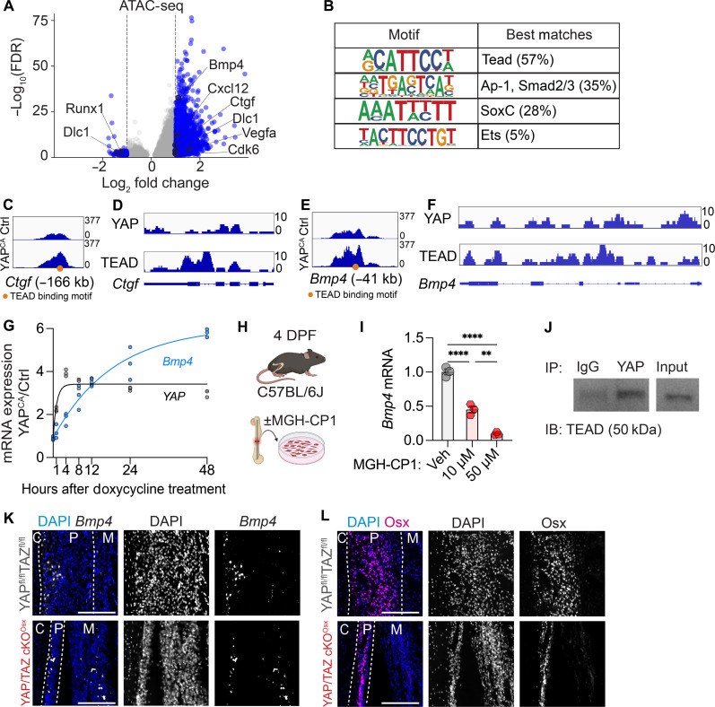
Bone fracture repair initiates by periosteal expansion. The periosteum is a bilayered tissue composed of inner cambium and outer fibrous layers. Typically quiescent, periosteal progenitor cells proliferate upon fracture; however, the underlying transcriptional mechanisms remain unclear. Here, we show that deletion of the transcriptional regulators, yes-associated protein (YAP) and transcriptional coactivator with PDZ binding motif (TAZ), from Osterix-expressing cells, which reside in the cambium, impairs periosteal expansion. YAP activation increases chromatin accessibility, preferentially at TEA domain transcription factor (TEAD) binding sites, and regulates both cell-intrinsic and cell-extrinsic cellular functions. We identify bone morphogenetic protein 4 (Bmp4) as a YAP-TEAD target gene expressed in the cambium. In YAP/TAZ knockout mice, BMP4 delivery increased periosteal expansion through matrix accumulation and fibrous layer cell proliferation. Conversely, in wild-type mice, BMP4 delivery increased osteogenic activity and angiogenesis. Together, these data identify YAP-mediated transcriptional programs that promote layer-specific periosteal expansion.


Figures

Fig. 1.
Fig. 1.. YAP/TAZ deletion from Osx-expressing cells impairs periosteal expansion.
(A) Schematic for evaluation of periosteal expansion at 4 DPF. (B) Timeline for mouse femoral osteotomy, followed by EdU injections at 4 DPF, 3 hours before euthanasia. (C) Staining of nuclei (DAPI), proliferating cells (EdU), and YAP protein. Quantification of (D) YAP+ cells per length of the periosteum, (E) percentage of YAP+ cells in the periosteum, (F) average periosteal thickness, and (G) number of cells per length of the periosteum after Osx-conditional YAP/TAZ deletion. (H) Staining of nuclei (DAPI), proliferating cells (EdU), and Osx protein. Quantification of (I) percentage of EdU+ cells, (J) percentage of proliferative YAP+ cells, (K) Osx+ cells per length of the periosteum, and (L) percentage of Osx+ cells in the periosteum after Osx-conditional YAP/TAZ deletion. All scale bars are 100 μm. C, cortical bone; P, periosteum; M, muscle. Gray bars indicate levels in uninjured bones in WT mice. *P < 0.05 and **P < 0.01.
Fig. 2.
Fig. 2.. Establishment of a gain-of-function model to identify early YAP target genes.
(A) Schematic for femoral osteotomies and periosteal cell isolation followed by doxycycline treatment in vitro. Quantification of mRNA for YAP (B), Cyr61 (C), and Ctgf (D). (E) Immunoblot for YAP, TAZ, and β-actin protein. (F) Immunofluorescence staining for DAPI, YAP, and EdU. (G) Quantification of the fraction of EdU+ cells after 24 hours of YAPCA expression. (H) Schematic for periosteal cell isolation and RNA/protein isolation after 4 and 8 hours of YAPCA expression. Quantification of mRNA of YAP (I), Cyr61 (J), and CTGF (K). (L) Western blot quantification of YAP/TAZ and actin protein levels. Molecular weights are in kDa. (M) Immunofluorescence staining for DAPI, EdU, and YAP 8 hours after YAPCA expression. (N) Percentage of EdU+ cells after 8 hours of YAPCA expression. Scale bars, 10 μm. *P < 0.05, ***P < 0.001, and ****P < 0.0001.
Fig. 3.
Fig. 3.. YAP regulates cell-intrinsic and cell-extrinsic pathways that drive periosteal expansion.
(A) Volcano plot indicating important genes up- or down-regulated after 8 hours of YAPCA expression. Blue dots indicate genes with −0.3 > log2 fold change > 0.3 and P value less than 0.05. RNA-seq, RNA sequencing. (B) Heatmap indicating enrichment scores for 261 selected pathways that are dysregulated by YAPCA that could play a role in periosteal expansion. Individual enrichment scores of (C) cell-intrinsic pathways and (D) cell-extrinsic pathways. *Padj < 0.05. ECM, extracellular matrix.
Fig. 4.
Fig. 4.. YAP/TAZ deletion from Osx-expressing cells affects both cambium and fibrous layers.
(A and C) Schematic demonstrating spatial distribution of cambium (Osx+) and fibrous (Osx) layers in the periosteum. (B and D) Quantification of fluorescence intensity of Osx and Cre:GFP in WTfl/fl and YAP/TAZcKOOsx mice, respectively. GFP, green fluorescent protein. (E) Staining for Osx protein, EdU, and DAPI. Dotted lines indicate the periosteum and the separation between the Osx+ cambium and Osx fibrous layers. (F) Quantification of cambium and fibrous layer thickness. (G) Percentage of proliferative EdU+ cells in each layer. All scale bars are 100 μm. *P < 0.05.
Fig. 5.
Fig. 5.. YAP transcribes BMP4 via TEAD in periosteal cells.
(A) Volcano plot of differentially accessible chromatin sites after 8 hours of YAPCA expression. Log2 fold change > 2 indicates more accessible chromatin after YAPCA expression. (B) Motif analysis on differentially accessible chromatin loci. (C) Chromatin accessibility at locus proximate to Ctgf. (D) Chromatin immunoprecipitation of CTGF by YAP and TEAD in human cells. (E) Chromatin accessibility at locus proximate to Bmp4. (F) Chromatin immunoprecipitation of BMP4 by YAP and TEAD in human cells. (G) Temporal dynamics of Yap and Bmp4 expression after 1, 4, 8, 12, 24, and 48 hours of doxycycline treatment. (H) Schematic for femoral osteotomies and periosteal cell isolation followed by MGH-CP1 treatment in vitro. (I) Bmp4 mRNA expression after 24 hours of treatment with 10 µm or 50 µm of MGH-CP1 in vitro. (J) Coimmunoprecipitation of TEAD with YAP in fracture-activated periosteal cells. IP, immunoprecipitation; IB, immunoblotting. (K) RNA in situ hybridization for Bmp4 in the periosteum of WTfl/fl and YAP/TAZcKOOsx mice. (L) Staining for Osx protein and DAPI in adjacent sections to sections in (K). Dotted lines indicate the periosteum. Scale bars, 100 μm. **P < 0.01 and ****P < 0.0001.
Fig. 6.
Fig. 6.. In vivo delivery of BMP4 increases the expansion of the fibrous layer in YAP/TAZ cKOOsx mice.
(A) Timeline of in vivo recombinant mouse BMP4 (rmBMP4) delivery for 4 days after fracture in YAP/TAZ cKOOsx mice. (B) Immunofluorescence staining of Osx protein with DAPI and EdU. Dotted lines indicate the periosteum and the separation between the Osx+ cambium and Osx fibrous layers. (C) Average periosteal thickness. (D) Percentage of proliferative EdU+ cells in the periosteum. (E) Quantification of cambium and fibrous layer thickness. (F) Percentage of proliferative EdU+ cells in each layer. Scale bars, 100 μm. *P < 0.05, **P < 0.01.
Fig. 7.
Fig. 7.. BMP4 delivery induces collagen matrix production in YAP/TAZ cKOOsx mice.
(A) ALP activity in the periosteum of YAP/TAZ cKOOsx mice treated with saline or BMP4 at 4 DPF. (B and C) Quantification of ALP intensity (B) and ALP+ area fraction (C). (D) Blood vessels marked by endomucin (Emcn) staining. (E) Frequency distribution of blood vessel loop size. (F) SHG imaging of collagen matrix content and organization. (G) Quantification of SHG signal. Scale bars, 100 μm. *P < 0.05.
Fig. 8.
Fig. 8.. In vivo delivery of BMP4 does not affect periosteal expansion in WT YAPfl/flTAZfl/fl mice.
(A) Timeline of in vivo recombinant mouse BMP4 delivery for 4 days after fracture in WT YAP/TAZfl/fl mice. (B) Immunofluorescence staining of Osx protein with DAPI and EdU. Dotted lines indicate the periosteum and the separation between the Osx+ cambium and Osx fibrous layers. (C) Average periosteal thickness. (D) Percentage of proliferative EdU+ cells in the periosteum. (E) Quantification of cambium and fibrous layer thickness. (F) Percentage of proliferative EdU+ cells in each layer. Scale bars, 100 μm.
Fig. 9.
Fig. 9.. BMP4 delivery increases osteogenesis and angiogenesis in WT YAPfl/flTAZfl/fl mice.
(A) ALP activity in the periosteum of YAPfl/flTAZfl/fl mice treated with saline or BMP4 at 4 DPF. (B and C) Quantification of ALP intensity (B) and ALP+ area fraction (C). (D) Blood vessels marked by endomucin staining. (E) Frequency distribution of blood vessel loop size. K-S, Kolmogorov-Smirnov. (F) SHG imaging of collagen matrix content and organization. (G) Quantification of SHG signal. Scale bars, 100 μm. *P < 0.05, **P < 0.01.

Update of

References

    1. Duhamel du Monceau H. L., Stack T. IV, Observations and experiments with madder-root, which has the faculty of tinging the bones of living animals of a red colour, by M. Du Hamel du Monceau, F.R.S. & c. communicated in a letter to Sir Hans Sloane, Bart. Pr. R.S. Translated from the French by T.S. M.D.F.R.S. Phil. Trans. R. Soc. 41, 390–406 (1740).
    1. Utvag S. E., Grundnes O., Reikeraos O., Effects of periosteal stripping on healing of segmental fractures in rats. J. Orthop. Trauma 10, 279–284 (1996). - PubMed
    1. Nakashima K., Zhou X., Kunkel G., Zhang Z., Deng J. M., Behringer R. R., de Crombrugghe B., The novel zinc finger-containing transcription factor osterix is required for osteoblast differentiation and bone formation. Cell 108, 17–29 (2002). - PubMed
    1. Kaback L. A., Soung D. Y., Naik A., Smith N., Schwarz E. M., O'Keefe R. J., Drissi H., Osterix/Sp7 regulates mesenchymal stem cell mediated endochondral ossification. J. Cell. Physiol. 214, 173–182 (2008). - PubMed
    1. Hixon K. R., McKenzie J. A., Sykes D. A. W., Yoneda S., Hensley A., Buettmann E. G., Zheng H., Skouteris D., McAlinden A., Miller A. N., Silva M. J., Ablation of proliferating osteoblast lineage cells after fracture leads to atrophic nonunion in a mouse model. J. Bone Miner. Res. 36, 2243–2257 (2021). - PMC - PubMed

MeSH terms