. 2025 Aug;599(16):2301-2317.
doi: 10.1002/1873-3468.70129. Epub 2025 Aug 6.

The cytochrome oxidase defect in ISC-depleted yeast is caused by impaired iron-sulfur cluster maturation of the mitoribosome assembly factor Rsm22

Affiliations

The cytochrome oxidase defect in ISC-depleted yeast is caused by impaired iron-sulfur cluster maturation of the mitoribosome assembly factor Rsm22

Ulrich Mühlenhoff et al. FEBS Lett. 2025 Aug.

Abstract

Mitochondria contain the bacteria-inherited iron-sulfur cluster assembly (ISC) machinery to generate cellular iron-sulfur (Fe/S) proteins. Mutations in human ISC genes cause severe disorders with a broad clinical spectrum and are associated with strong defects in mitochondrial Fe/S proteins, including respiratory complexes I-III. For unknown reasons, complex IV (aka cytochrome c oxidase), a non-Fe/S, heme-containing enzyme, is severely affected. Using yeast as a model, we show that depletion of Rsm22, the counterpart of the human mitoribosome assembly factor METTL17, phenocopies the defects observed upon impairing late-acting ISC proteins, that is, diminished activities of mitoribosomal translation and respiratory complexes III and IV. Rsm22 binds Fe/S clusters in vivo, thereby satisfactorily explaining the defect of respiratory complex IV in ISC-deficient cells, because this complex contains three mitochondrial DNA-encoded subunits. Impact statement Defects in mitochondrial Fe/S protein biogenesis also impact respiratory complex IV (COX), even though it lacks Fe/S clusters. Here, we show that the mitoribosome assembly factor Rsm22 binds Fe/S clusters in vivo. Rsm22 maturation defects impair mitoribosomal protein translation including COX subunits, explaining the COX defects in Fe/S cluster-deficient cells.

Keywords: biogenesis; cytochromes; iron–sulfur protein; mitochondrial DNA; mitochondrial ribosomes; respiratory chain complexes; translation.


Figures

Fig. 1
Fig. 1
Rapid loss of cytochrome oxidase activity upon depletion of Isa1 and Isa2 in S. cerevisiae. Wild‐type (WT), Gal‐ISA1 and Gal‐ISA12 cells (W303‐1A background) were initially cultivated in YPGal medium, and then inoculated at OD600nm = 0.1 in rich YPD medium. After 4, 16, 40, and 64 h, enzyme activities of aconitase (A) and complex IV (COX) (B) were determined in cell extracts. At 16‐h and 40‐h time points, cells were transferred into fresh YPD medium. Date are presented relative to the activity of malate dehydrogenase (MDH). Dotted lines indicate the SD (n ≥ 4).
Fig. 2
Fig. 2
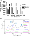
Impaired COX activity upon depletion of Isa1 is not caused by a heme or complex III defect. Mitochondria were isolated from wild‐type (WT), Gal‐ISA1 (ISA1↓; depleted for 40 h), cyt2Δ, and rip1Δ cells (the latter two being deletion mutants with a defective respiratory complex III) after cultivation in YPD medium. Mitochondria were analyzed for (A) enzyme activities of respiratory complexes II (SDH (DCPIP)), II and III (SDH (CytC)), and IV (COX) relative to malate dehydrogenase (MDH), or (B) reduced‐minus‐oxidized cytochrome spectra. The peaks of the various cytochrome types are indicated. Depleted Gal‐YFH1 cells (frataxin) served as a control. The individual spectra were offset for better visualization. (C) Ferrochelatase activities of mitochondria isolated from wild‐type or depleted Gal (↓) yeast strains of the indicated genes were determined by following the fluorescence emission at 588 nm (excitation at 418 nm) associated with the insertion of Zn2+ into protoporphyrin IX. (D) Levels of the indicated proteins from mitochondria of part A were determined by Western blotting. Porin served as a loading control. The apparent molecular masses of marker proteins are indicated in kDa. Error bars indicate the SD (n ≥ 3).
Fig. 3
Fig. 3
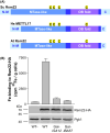
The mitoribosomal assembly factor Rsm22 binds a Fe/S cluster in a late‐acting ISC‐dependent fashion. (A) Cartoon for the domain structure of fungal, vertebrate, and plant members of the Rsm22/METTL17 protein family [73]. Even though the overall sequence similarity is low in eukaryotes, the proteins share a middle methyltransferase‐like (MTase‐like) domain and an oligonucleotide‐binding fold (OB‐fold) within a conserved C‐terminal segment (dark blue). The N‐terminal non‐conserved region contains a mitochondrial targeting sequence (N‐M), while the C termini (blue C) are quite variable. For detailed alignments see Fig. S1A–C. At, A. thaliana; Hs, H. sapiens; Sc, S. cerevisiae. (B) Wild‐type (WT), Gal‐ISA12, and Gal‐IBA57 cells overproducing C‐terminally HA‐tagged Rsm22 from vector p424‐TDH3 were radiolabeled in vivo with 55Fe in iron‐poor SD minimal medium. Wild‐type cells carrying an empty plasmid (WT‐) served as background control. Before the radiolabeling, Gal‐ISA12 and Gal‐IBA57 cells were depleted by cultivation in SD medium for 64 h. Cell extracts were prepared, Rsm22‐HA was immunoprecipitated with α‐HA antibodies, and 55Fe associated with the immunobeads was quantified by scintillation counting. Protein levels of Rsm22‐HA in radiolabeled extracts was assessed by immunostaining with α‐HA antibodies (bottom part). The cytosolic protein Pgk1 served as a loading control. Molecular masses of marker proteins are given in kDa. Error bars indicate the SD (n ≥ 6).
Fig. 4
Fig. 4
Depleted Gal‐RSM22, Gal‐MRPS5, and Gal‐ISA12 cells retain mtDNA. The indicated yeast strains were cultivated for 40 h in SD minimal medium, in order to deplete the respective Gal‐strains to critical levels (Fig. 1; Fig. S2). Cells were cultivated to mid‐log phase in SD medium, stained with DAPI, and analyzed in a DMI6000B fluorescence microscope at 100‐fold magnification (scale bars are 10 μm). W303‐1A and W303‐1A rho0 cells served as controls. Due to different staining efficiencies, the exposure times for the different strains vary slightly.
Fig. 5
Fig. 5
Rsm22, Isa1, and Isa2 are required for mitochondrial translation. (A) Top: Wild‐type (WT), Gal‐RSM22, Gal‐MRPS5, isa12Δ, and Gal‐ISA12 cells were subjected to 35S‐Met/Cys radiolabeling in the presence of the cytosolic protein synthesis inhibitor cycloheximide. Before the radiolabeling, respective proteins were depleted in Gal strains for 40 h in SD minimal medium. Cells were washed and extracts subjected to SDS/PAGE and blotting to a nitrocellulose membrane. A representative autoradiogram of the 35S‐labeled translation products is shown. Strain isa12Δ is devoid of mtDNA (rho0 cells) and thus served as a control for specific labeling of mtDNA‐derived translation products. Bottom: A protein stain with fast green dye (FCF) served as a loading control. (B) The indicated cells were cultivated in YPD medium for 40 h, and mitochondria were isolated. Levels of the indicated proteins (mt, mtDNA‐encoded) were determined by Western blotting. A non‐specific cross‐reactive protein band from Cox1 antibody staining served as the loading control. The apparent molecular masses of marker proteins are indicated in kDa.
Fig. 6
Fig. 6
Cells depleted of Rsm22 lack complex III and IV activities but retain heme. (A) Mitochondria were isolated from wild‐type (WT) and depleted (↓) Gal‐RSM22 or Gal‐MRPS5 cells. Enzyme activities were measured for respiratory complex II (SDH (DCPIP)), complex II and III (SDH/CytC), and complex IV (COX) relative to the activity of malate dehydrogenase (MDH). Aconitase (Aco) activity served as a control. Error bars indicate the SD (n ≥ 4). (B) Reduced‐minus‐oxidized cytochrome spectra were recorded using 1 mg mitochondrial protein from the indicated (depleted) strains. The absorption peaks of the various cytochrome types are indicated. The individual spectra were offset for better visualization.
Fig. 7
Fig. 7
Model for the deficiency of cytochrome oxidase in ISC‐depleted mitochondria. The mitochondrial ISC machinery (yeast nomenclature) synthesizes [2Fe‐2S] and [4Fe‐4S] clusters in several consecutive steps requiring core ISC proteins, transfer proteins, and late‐acting ISC proteins (for details on the ISC system and nomenclature see Introduction and Ref. [5]). Failure of the late‐acting ISC system leads to selective impairment of [4Fe‐4S] protein assembly such as aconitase (Aco1) or respiratory complex II, while [2Fe‐2S] proteins such as Rieske Fe/S protein of complex III are still generated. As shown in this work, the mtSSU assembly protein Rsm22 holds a [4Fe‐4S] cluster in vivo, like its mammalian counterpart METTL17 [69, 70]. Defective Fe/S maturation of this protein as a consequence of ISC defects (red cross) leads to impaired maturation of Rsm22, and as a consequence, defective mitoribosomal assembly and protein translation (red dotted arrows). This diminishes all mtDNA‐encoded proteins with the consequence of a respiratory defect because functional OxPhos complexes III–V can no longer be assembled. The model explains why defects in mitochondrial Fe/S cluster assembly indirectly, via Rsm22, lead to a complex IV (COX) defect, even though this enzyme does not contain a Fe/S cluster. Depletion of Rsm22 does not affect complex II (SDH) activity with its three Fe/S clusters because the enzyme contains only nucleus‐encoded proteins. Further details see text. IMS, intermembrane space; red and yellow circles, iron and sulfur ions.

Similar articles

References

    1. Brischigliaro M, Sierra‐Magro A, Ahn A and Barrientos A (2024) Mitochondrial ribosome biogenesis and redox sensing. FEBS Open Bio 14, 1640–1655. doi: 10.1002/2211-5463.13844 - DOI - PMC - PubMed
    1. Rackham O, Saurer M, Ban N and Filipovska A (2025) Unique architectural features of mammalian mitochondrial protein synthesis. Trends Cell Biol 35, 11–23. doi: 10.1016/j.tcb.2024.05.001 - DOI - PubMed
    1. Ben Zichri‐David S, Shkuri L and Ast T (2025) Pulling back the mitochondria's iron curtain. NPJ Metab Health Dis 3, 6. doi: 10.1038/s44324-024-00045-y - DOI - PMC - PubMed
    1. Dutt S, Hamza I and Bartnikas TB (2022) Molecular mechanisms of iron and heme metabolism. Annu Rev Nutr 42, 311–335. doi: 10.1146/annurev-nutr-062320-112625 - DOI - PMC - PubMed
    1. Lill R and Freibert SA (2020) Mechanisms of mitochondrial iron‐sulfur protein biogenesis. Annu Rev Biochem 89, 471–499. doi: 10.1146/annurev-biochem-013118-111540 - DOI - PubMed

MeSH terms

LinkOut - more resources