. 2025 Aug 22;12(8):ENEURO.0204-25.2025.
doi: 10.1523/ENEURO.0204-25.2025. Print 2025 Aug.

Oxytocin Receptor Expression and Activation in Parasympathetic Brainstem Cardiac Vagal Neurons

Affiliations

Oxytocin Receptor Expression and Activation in Parasympathetic Brainstem Cardiac Vagal Neurons

Xin Wang et al. eNeuro. .

Abstract

Autonomic imbalance-particularly reduced activity from brainstem parasympathetic cardiac vagal neurons (CVNs)-is a major characteristic of many cardiorespiratory diseases. Therapeutic approaches to selectively enhance CVN activity have been limited by the lack of defined, translationally relevant targets. Previous studies have identified an important excitatory synaptic pathway from oxytocin (OXT) neurons in the paraventricular nucleus of the hypothalamus to brainstem CVNs, suggesting that OXT could provide a key selective excitation of CVNs. In clinical studies, intranasal OXT has been shown to increase parasympathetic cardiac activity, improve autonomic balance, and reduce obstructive event durations and oxygen desaturations in obstructive sleep apnea patients. However, the mechanisms by which activation of hypothalamic OXT neurons, or intranasal OXT, enhance brainstem parasympathetic cardiac activity remain unclear. CVNs are located in two cholinergic brainstem nuclei: nucleus ambiguus (NA) and dorsal motor nucleus of the vagus (DMNX). In this study, we characterize the colocalization of OXT receptors (OXTRs) in both CVNs and non-CVN cholinergic neurons in the male and female mouse NA and DMNX nuclei. We found that OXT receptors are highly expressed in CVNs in the DMNX, but not in the NA. OXT increases the firing of DMNX CVN, with no effect on NA CVNs. Selective chemogenetic excitation of OXTR+ CVNs in the DMNX-achieved by a combination of Cre- and flp-dependent DREADD expression-evoked a rapid and sustained bradycardia. These findings suggest that activation of DMNX CVNs expressing OXTR with oxytocin may represent a novel translational therapeutic target for restoring autonomic balance in cardiorespiratory disorders.

Keywords: DMNX and NA; DREADDs; autonomic regulation; cardiac vagal neurons; chemogenetic; oxytocin receptors.


Conflict of interest statement

The authors declare no competing financial interests.

Figures
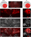
Figure 1.
Figure 1.
Percentage of CVNs-ChAT neurons in the DMNX and NA in OXTR-Cre- ChR2-eYFP male and female mice. Top middle, An example confocal image showing overview of brainstem containing CVNs (red) in the DMNX and NA. The DMNX data presented on the left and NA on the right panel. The top two Venn diagrams showing CVNs account for the percentage population of the cholinergic neurons in the DMNX (left) and NA (right). Example confocal images showing the CVNs in red (top), ChAT in gray (middle), and merged CVNs and ChAT neurons light red (bottom) in the DMNX and NA, respectively. Scale bar, 50 µm. For this and all the following figures: 4V, the 4th ventricle; XII MNs, hypoglossal motor neurons; CVNs, cardiac vagal neurons; ChAT, cholinergic neurons.
Figure 2.
Figure 2.
Colocalization of OXTR+ neurons with CVNs and non-CVN ChAT cells in the DMNX. Top right, An example of a low-resolution confocal image showing overview of brainstem dorsal side DMNX area containing CVNs (red), ChAT (gray), and OXTR+ (green). Top left two Venn diagrams showing the percentage of CVNs and non-CVN-ChAT neurons containing OXTR, respectively. The middle panel represents CVNs colocalization with OXTR+ neurons: the confocal images mark CVNs in red (left), OXTR+ in green (middle), and CVNs merged with OXTR+ labeling (right); scale bar, 20 µm. The bottom panel illustrates the OXTR+ neurons colocalized with ChAT neurons that are not CVNs. Typical confocal images illustrate ChAT neurons in gray (left), OXTR+ neurons in green (middle), and merged OXTR+ and ChAT neurons (right); scale bar, 10 µm. OXTR, oxytocin receptor.
Figure 3.
Figure 3.
The OXTR+ neurons colocalization with CVNs and non-CVN ChAT cells in the NA. A: The low-resolution confocal image showing overview of a brainstem containing CVNs (red), ChAT (gray), and OXTR+ (green) in the NA. The middle panel reveals the percentage of CVNs containing OXTR+ (Venn diagram on the left), confocal images represent CVNs in red (2nd left), OXTR+ neurons in green (2nd from right), and colocalized OXTR+ neuron with CVNs (right). The bottom panel denotes the percentage of non-CVN ChAT neurons containing OXTR+ (Venn diagram on the left), confocal images showing ChAT in gray (2nd left), OXTR+ neurons in green (2nd from right), and colocalized OXTR+ neuron with non-CVN ChAT neurons (right). Scale bar, 50 µm.
Figure 4.
Figure 4.
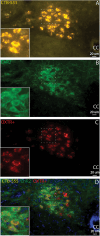
ChR2-eGFP-expressing neurons are correlated with active OXTR mRNA transcription in adult crossbred OXTR+ Cre × floxed ChR2-eGFP animals. A, CTB-555 labeled CVNs (gold); B, ChR2-eGFP-labeled OXTR+ neurons and fibers (green); C, OXTR mRNA+ cells (red); and D, all of OXTR+-Cre ChR2-positive neurons (green) colocalized with the OXTR mRNA probe (red). Insert, focus on the dash line marked area. cc, central canal.
Figure 5.
Figure 5.
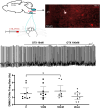
Administration of OXT enhanced CVNs action potential in the brainstem DMNX containing slice. Top left, Diagram illustrating procedure of retrograde labeling CVNs in the brainstem; top right, an example confocal image presenting CTB-555-labeled CVNS (red) and patched CVN neuron (red cell filled with biocytin in white); middle, typical spike trace showing OXT increased CVN neuron action potential firing rate; bottom bar graph presenting original data with 95% CI (n = 9 neurons from 7 animals). Data analyzed using RM one-way ANOVA with Dunnett's post hoc. *p < 0.05.
Figure 6.
Figure 6.
Chemogenetic activation of OXTR+ CVNs increases parasympathetic activity to the heart. Upper left diagram illustrating procedure of two-step viral vector injection to specifically identify and activate CVNs that project to the heart; top right, an example confocal image showing cells with mCherry expression in the brainstem slice (red); bottom left diagram illustrating plethysmography and ECG recording and bottom right presenting original data and statistic results. Two-way repeated-measures ANOVA with Tukey's post hoc **p < 0.001, ***p < 0.0001.

Update of

References

    1. Charpak S, Armstrong WE, Muhlethaler M, Dreifuss JJ (1984) Stimulatory action of oxytocin on neurones of the dorsal motor nucleus of the vagus nerve. Brain Res 300:83–89. 10.1016/0006-8993(84)91342-8 - DOI - PubMed
    1. Cheng Z, Powley TL, Schwaber JS, Doyle FJ 3rd (1999) Projections of the dorsal motor nucleus of the vagus to cardiac ganglia of rat atria: an anterograde tracing study. J Comp Neurol 410:320–341. 10.1002/(SICI)1096-9861(19990726)410:2<320::AID-CNE12>3.0.CO;2-5 - DOI - PubMed
    1. Cheng Z, Zhang H, Guo SZ, Wurster R, Gozal D (2004) Differential control over postganglionic neurons in rat cardiac ganglia by NA and DmnX neurons: anatomical evidence. Am J Physiol Regul Integr Comp Physiol 286:R625–R633. 10.1152/ajpregu.00143.2003 - DOI - PubMed
    1. Cheshire WP (2013) Highlights in clinical autonomic neurosciences: sudden unexpected death in epilepsy. Auton Neurosci 179:5–8. 10.1016/j.autneu.2013.07.005 - DOI - PubMed
    1. Coverdell TC, Abraham-Fan RJ, Wu C, Abbott SBG, Campbell JN (2022) Genetic encoding of an esophageal motor circuit. Cell Rep 39:110962. 10.1016/j.celrep.2022.110962 - DOI - PMC - PubMed

LinkOut - more resources