. 2025 Aug 6;11(1):109.
doi: 10.1038/s41537-025-00654-6.

Temporal characteristics of hemodynamic responses during active and passive hand movements in schizophrenia spectrum disorder

Affiliations

Temporal characteristics of hemodynamic responses during active and passive hand movements in schizophrenia spectrum disorder

Harun A Rashid et al. Schizophrenia (Heidelb). .

Abstract

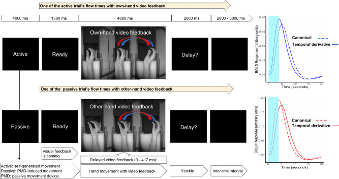
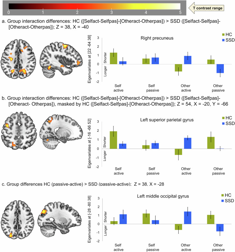
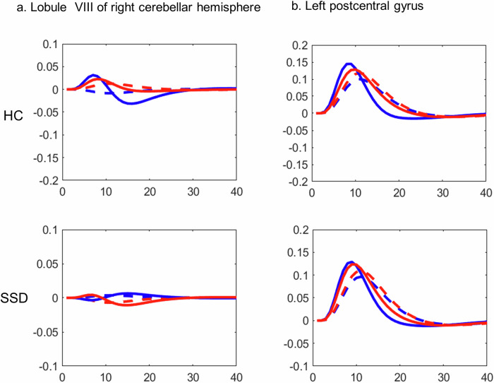
In healthy individuals, active hand-movements typically elicit earlier neural processing than passive one, reflected by more positive contrast estimates of the first-order temporal derivative (TD) of hemodynamic response function (HRF) in functional MRI (fMRI) analyses. This temporal advantage might be due to prior movement-awareness and predictive mechanisms that support self-other distinction. However, it is unknown whether impaired predictive mechanisms in Schizophrenia Spectrum Disorder (SSD) influence earlier neural processing. Patients with SSD (n = 20) and healthy controls (HC; n = 20) performed active and passive hand movements, while detected delays in video feedback of their own or another person's hand. The recorded fMRI data were analysed applying TD to examine timing and second-order dispersion derivative (DD) to evaluate duration of neural responses. Compared to HC, patients with SSD exhibited delayed BOLD responses during active vs. passive movements in the right caudate nucleus, lobule VIII of right cerebellar hemisphere, left superior temporal gyrus, left postcentral gyrus, left thalamus, and left putamen/insula. Furthermore, during active movement with own hand feedback, patients with SSD showed delayed activation in the bilateral putamen and insula. Delayed insula/putamen responses' were associated with symptom severity. However, these exploratory findings remain not significant after correction for multiple comparisons and attenuated with Spearman's-rank correlations. Delayed BOLD responses in patients with SSD, particularly in the right cerebellar lobule VIII, left thalamus, and bilateral insula/putamen may contribute to disturbances in the sense of agency. Altered timing/duration of neural responses reflects new insight underlying deficits in predictive and feedback-monitoring mechanisms in SSD.


Conflict of interest statement

Competing interests: The authors declare no competing interests.

Figures

Fig. 1
Fig. 1. Experimental design.
A video demonstration of the hand movement with feedback is available at 10.5281/zenodo.2621302. Each run (48 trials), had two blocks, the block for active movement were beginning with a cue “Active”, and for passive movement with a cue “Passive”. In the beginning of each run participants were instructed to perform hand movement by themselves when cued “Active” (24 trials), or to relax the hand but keep holding the PMD handle when cued “Passive” (24 trials) and let their hand be moved by the PMD. Each trial commenced with a “Ready” cue, followed by visual feedback for movement planning and execution, culminating with a question “Delay?”. The black screen with variable duration (2000 - 5000 ms) is shown during inter trial interval. In the beginning, the hand video feedback was mostly static (1st hand) since the subject was instructed to move when ready to perform the movement. The movement onset from the left (2nd hand) moves to the right (3rd hand), then the hand moves it back to the left (from 3rd hand to the 2nd hand) position. In the video feedback, only the right-hand movement was displayed, here 3 different hand positions are shown just to visualize the whole process of planning and execution direction, separately. Considering male participants for this figure, the upper row shows the sequence of a trial with active and own-hand video feedback, while the lower row shows another trial with the passive and other hand (pre-recorded image from the female). In the case of a female subject, the other hand video is shown from a male hand (pre-recorded image). Self-other hand was displayed randomly across the trial and run. On the right, the expected active and passive movement specific canonical hemodynamic response function and temporal derivative of BOLD responses are shown (the 4000 ms period of hand movement with feedback stimulus marked with light blue).
Fig. 2
Fig. 2. Timing of neural activation in active compared to passive condition.
a Earlier activation in healthy control (HC) shown at Z = 0, Y = 0, X = 14; (b) earlier activation in schizophrenia spectrum disorder (SSD) shown at Z = 0, Y = 0, X = 14; (c). common earlier activated brain areas between HC and SSD at Z = 60, Y = 0, X = 26, (d) earlier activation specific to HC compared to SSD shown at Z = 8, X = 10; (e) interaction related earlier activation in HC compared SSD patients shown at Z = 14, Y = −18, X = −28; f active with own hand feedback specific earlier activated brain area in HC compared to SSD patients shown at Z = 10, X = −36. Positive eigenvariates reflect earlier activation while negative reflects later activation timing in the bar graph. HC healthy control, (n = 20); SSD schizophrenia spectrum disorder, (n = 20).
Fig. 3
Fig. 3. Duration of neural activation in active compared to passive and vice versa.
a Group interaction: group (HC > SSD) X movement (active>passive) X feedback (own>other) hand shown at Z = 38, X = −40. b Group interaction masked by HC X movement (active>passive) X feedback (own>other) shown at Z = 54, X = −20. c Group differences between passive and active movement regardless of feedback condition are shown at Z = 38, X = −28. Positive eigenvariates reflect shorter activation durations, while negative ones reflect longer activation durations in the bar graph. HC healthy control, (n = 20); SSD schizophrenia spectrum disorder, (n = 20).
Fig. 4
Fig. 4. Latency functions (LF), from TD and from the canonical hemodynamic response function (HRF) in healthy control (upper row) and in schizophrenia spectrum disorder (SSD) in the lower row.
a LF at the lobule VIII of the right cerebellar hemisphere, and (b) LF at the left post central gyrus. Active: canonical (dashed blue), HRF with temporal derivative (solid blue); Passive: canonical (dashed red), HRF with temporal derivative (solid red). HC healthy control, (n = 20); SSD schizophrenia spectrum disorder, (n = 20).

Similar articles

References

    1. Yao, B. et al. Structural thalamofrontal hypoconnectivity is related to oculomotor corollary discharge dysfunction in schizophrenia. J. Neurosci.39, 2102–2113 (2019). - PMC - PubMed
    1. Voss, M. et al. Altered awareness of action in schizophrenia: a specific deficit in predicting action consequences. Brain133, 3104–3112 (2010). - PubMed
    1. Uhlmann, L., Pazen, M., Van Kemenade, B. M., Kircher, T. & Straube, B. Neural correlates of self-other distinction in patients with schizophrenia spectrum disorders: the roles of agency and hand identity. Schizophr. Bull.47, 1399–1408 (2021). - PMC - PubMed
    1. Thakkar, K. N. & Rolfs, M. Disrupted corollary discharge in schizophrenia: evidence from the oculomotor system. Biol. Psychiatry Cogn. Neurosci. Neuroimaging4, 773–781 (2019). - PMC - PubMed
    1. Spering, M., Dias, E. C., Sanchez, J. L., Schutz, A. C. & Javitt, D. C. Efference copy failure during smooth pursuit eye movements in schizophrenia. J. Neurosci.33, 11779–11787 (2013). - PMC - PubMed

LinkOut - more resources