

Diffusion tensor imaging reveals myocardial architectural differences between porcine and primate hearts with potential implications for cardiac xenotransplantation
- PMID: 40770390
- PMCID: PMC12329012
- DOI: 10.1038/s41598-025-14368-3
Diffusion tensor imaging reveals myocardial architectural differences between porcine and primate hearts with potential implications for cardiac xenotransplantation
Abstract
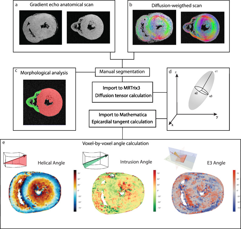
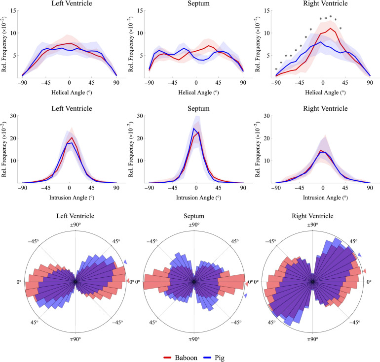
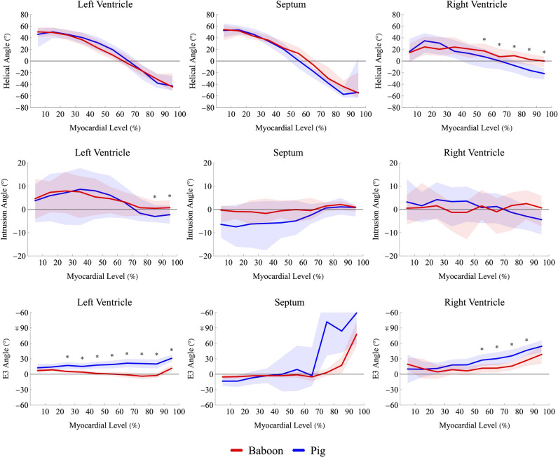
Although pig-to-baboon cardiac xenotransplantation has become increasingly successful, challenges remain in its clinical translation, particularly in addressing xenograft overgrowth. While several causes can be managed through genetic modifications and hemodynamic control, growth persists. Research on myocardial architecture and interspecies variation is limited. This study employs diffusion tensor imaging to probe the orientation of cardiomyocytes and their aggregations and aims to investigate whether these parameters may act as intrinsic factors, contributing to xenograft overgrowth in a setting of extrinsic hemodynamic mismatch. Five pig and five baboon ex-vivo hearts were compared by cardiac diffusion tensor magnetic resonance imaging. Myocardial architecture was assessed by quantifying helical, intrusion and E3-angles in left ventricle, septum and right ventricle. Notable differences were found in E3-angles of the left ventricle. The E3 angle was closer to 0° throughout the baboon myocardium. The epicardial E3-angle differed by -9°, midwall by -17.1°, and endocardial by -23.7° The myocardial architecture observed in baboon hearts may support a greater contractional deformation, potentially reflecting an enhanced contractile potential as compared to the porcine heart. Further ex- and in-vivo investigation of both pre- and post-transplantation animals is warranted to assess the exact functional implications of these myocardial architecture differences.
© 2025. The Author(s).
Conflict of interest statement
Declaration. Competing interests: The authors declare no competing interests.
Figures

Similar articles
-
Quantitative Proteomic Analysis of Cardiac Xenograft Failure in a Pig-to-Non-Human Primate Model Identifies NF-κB as a Critical Immunomodulatory Target.Xenotransplantation. 2025 May-Jun;32(3):e70040. doi: 10.1111/xen.70040. Xenotransplantation. 2025. PMID: 40375624 Free PMC article.
-
Distinguishing shear and tensile myocardial wall stiffness using ex vivo anisotropic Magnetic Resonance Elastography.Acta Biomater. 2025 Aug;202:276-291. doi: 10.1016/j.actbio.2025.06.031. Epub 2025 Jun 18. Acta Biomater. 2025. PMID: 40541764
-
[Volume and health outcomes: evidence from systematic reviews and from evaluation of Italian hospital data].Epidemiol Prev. 2013 Mar-Jun;37(2-3 Suppl 2):1-100. Epidemiol Prev. 2013. PMID: 23851286 Italian.
-
Short-Term Memory Impairment.2024 Jun 8. In: StatPearls [Internet]. Treasure Island (FL): StatPearls Publishing; 2025 Jan–. 2024 Jun 8. In: StatPearls [Internet]. Treasure Island (FL): StatPearls Publishing; 2025 Jan–. PMID: 31424720 Free Books & Documents.
-
The Black Book of Psychotropic Dosing and Monitoring.Psychopharmacol Bull. 2024 Jul 8;54(3):8-59. Psychopharmacol Bull. 2024. PMID: 38993656 Free PMC article. Review.
References
-
- Swindle, M. & Smith, A. C. Comparative anatomy and physiology of the pig. Scandinavian J. Lab. Anim. Sci.25(11), 21 (1998).
-
- Jensen, B. et al. Trabeculations of the porcine and human cardiac ventricles are different in number but similar in total volume. Clin. Anat.37(4), 440–454. 10.1002/ca.24135 (2024). - PubMed
-
- Ibrahim, Z. et al. Selected physiologic compatibilities and incompatibilities between human and porcine organ systems. Xenotransplantation13(6), 488–499. 10.1111/j.1399-3089.2006.00346.x (2006). - PubMed
MeSH terms
Grants and funding
LinkOut - more resources
Full Text Sources
Medical
