

Fibrinogen contributes to myelin deficit and cognitive impairment in aged mice after anesthesia and surgery
- PMID: 40770921
- PMCID: PMC12331660
- DOI: 10.1177/0271678X251338953
Fibrinogen contributes to myelin deficit and cognitive impairment in aged mice after anesthesia and surgery
Abstract
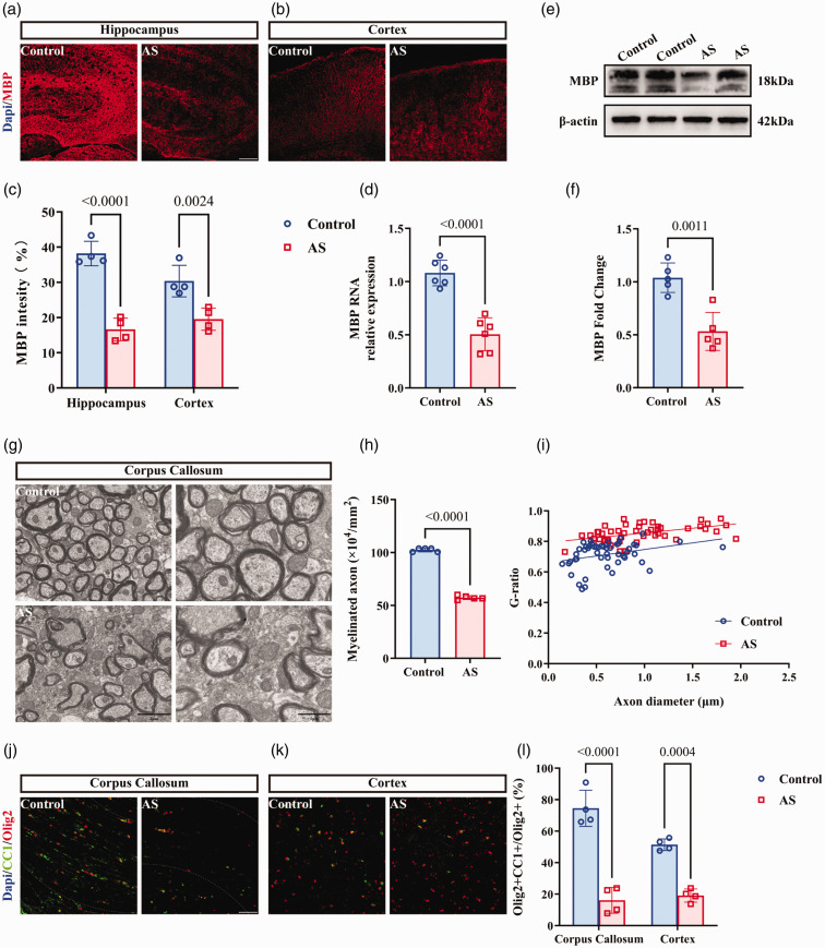
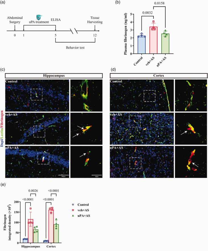
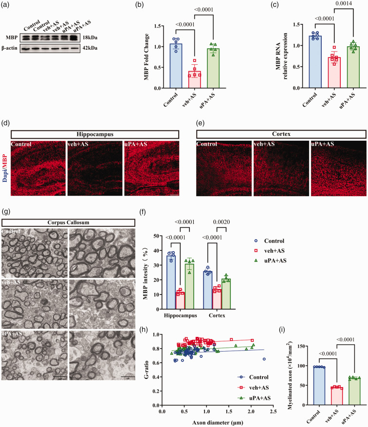
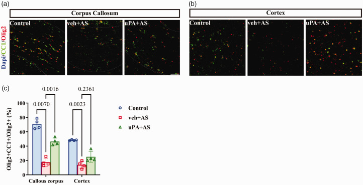
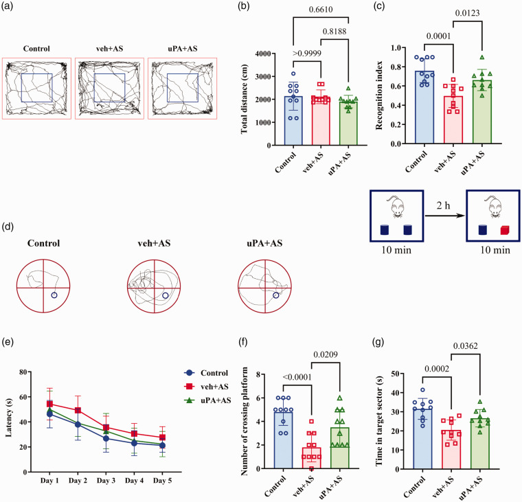
Perioperative neurocognitive disorder (PND) is a common complication of anesthesia and surgery, which is more prevalent in elderly patients. Fibrinogen is known to contribute to the pathophysiology of neurodegenerative disorders. This study investigated whether fibrinogen induces myelin deficit and cognitive impairment in aged mice after anesthesia and surgery. Here, abdominal surgery was performed on 17-month-old C57BL/6 mice to establish a PND model. Following anesthesia and surgery, cognitive function and exploratory locomotion of mice were assessed using behavioral tests. We used in vivo two-photon brain microscopy to track the perivascular accumulation of blood-derived fibrinogen in the central nervous system (CNS). Immunostaining, electron microscopy (EM), and western blotting were used to measure myelin sheath density and oligodendrocyte alterations, and inflammatory markers were determined by reverse transcription-quantitative polymerase chain reaction (RT-qPCR). In the current study, we found that fibrinogen deposited in the CNS after blood-brain barrier (BBB) disruption, induces oligodendrocyte loss, myelin deficits and causes behavioral abnormalities in PND model. Fibrinogen depletion could reverse myelin deficits and cognitive function which induced by anesthesia and surgery. In summary, our data support that fibrinogen is a key determinant in the early pathogenesis of PND.
Keywords: Perioperative neurocognitive disorder; blood-brain barrier; fibrinogen; myelination; oligodendrocyte.
Conflict of interest statement
The author(s) declared no potential conflicts of interest with respect to the research, authorship, and/or publication of this article.
Figures


Similar articles
-
Gamma Oscillation Disruption Induced By Microglial Activation Contributes to Perioperative Neurocognitive Disorders in Aged Mice.J Mol Neurosci. 2025 Aug 2;75(3):97. doi: 10.1007/s12031-025-02380-1. J Mol Neurosci. 2025. PMID: 40753364
-
CCL2 Inhibitor Bindarit Improve Postoperative Cognitive Function by Attenuating Pericyte Loss-Related Blood-Brain Barrier Disruption and Neuroinflammation.Mediators Inflamm. 2025 Jun 12;2025:7248780. doi: 10.1155/mi/7248780. eCollection 2025. Mediators Inflamm. 2025. PMID: 40548298 Free PMC article.
-
Signs and symptoms to determine if a patient presenting in primary care or hospital outpatient settings has COVID-19.Cochrane Database Syst Rev. 2022 May 20;5(5):CD013665. doi: 10.1002/14651858.CD013665.pub3. Cochrane Database Syst Rev. 2022. PMID: 35593186 Free PMC article.
-
Intravenous versus inhalational maintenance of anaesthesia for postoperative cognitive outcomes in elderly people undergoing non-cardiac surgery.Cochrane Database Syst Rev. 2018 Aug 21;8(8):CD012317. doi: 10.1002/14651858.CD012317.pub2. Cochrane Database Syst Rev. 2018. PMID: 30129968 Free PMC article.
-
GABAB receptor activation contributes to post-surgery cognitive impairments in mice by inducing hippocampal BDNF hypermethylation.Ann Med. 2025 Dec;57(1):2536221. doi: 10.1080/07853890.2025.2536221. Epub 2025 Jul 22. Ann Med. 2025. PMID: 40693442 Free PMC article.
References
-
- Luo ALin, Yan J, Tang XLe, et al. Postoperative cognitive dysfunction in the aged: the collision of neuroinflammaging with perioperative neuroinflammation. Inflammopharmacology 2019; 27: 27–37. - PubMed
-
- Punjasawadwong Y, Chau-In W, Laopaiboon M, et al. Processed electroencephalogram and evoked potential techniques for amelioration of postoperative delirium and cognitive dysfunction following non-cardiac and non-neurosurgical procedures in adults. Cochrane Database Syst Rev 2018; 5: Cd011283. - PMC - PubMed
LinkOut - more resources
Full Text Sources
