

Efficacy of Combined Use of PELNAC and Platelet-rich Plasma in Promoting Wound Healing
- PMID: 40771260
- PMCID: PMC12327585
- DOI: 10.1097/GOX.0000000000007062
Efficacy of Combined Use of PELNAC and Platelet-rich Plasma in Promoting Wound Healing
Abstract
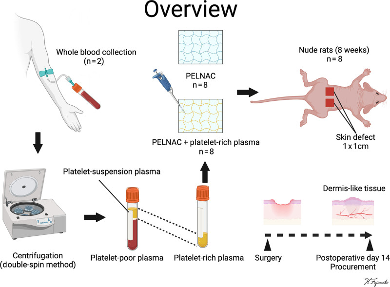
Background: Artificial dermis is useful for treating full-thickness skin defects but requires a long time to form dermis-like granulation tissue. This study investigated whether platelet-rich plasma (PRP) combined with PELNAC, a collagen-based artificial dermis, promotes wound healing and examined the underlying mechanism.
Methods: Two 1-cm² full-thickness skin defects were created in 8-week-old male nude rats (n = 8). One defect received saline-impregnated PELNAC (control group); the other received PRP-impregnated PELNAC (combination group). Cytokine levels in PRP were measured. Wound area measurements and histological and immunohistochemical evaluations (transforming growth factor-β1 [TGF-β1], vascular endothelial growth factor [VEGF], α-smooth muscle actin, and von Willebrand factor) were performed on day 14.
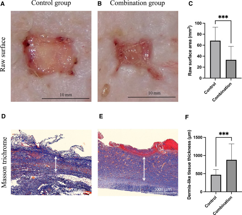
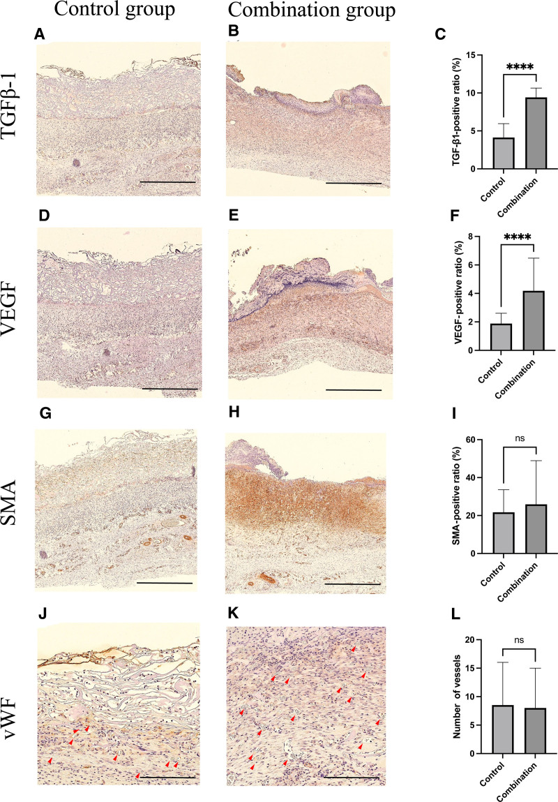
Results: Compared with the control group, the combination group showed a significantly reduced wound area (32.4 ± 14.89 versus 68.3 ± 14.9 mm², P < 0.001) and increased dermis-like granulation tissue thickness (947 ± 219 versus 448 ± 165 μm, P < 0.001). Expression of TGF-β1 (8.9% ± 1.7% versus 4.2% ± 1.2%, P < 0.0001) and VEGF (4.4% ± 1.2% versus 1.9% ± 0.5%, P < 0.0001) was significantly higher in the combination group, whereas α-smooth muscle actin-positive cells and von Willebrand factor-positive blood vessels showed no significant differences.
Conclusions: The combined use of PELNAC and PRP accelerated dermis-like granulation tissue formation through increased expression of VEGF and transforming growth factor-β1, significantly improving wound healing. This combination therapy could be easily applied in clinical practice and may contribute to shortening healing times for deep dermal defects.
Copyright © 2025 The Authors. Published by Wolters Kluwer Health, Inc. on behalf of The American Society of Plastic Surgeons.
Conflict of interest statement
The authors have no financial interest to declare in relation to the content of this article. This study was supported by the GUNZE Foundation (Kyoto, Japan).
Figures
Similar articles
-
[Application study of platelet-rich plasma combined with arterial supercharging technique to enhance survival of ischemic cross-body region skin flaps in rabbits].Zhongguo Xiu Fu Chong Jian Wai Ke Za Zhi. 2025 Jul 15;39(7):873-880. doi: 10.7507/1002-1892.202504080. Zhongguo Xiu Fu Chong Jian Wai Ke Za Zhi. 2025. PMID: 40659592 Free PMC article. Chinese.
-
[Experimental study on promotion of skin radiation damage repair by icarin via HIF-2α/VEGF/Notch pathway to enhance the paracrine function of adipose-derived stem cells].Zhongguo Xiu Fu Chong Jian Wai Ke Za Zhi. 2025 Jul 15;39(7):881-890. doi: 10.7507/1002-1892.202503089. Zhongguo Xiu Fu Chong Jian Wai Ke Za Zhi. 2025. PMID: 40659593 Free PMC article. Chinese.
-
[Influence and mechanism of extracellular vesicles derived from human dermal papilla cells on skin fibrosis in mice].Zhonghua Shao Shang Yu Chuang Mian Xiu Fu Za Zhi. 2025 Jun 20;41(6):559-568. doi: 10.3760/cma.j.cn501225-20240925-00348. Zhonghua Shao Shang Yu Chuang Mian Xiu Fu Za Zhi. 2025. PMID: 40588404 Free PMC article. Chinese.
-
Platelet-rich therapies for musculoskeletal soft tissue injuries.Cochrane Database Syst Rev. 2013 Dec 23;(12):CD010071. doi: 10.1002/14651858.CD010071.pub2. Cochrane Database Syst Rev. 2013. Update in: Cochrane Database Syst Rev. 2014 Apr 29;(4):CD010071. doi: 10.1002/14651858.CD010071.pub3. PMID: 24363098 Updated.
-
Platelet-rich therapies for musculoskeletal soft tissue injuries.Cochrane Database Syst Rev. 2014 Apr 29;2014(4):CD010071. doi: 10.1002/14651858.CD010071.pub3. Cochrane Database Syst Rev. 2014. PMID: 24782334 Free PMC article.
References
-
- Tonnesen MG, Feng X, Clark RAF. Angiogenesis in wound healing. J Investig Dermatol Symp Proc. 2000;5:40–46. - PubMed
-
- Singer AJ, Clark RAF. Cutaneous wound healing. N Engl J Med. 1999;341:738–746. - PubMed
-
- Werner S, Grose R. Regulation of wound healing by growth factors and cytokines. Physiol Rev. 2003;83:835–870. - PubMed
-
- Carmeliet P. Mechanisms of angiogenesis and arteriogenesis. Nat Med. 2000;6:389–395. - PubMed
-
- Lucich EA, Rendon JL, Valerio IL. Advances in addressing full-thickness skin defects: a review of dermal and epidermal substitutes. Regen Med. 2018;13:443–456. - PubMed
LinkOut - more resources
Full Text Sources
Research Materials