Review
. 2025 Jul 23:16:1594118.
doi: 10.3389/fendo.2025.1594118. eCollection 2025.

Phosphomannomutase 2-congenital disorder of glycosylation: exploring the role of N-glycosylation on the endocrine axes

Affiliations
Review

Phosphomannomutase 2-congenital disorder of glycosylation: exploring the role of N-glycosylation on the endocrine axes

Giulia Del Medico et al. Front Endocrinol (Lausanne). .

Abstract

Congenital disorders of glycosylation (CDG) are a heterogeneous group of inborn errors of metabolism caused by impaired protein glycosylation. Among these, PMM2-CDG, caused by defective phosphomannomutase 2 activity and affecting protein N-glycosylation, is the most prevalent. As glycoproteins are involved in almost every physiological process, the clinical manifestations in PMM2-CDG are diverse and multisystemic. In the endocrine system, glycoproteins are present in every axis, acting as hormones, prohormones, receptors, enzymes, and transport proteins. Hypoglycosylation can alter hormonal function on multiple levels. As a result, endocrinopathies are frequently part of the clinical spectrum of PMM2-CDG, particularly hypergonadotrophic hypogonadism and pubertal abnormalities in female patients. Symptoms of endocrine involvement, especially hyperinsulinemic hypoglycemia and failure to thrive during infancy, can be the presenting sign of the disease. The clinical spectrum of PMM2-CDG endocrinopathy is variable; for example, thyroid involvement can range from isolated transitory hyperthyrotropinemia to clinical hypothyroidism. Some endocrine abnormalities, such as adrenal insufficiency, are uncommon and probably underdiagnosed in PMM2-CDG. The new insights into the role of N-glycosylation on the endocrine system over the past twenty years have deepened our understanding of this complex disorder and should enable us to improve and personalize the clinical management of these patients.

Keywords: N-glycosylation; congenital disorder of glycosylation; endocrine dysfunction; hypergonadotropic hypogonadism; hypoglycosylation.


Conflict of interest statement

The authors declare that the research was conducted in the absence of any commercial or financial relationships that could be construed as a potential conflict of interest.

Figures
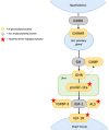
Figure 1
Figure 1
N-glycosylation in the GH-IGF1 axis. The GHRH, produced by the hypothalamus, stimulates its glycosylated receptor, GHRHR, on the anterior pituitary gland to release GH. GH binds to GHR on target tissues, including the liver. GHR contains N-linked glycosylation sites in the extracellular domain, which can be cleaved functioning as GHBP. Impact of hypoglycosylation on GHR and GHBP is unknown. In the hepatocytes, proIGF1Ea is the intracellular prohormone of IGF1. When the Ea domain of the prohormone is hypoglycosylated, mature IGF1 secretion is significantly reduced. In the circulation, IGFBP3 and ALS bind circulating IGF1 in a ternary complex. Hypoglycosylation destabilizes the complex reducing the half-life of circulating IGF1. On target tissues, IGF1 binds to IGF1R, which requires N-glycosylation to properly mature and translocate to the cell surface. Red stars indicate proteins whose function is impaired when hypoglycosylated. [ALS, acid-labil subunit; IGFBP3, Insulin-like growth factor-binding protein 3; GHR, growth hormone receptor; GHRH, growth hormone releasing hormone; GHRHR, growth hormone releasing hormone receptor; GHBP, growth hormone binding protein; IGF1R, insulin-like growth factor-1 receptor; IGF1, insulin-like growth factor-1; GH, growth hormone].
Figure 2
Figure 2
N-glycosylation in the Gonadal axis. GnRH is secreted by the hypothalamus and stimulates the anterior pituitary through GnRHR to release FSH and LH. These N-glycosylated gonadotropins act on their respective N-glycosylated receptors (FSHR and LHR) located in the gonads. In the ovaries, FSH promotes estradiol (E2) production and follicular development, while LH regulates ovulation and luteinization. In the testes, FSH acts on Sertoli cells to support spermatogenesis, whereas LH stimulates Leydig cells to produce testosterone. Glycosylation is essential for the proper folding, trafficking, and function of FSH, FSHR and LHR, while it has less impact on LH bioactivity. Red stars indicate proteins whose function is impaired when hypoglycosylated. [GnRH, gonadotropin-releasing hormone; FSH, follicle-stimulating hormone; LH, luteinizing hormone; FSHR, FSH receptor; LHR, LH receptor].
Figure 3
Figure 3
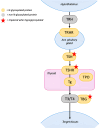
N-glycosylation in the Hypothalamus-Pituitary-Thyroid axis. TRH stimulates the pituitary gland to release TSH, which binds to TSHR in the thyroid. Hypoglycosylation of TSH and TSHR impairs binding and signal transduction. The thyroid produces T3 and T4 from Tg, this process requires multiple enzymes, including TPO. Impact of hypoglycosylation on Tg and TPO is unknown. Once released in the circulation T3 and T4 bind to TBG to reach target tissues. Hypoglycosylation of TBG reduces its half-life but doesn’t seem to affect its ability to bind thyroid hormones. Red stars indicate proteins whose function is impaired when hypoglycosylated. [TRH, thyrotropin releasing hormone; TSH, thyrotropin; TSHR, thyrotropin receptor; TBG, thyroxine-binding globulin; Tg, thyroglobulin; TPO, thyroperoxidase; T4, thyroxine; T3, triiodothyronine].
Figure 4
Figure 4
N-glycosylation in the Insulin axis. In presence of euglycemia, excess of insulin release is prevented by beta-cell depolarization, which is obtained through the ATP-dependent potassium-channels (KATP). KATP is a complex which includes SUR1 and Kir6.2. Hypoglycosylation of SUR1 could impair KATP’s function leading to excessive insulin release and hypoglycemia. On target cells insulin binds to Insulin R. The impact of hypoglycosylation of this receptor is still unknown. [SUR1, sulfonylurea 1 receptor; Insulin R, insulin receptor].
Figure 5
Figure 5
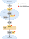
N-glycosylation in the Hypothalamus-Pituitary-Adrenal axis. CRH activates CRHR1 on the pituitary gland to stimulate ACTH’s production and secretion. Hypoglycosylation of CRHR1 impairs binding to CRH and signal transduction. In the pituitary, POMC is cleaved by PC1/3 to release BLPH and ACTH. PC1/3 activity could be impaired by hypoglycosylation, leading to decreased ACTH secretion. ACTH stimulates cortisol’s secretion on the adrenal gland by binding MC2R and the activation of the receptor depends on its glycosylation state. In the circulation, cortisol binds to CBG. Dysfunction of CBG due to hypoglycosylation decreases circulating cortisol levels. Red stars indicate proteins whose function is impaired when hypoglycosylated. [CRH, corticotropin-releasing hormone; CRHR1, corticotropin-releasing hormone receptor 1; PC1/3, prohormone convertase 1/3; POMC, proopiomelanocortin; beta-lipotropic hormone; ACTH, adrenocorticotropin hormone; MC2R, melanocortin 2 receptor; CBG, corticosteroid-binding globulin].
Figure 6
Figure 6
N-glycosylation in lipid metabolism. When intracellular concentration of sterols decreases, SREBP2 is released from the INSIG1-SCAP complex and, after proteolytic activation, translocates into the nucleus where it induces the transcription of genes involved in cholesterol synthesis, including the LDLR gene. LDLR removes apoB-containing lipoproteins from circulation until the intracellular concentration of sterols is restored. Hypoglycosylation of INSIG1 could impair its binding to SREBP2, leading to an upregulation of LDLR and increased clearance of apoB-containing lipoproteins. [INSIG1, insulin-induced gene 1; SCAP, SREBP cleavage-activating protein; SREBP2, sterol regulatory element-binding protein 2; LDL, low-density lipoprotein; LDLR, low-density lipoprotein cholesterol receptor].

Similar articles

References

    1. Altassan R, Péanne R, Jaeken J, Barone R, Bidet M, Borgel D, et al. International clinical guidelines for the management of phosphomannomutase 2-congenital disorders of glycosylation: Diagnosis, treatment and follow up. J Inherit Metab Dis. (2019) 42:5–28. doi: 10.1002/JIMD.12024, PMID: - DOI - PubMed
    1. Tian Y, Zhang H. Characterization of disease-associated N-linked glycoproteins. Proteomics. (2013) 13:10. doi: 10.1002/PMIC.201200333, PMID: - DOI - PMC - PubMed
    1. Vaes L, Rymen D, Cassiman D, Ligezka A, Vanhoutvin N, Quelhas D, et al. Genotype-phenotype correlations in pmm2-cdg. Genes (Basel). (2021) 12. doi: 10.3390/GENES12111658, PMID: - DOI - PMC - PubMed
    1. Péanne R, de Lonlay P, Foulquier F, Kornak U, Lefeber DJ, Morava E, et al. Congenital disorders of glycosylation (CDG): Quo vadis? Eur J Med Genet. (2018) 61:643–63. doi: 10.1016/J.EJMG.2017.10.012, PMID: - DOI - PubMed
    1. Muthusamy K, Perez-Ortiz JM, Ligezka AN, Altassan R, Johnsen C, Schultz MJ, et al. Neurological manifestations in PMM2-congenital disorders of glycosylation (PMM2-CDG): Insights into clinico-radiological characteristics, recommendations for follow-up, and future directions. Genet Med. (2024) 26. doi: 10.1016/j.gim.2023.101027, PMID: - DOI - PubMed

MeSH terms

Substances